

Introduction
Paramedics are often required to perform endotracheal intubation in challenging conditions. They must overcome difficulty with airway visualization due to vomitus, blood and secretions, suboptimal patient positioning, and other challenges associated with the prehospital environment. These conditions may increase the risk of endotracheal tube (ETT) misplacement or dislodgment.Reference Katz and Falk 1 - Reference Zadel, Strnad, Prosen and Mekis 3 Previous studies have identified rates of misplaced or dislodged prehospital placed ETTs, ranging from 4.8%-25.0%.Reference Katz and Falk 1
All modalities to verify ETT location have limitations. According to the 2010 Advanced Cardiovascular Life Support (ACLS) guidelines, quantitative continuous waveform capnography is currently recommended as the gold standard to confirm correct ETT placement.Reference Neumar, Otto and Link 2 Capnography, however, has its limitations in the setting of cardiac arrest, low pulmonary blood flow, and requires ventilation. The 2015 ACLS guidelines update has recommended ultrasound (US) as an additional method to confirm ETT placement.Reference Link, Berkow and Kudenchuk 4 However, there is lack of research that evaluates prehospital providers’ ability to use US to verify tube placement.
Confirmation of correct ETT placement with US has been shown to be superior to auscultation and as sensitive as capnography in controlled settings.Reference Chou, Tseng and Wang 5 - Reference Drescher, Conard and Schamban 7 Real-time US guided ETT placement during intubation can aid in the identification of proper tube placement with greater sensitivity and specificity than post-intubation images alone.Reference Chou, Tseng and Wang 5 , Reference Drescher, Conard and Schamban 7 - Reference Werner, Smith, Goldstein, Jones and Cydulka 13 Previous studies have demonstrated high sensitivity and specificity with US verification of ETT location when performed by Emergency Medicine physicians and anesthesiologists in cadaver models, in the operating room (OR) and in acutely ill patients in the emergency department (ED) and intensive care unit.Reference Chou, Tseng and Wang 5 , Reference Milling, Jones and Khan 6 , Reference Chou, Dickman and Tsou 8 , Reference Hoffmann, Gullett and Hill 11 , Reference Werner, Smith, Goldstein, Jones and Cydulka 13 - Reference Ma, Davis, Schmitt, Vilke, Chan and Hayden 18
To the best of the authors’ knowledge, there are no known studies that investigate paramedic use of trans-tracheal US to verify ETT placement in the prehospital environment. This study evaluates paramedics’ ability to verify ETT location with US, and particularly, identify esophageal intubations.
Methods
Study Design and Setting
This was a prospective, observational study to assess paramedic ability to confirm correct ETT placement and identify inadvertent esophageal intubations with US. The study took place at the University at Buffalo (Buffalo, New York USA) anatomy lab. The University’s Health Sciences Institutional Review Board approved this study. All subjects provided written informed consent prior to participation in the study. The use of cadavers was permitted under their unrestricted donation to the University’s Anatomical Gift Program.
Participants
Fifty-eight paramedics with current New York State certification volunteered to participate in the study. One paramedic was excluded due to the inability to complete the study. Fifty-seven paramedics were ultimately enrolled. Subjects were not compensated for their involvement. Participants completed a survey of their paramedic intubation experience over the previous 12 months, employment history, and previous exposure to US training during study enrollment.
Cadaver Model
Twenty-five recently deceased, un-embalmed cadavers from the University Anatomical Gift Program were utilized for the study. Cadavers were removed from refrigerators the morning of each study session. Study investigators manually stretched cadaver jaws and necks to relax the musculature and break the rigor.Reference Perper 19 The cadaver oropharynx was cleared of debris and fluid with a portable suction device prior to the study. Cadavers with an obvious neck mass, previous surgery, or trauma were not used in the study.
Twenty-five cadavers were used. One training cadaver and four study cadavers were allocated for each study session. Some cadavers were re-used during consecutively scheduled study sessions.
Study Sessions
Five cadavers were contributed for each study session by the anatomy lab staff according to cadaver availability. Four cadavers were assigned for the study and one cadaver set aside for US training and a skills session. The designated training cadaver was not used for study data to prevent introduction of any bias from study participants becoming familiar with the cadaver’s airway during data collection.
Ultrasound Training and Skills Session
All subjects were exposed to a 15-minute lecture and practical US training session provided by the same investigator (PCL), an Emergency Ultrasound Fellowship trained emergency physician. The didactic lecture described the US technique, US images, and artifacts of normal airway, trachea, and esophagus with and without tubes. A separate training cadaver, not incorporated in the study data collection, was used for the practical US session.
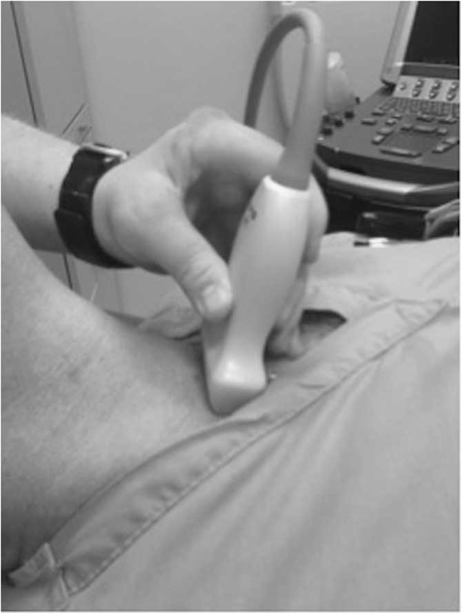
Study investigators placed a 7.0 cuffed ETT (Cardinal Health; Waukegan, Illinois USA) in the cadavers. Endotracheal tubes were placed by direct laryngoscopy in the trachea distal to the vocal cords. Esophageal tubes were placed by direct laryngoscopy in the esophagus. The US machine (Sonosite; Bothell, Washington USA) was placed to the left of the cadaver and subject. A 10MHz linear US transducer was placed transversely on the cadaveric neck, just proximal to the suprasternal notch, with the transducer marker pointed to the subject’s left side (Figure 1). All study participants performed an US examination on the training cadaver’s neck. Subjects identified a tracheal intubation and an esophageal intubation with US. All subjects were required to identify the trachea, esophagus, and tube location with US for each encounter.
Linear Ultrasound Transducer Placed Transversely on the Anterior Neck, Proximal to the Suprasternal Notch. Note: The ultrasound machine and transducer marker are directed to the sonographer’s left side.

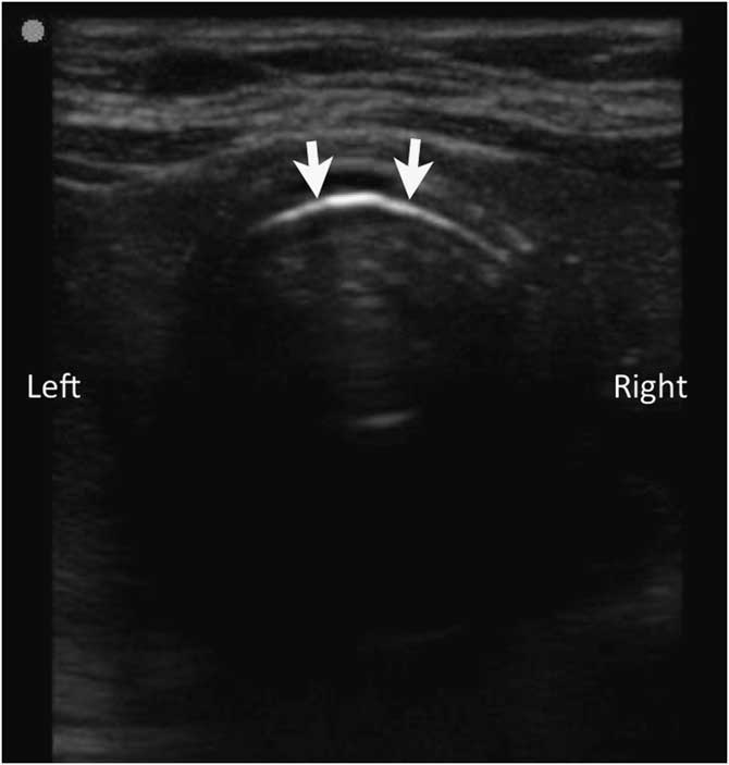
The trachea was identified with US as a hyperechoic line in the anterior neck. Reverberation artifact was noted posteriorly (Figure 2).Reference Chou, Tseng and Wang 5 , Reference Hoffmann, Gullett and Hill 11 , Reference Chou, Chong and Sim 16 An esophagus was identified by an anechoic area or by a multi-layered, elliptical structure to the left of the trachea.Reference Chou, Tseng and Wang 5 , Reference Hoffmann, Gullett and Hill 11 , Reference Chou, Chong and Sim 16 An endotracheal intubation was identified as a hyperechoic double line within the trachea with one air-mucosa interface and an empty esophagus (Figure 3).Reference Chou, Tseng and Wang 5 , Reference Hoffmann, Gullett and Hill 11 An inadvertent esophageal intubation was identified by the presence of the “double trachea” or “double tract” sign and two air-mucosa interfaces: one in the esophagus and one in the trachea (Figure 4).Reference Chou, Tseng and Wang 5 , Reference Hoffmann, Gullett and Hill 11 , Reference Chou, Chong and Sim 16 Subjects were able to manipulate the tube with a gentle twisting motion in a clockwise/counter-clockwise rotation after US identification to further verify tube location within the trachea or esophagus.
Ultrasound Image of the Normal Trachea without Endotracheal Tube. Note: The hyperechoic line is the air-mucosa interface (arrows).

The “Double Trachea” or “Double Tract” Sign. Note: Ultrasound image of the trachea (arrows) with endotracheal tube (arrowheads). One air-mucosa (A-M) interface is present. Abbreviation: ETT, endotracheal tube.

Ultrasound Image of an Inadvertent Esophageal Intubation to the Left of the Trachea. Note: Two air-mucosa (A-M) interfaces are present.

Ultrasound Identification of Pre-Placed Tubes
Study investigators randomly intubated the four study cadavers by direct laryngoscopy and US guidance into the trachea or esophagus. The cadaver’s face, including the tube, was covered with a cloth to prevent any additional information of tube location. Paramedics were blinded during tube placement by investigators and from other paramedic study sessions. Paramedics diagnosed whether a tracheal or esophageal intubation had occurred using US. Time to diagnosis was recorded. Time began when the transducer was placed on the cadaver neck and ended once the subject had a verbal diagnosis of tube location. Paramedic confidence level, on a Likert scale of one to ten (one=not confident; ten=extremely confident), regarding US diagnosis of tube placement was ascertained. The subject was then able to manipulate the tube under US guidance to further verify tube location. Paramedic diagnosis and confidence level post-tube manipulation was recorded.
Statistical Methods
The ETT placement success rates were calculated by dividing the number of correctly placed tubes by the total number of attempts. T-tests were used to compare mean diagnosis times for esophageal and tracheal tubes. Logistic regression was used to assess the effects of paramedic employment history, intubation experience over the previous 12 months, and previous exposure to US training on US identification of tube placement. All analyses were performed using SAS version 9.4 (SAS Institute; Cary, North Carolina USA).
Results
Twelve 2.5-hour study sessions were conducted over five days from March 2014 through December 2015. Fifty-eight paramedics were enrolled in the study. One paramedic was excluded due to incomplete data collection. Forty-eight subjects were male (84.0%). Mean age was 39 years (range: 22-62 years). Median experience as a paramedic was seven years (range: seven months-39 years). Two subjects self-reported no intubation experience over the past 12 months. Fifty subjects self-reported one to 25 intubations. Five subjects self-reported 26-50 intubations.
Ultrasound Identification of Tube Location
A total of 228 intubations occurred. Study investigators randomly placed 113 tubes in the trachea and 115 tubes in the esophagus with direct laryngoscopy and US guidance. Two study investigators verified tube location prior to paramedic assessment. Paramedics correctly identified tube location with US in 158 cases (69.3%). Paramedics correctly identified 98 of 113 tracheal tubes and 60 of 115 esophageal tubes with US. The sensitivity and specificity of identifying esophageal tube location with US was 52.2% and 86.7%, respectively. Mean time to diagnose tube location with US was 40.1 (SD=27.2) seconds. Mean time to diagnose esophageal intubations was 35.4 seconds compared to the mean time to diagnose tracheal intubations at 44.9 seconds. Esophageal intubations were diagnosed 9.47 seconds faster than tracheal intubations (P=.0083). Further manipulation of the tube with US resulted in paramedics correctly identifying tube location in 194 (85.1%) cases. Paramedics correctly identified 94 of 113 tracheal tubes and 100 of 115 esophageal tubes with a sensitivity and specificity of 87.0% and 83.2%, respectively. After tube manipulation with US, paramedics correctly identified 41 previously incorrectly identified esophageal intubations. One paramedic incorrectly identified one previously correctly identified esophageal intubation. Tube manipulation with US significantly improved sensitivity (P<.0001) from 52.2% (95% confidence interval [CI], 43.0-61.0) to 87.0% (95% CI, 81.0-93.0). Specificity decreased from 86.7% (95% CI, 81.0-0.9) to 83.2% (95% CI, 0.8-0.9). However, this decrease was not significant (P=.45). The positive predictive value (PPV) of the identification of esophageal intubations with US was 56.7%. The PPV with tube manipulation and US was 63.3% (P=.36). The negative predictive value (NPV) of the identification of esophageal intubations with US was 95.1%. The NPV with tube manipulation and US was 93.7% (P=.61). Years of paramedic experience, previous number of intubations, and previous US course exposure did not correlate with ability to identify tube location with US.
Discussion
Continuous waveform capnography is currently the gold standard for confirmation of ETT placement.Reference Neumar, Otto and Link 2 This modality is often unavailable in the prehospital setting and reliability is variable in low perfusion states of cardiac arrest. Ultrasonography has become an extension of tools available to confirm ETT placement according to changes in the 2015 ACLS guidelines.Reference Link, Berkow and Kudenchuk 4 It is a natural progression that this tool be extended to the prehospital setting.
Several studies demonstrate the ability of prehospital personnel to obtain and interpret basic US studies. Heegard, et al describe a 6-hour point-of-care US (POCUS) orientation (three hours of didactics, three hours of hands-on practice) to train paramedics to perform focused assessment with sonography for trauma (FAST) and abdominal aorta exams.Reference Heegaard, Hildebrandt, Spear, Chason, Nelson and Ho 20 Similarly, a systematic review by McCallum, et al concluded that paramedics can effectively learn the FAST exam and interpretation in six to eight hours and can detect pleural effusions with as little as 25 minutes of training.Reference McCallum, Vu, Sweet and Kanji 21
There have been many small- to medium-sized studies that have evaluated US detection of ETT placement with Emergency Medicine physician sonographers. Despite the breadth of studies, there is not a consensus regarding visualizing ETT placement dynamically during intubation versus visualization post-intubation. The systematic review performed by Chou, et al demonstrates no significant difference between both static and dynamic ETT confirmation.Reference Chou, Tseng and Wang 5 , Reference Chou, Chong and Sim 16 Tessaro, et al found a high sensitivity for ETT placement when temporarily adding saline to the ETT cuff to increase visualization.Reference Tessaro, Salant, Arroyo, Haines and Dickman 22 This study is the first of its kind to evaluate static images followed by ETT tube manipulation via gentle twisting motion of the ETT tube. As opposed to US ETT studies in the ED and the OR, there are very few studies performed in the prehospital setting using US to confirm ETT placement. There is one study using prehospital patients to confirm ETT placement; however, this study utilizes an emergency physician sonographer rather than paramedics.Reference Zadel, Strnad, Prosen and Mekis 3 This study is the first of its kind to utilize paramedic novice sonographers.
Limitations
Limitations of this study include those that are inherent in cadaver studies. Anatomic variants produced more challenging models, including one cadaver with previous neck surgery. Molding of the airway or creation of false passages by repeat intubations and further introduction of air into static tissue may have limited the ability for correct identification of the ETT with US. These limitations were mitigated with a large pool of cadaver models to reduce increased manipulation. While this study’s sensitivity of 87.0% and specificity of 83.2% is lower than previously performed studies, this is likely reflected by the use of a large pool of paramedic novice sonographers. Sensitivity and specificity may improve with additional sonographic training.
Previous studies using the “double trachea” or “double tract” signs have shown 100.0 % sensitivity and 85.7%-91.0 % specificity for esophageal intubation. The “double trachea” or “double tract” signs had a PPV of 91.0%-98.8 % and NPV of 99.0%-100.0% for the correct identification of esophageal intubation. The PPV for an empty esophagus detected for tracheal intubation was 100.0 %.Reference Chou, Tseng and Wang 5 , Reference Hoffmann, Gullett and Hill 11 , Reference Chou, Chong and Sim 16
One case report of prehospital use of lung US to verify ETT placement and one study of emergency physician use of US to verify ETT in the prehospital environment were encountered.Reference Zadel, Strnad, Prosen and Mekis 3 , Reference Brun, Bessereau, Cazes, Querellou and Chenaitia 23
Conclusion
Paramedics can readily identify esophageal intubations with US. Further gentle manipulation of the tube in a twisting motion with US guidance improves the accuracy of tube location.
Acknowledgements
The authors gratefully acknowledge Kayla Graves; Kaori Tanaka, DO; Johanna Innes, MD; and Christopher Tanski, MD for assistance with the anatomy lab setup and research sessions. Author appreciation to Cheryl Sanchez for her assistance with the IRB. Author gratitude to Ray Dannenhoffer, PhD and the University at Buffalo Anatomical Gift Program for their assistance with this study.