Introduction
Phosphatidylcholine (PC) is the most abundant phospholipid in mammalian cells and plays an important structural role in cellular membranes (Vance & Vance, Reference Vance and Vance2004; Jackowski & Fagone, Reference Jackowski and Fagone2005). The CDP-choline pathway (Kennedy pathway) is the principal route for PC biosynthesis, involving three steps. Choline is an essential nutrient in mammals: it is transported into the cell and phosphorylated by choline kinase (CK). CTP:phosphocholine cytidylyltransferase (CCT) catalyses the condensation of CTP and phosphocholine to form CDP-choline, which in turn donates the phosphocholine moiety to diacylglycerol to form PC in a reaction catalysed by CDP-choline:diacylglycerol cholinephosphotransferase (Vance & Vance, Reference Vance and Vance2004). CTP:phosphocholine cytidylyltransferase mediates the rate-limiting step in this pathway (Vance & Vance, Reference Vance and Vance2004; Jackowski & Fagone, Reference Jackowski and Fagone2005) and the regulation of its enzymatic activity is characterized by its association with membrane structures (Vance & Vance, Reference Vance and Vance2004; Jackowski & Fagone, Reference Jackowski and Fagone2005). A current model suggests that the bilayer curvature elastic stress is sensed by CCT and governs the degree of membrane association, thus providing a mechanism for both positive and negative regulation of activity (Attard et al., Reference Attard, Templer, Smith, Hunt and Jackowski2000; Davies et al., Reference Davies, Epand, Kraayenhof and Cornell2001).
Immediately following fertilization, vertebrate eggs undergo a series of divisions, pushing the embryo into a coordinated new plasma membrane biosynthesis. In general, in this first period, the embryo does not become larger and the cells become smaller with each division. Therefore, the synthesis and integrity of cellular membranes is likely to be an important controlling factor during embryogenesis of vertebrate animals (Gilbert, Reference Gilbert2000). In particular, it has been demonstrated in mouse that preimplantation embryos are capable of synthesizing phospholipids de novo and are equipped to assemble new intracellular membranes and plasma membrane (Pratt, Reference Pratt1980). Also, the specific role of Chka and pCyt1a has been evaluated by generating knock-out (KO) models. Chka −/− mice were recovered at the blastocyst stage, before embryonic day 7.5 (Wu et al., Reference Wu, Aoyama, Young and Vance2008), and Pcyt1a −/− mice failed to form blastocysts, underscoring the importance of PC and the Kennedy pathway during early development (Wang et al., Reference Wang, Magdaleno, Tabas and Jackowski2005). Wang et al. (Reference Wang, Magdaleno, Tabas and Jackowski2005) suggested that the ability of the KO preimplantation embryos to progress through the first few divisions is probably due to the expression of maternal CCT, which is present at high levels in mature eggs (Jackowski et al., Reference Jackowski, Rehg, Zhang, Wang, Miller, Jackson and Karim2004).
The characteristics of the mouse model complicate the analysis of enzyme levels during early development and related experiments remain to be done. Amphibians are the most eligible systems for these approaches due to advantageous external fertilization and development, abundant gametes and embryos for biochemical studies, ease of manipulation and identification of their developmental stages. Amphibians, like other vertebrates with external fertilization, contain yolk platelets. In particular, it has been found that in the amphibian Bufo arenarum about 80% of the total embryonic phospholipids are in the yolk platelet fraction, and PC and phosphatidylethanolamine (PE) are the major phospholipids in these structures. Moreover, 32P-labelling experiments have demonstrated that immediately after fertilization there is an active phospholipid metabolism (PC and PE synthesis), which continues at least until late gastrula (Alonso et al., Reference Alonso, Bonini de Romanelli and Bazan1982a).
The current work shows that CCT and CK are expressed abundantly in unfertilized eggs. The levels of these enzymes decline as development progresses and reach a minimum around the gastrula stage, after which they start to recover. Interestingly, the lower levels of expression mainly overlap with the mid-blastula transition (MBT). The cytosolic:total CCT protein ratio is constant during all the developmental stages analysed, indicating that the activity of CCT diminishes throughout development until the gastrula stage. However, the mass of PC and PE per egg/embryo is constant, suggesting that in B. arenarum eggs the net synthesis of phospholipids appears not to be essential during the first developmental stages, thanks to yolk lipid reservoirs present in the unfertilized eggs. To our knowledge, this study is the first time that CCT and CK protein levels have been studied during early development in vertebrates.
Materials and methods
Animals, gametes and embryos
Sexually mature B. arenarum specimens were collected in the neighbourhoods of Rosario city and kept in a moist dark chamber at 12°C until used. All experiments were performed in accordance with the Guide for the Care and Use of Laboratory Animals promulgated by the National Institute of Health (National Center for Research Resources), Bethesda, MD, USA).
Testes were dissected from male toads, and spermatozoa were obtained by mincing the organs in Ringer–Tris solution (0.11 M NaCl, 2 mM KCl, 1.4 mM CaCl2, 10 mM Tris, pH 7.2) at 4°C. The homogenate was filtered through gauze and centrifuged for 10 min at 130 g at 4°C to remove blood cells and tissue debris. The sperm suspension was centrifuged for 10 min at 650 g at 4°C. Pelleted spermatozoa were resuspended in Ringer–Tris solution.
Female specimens were kept in a moist chamber at 20–22°C for 1 day before stimulation with one homologous hypophysis homogenate injected intracoelomically. After 10–12 h, strings of eggs were ovulated or collected from ovisacs.
Just before fertilization, eggs were placed into 10 ml of 10% Ringer in a Petri dish, then 100 μl of the minced testes solution (~10 million sperm) was added to the unfertilized eggs, incubated for 30 min and finally washed with Ringer. These conditions typically result in 100% fertilization success rate.
Bufo arenarum embryo stages
Embryos were staged according to Del Conte & Sirlin (Reference Del Conte and Sirlin1952). At each stage studied, the removal of jelly coats was accomplished by immersion of egg/embryo strings in 1% sodium thioglycolate (pH 8) solution. Dejellied eggs/embryos were thoroughly rinsed with Ringer solution, frozen in liquid nitrogen and kept at –70°C.
Total membranes and cytosol isolation
Sub-cellular fractionation was performed according to the procedure described by Sato et al. (Reference Sato, Iwao, Fujimura, Tamaki, Ogawa, Iwasaki, Tokmakov, Hatano and Fukami1999) with modifications. All procedures were carried out at 4°C. Approximately 200 dejellied eggs/embryos were mixed with 2 ml of buffer A (20 mM Tris–HCl (pH 7.5), 1 mM EDTA, 1 mM EGTA, 1 mM Na3VO4) supplemented with 1% protein inhibitor cocktail (Sigma) and 2 mM PMSF and homogenized in a Teflon-glass potter. The homogenate was centrifuged 10 min at 1000 g (SS34 Rotor, Sorvall Instruments) to remove cellular debris and yolk platelets. The supernatant (total extract) was collected and centrifuged at 150,000 g for 1 h (Ti80 rotor, Beckman L-80 ultracentrifuge). After centrifugation, the clear supernatant was set aside as the cytosolic fraction. The pellet and fluffy part of the pellet were then carefully taken, resuspended with buffer A and centrifuged at 150,000 g for 30 min. The pellet fraction obtained was resuspended in 250 μl of buffer A plus 1% Triton X-100 and sonicated (total membrane fraction). Through this procedure, the efficiency was 7 ± 0.4 μg of cytosolic protein/embryo and 3.8 ± 0.4 μg of membrane protein/embryo. The total amount of protein per egg/embryo was 148.8 ± 5.8 μg. This amount and the yield of cytosolic and total membrane proteins were independent of the developmental stage.
Protein assays
Protein concentrations were determined according to Sedmak & Grossberg (Reference Sedmak and Grossberg1977), using bovine serum albumin as standard.
Electrophoresis, electrotransfer and western blot analysis
Sodium dodecyl sulphate polyacrylamide gel electrophoresis (SDS-PAGE) under denaturing conditions was performed essentially according to the method of Laemmli (Reference Laemmli1970). Samples were diluted with an appropriate volume of 6× sample buffer with 0.1 M dithiothreitol, boiled for 5 min, and loaded onto 12% acrylamide mini-gels with 5% stacking gel, and electrophoresed at 20 mA/gel (MiniProtean III Gel System, BioRad, Hercules, CA, USA). The apparent molecular weights were estimated with molecular mass standards (Amersham Pharmacia Biotech, Piscataway, NJ, USA). Gels were processed for Coomassie brilliant blue staining or electrotransferred to nitrocellulose membranes according to Towbin's method (Towbin et al., Reference Towbin, Staehelin and Gordon1979).
Membranes were blocked with TBS buffer supplemented with 3% w/v BSA and 0.1% v/v Tween 20. Membranes were then washed and incubated with a 1:500 dilution of the primary antibody for 1 h (anti-CCT rabbit polyclonal, kindly provided by Dr Suzanne Jackowski, Department of Infectious Diseases, St Jude Children's Research Hospital, Memphis, USA or anti-CK rabbit polyclonal (ChoK (H-210) Santa Cruz Biotechnology Inc., Dallas, TX, USA). After washing, membranes were incubated with the appropriate HRP-conjugated antibody, washed and developed using chemiluminescence and X-ray films. When membrane re-probing was necessary, 0.2 M NaOH treatment was used for stripping. Actin detection on membranes was performed to ensure equal loading. The antibody used was a rabbit polyclonal at a 1:600 dilution (sc-1616, Santa Cruz Biotechnology, Inc., Dallas, TX, USA) and the manufacturer's instructions were followed.
Exposed films were digitized and quantified using Gel-Pro Analyser 3.0 (Media Cybernetics). The integrated optical densities (IOD) corresponding to CK and CCT were normalized to their respective actin IODs. In the studies dealing with CCT sub-cellular distribution, the normalized CCT values were also normalized to the total protein content of the respective fraction (cytosol or total extracts) and then the ratio of cytosol: total extracts was calculated.
Lipid extraction and analysis
Lipids were extracted by the method of Bligh & Dyer (Iverson et al., Reference Iverson, Lang and Cooper2001), modified by us. Briefly, 25 eggs/embryos were homogenized in 1 ml of water using a Teflon potter. This homogenate was transferred to a glass tube and 5 ml of a solution 2:1 of methanol/chloroform was added. The tube was incubated overnight at –20°C. After centrifugation (5 min at 2800 rpm, SS34 rotor, Sorvall Instruments), a 1:1 chloroform/water solution was added to the supernatant and then vortexed. Following a second centrifugation step, the organic phase was collected, washed twice with 2 M KCl, then twice with water and finally dried under nitrogen. Afterwards, lipid samples were resuspended in 50 μl of chloroform and analysed by two-dimensional thin layer chromatography of organic phase on Silica Gel H layers (pore size 60 Å, Sigma Chemical Company, St Louis, USA) developed in chloroform/methanol/water (75:30:1.5, v/v) and chloroform/methanol/acetic acid/water (80:9:12:2, v/v). Phospholipids were revealed by staining with a CuSO4/H3PO4 solution (10% w/v CuSO4, 8% w/v H3PO4) (Christie, Reference Christie2003). Phosphatidylcholine and PE were identified by co-migrating lipid standards, quantified using ImagePro3 software (Media Cybernetics) and normalized to the number of eggs/embryos used.
Statistical analysis
Results are expressed as mean ± standard error of the mean (SEM). Data were analysed by parametric test, using Student's t-test and analysis of variance (ANOVA). Significance was declared at P-values ≤0.05.
mRNA expression analysis
RNA was isolated from B. arenarum unfertilized eggs using the TRIzol® Reagent (Invitrogen, Carlsbad, CA, USA) following the manufacturer's instructions. Purified RNA was incubated with RQ1 DNase (Promega Corporation, Madison, WI, USA). Total RNA was retro-transcribed with SuperScript II enzyme (Invitrogen, Carlsbad, CA, USA) and oligo(dT)16–18 primer. Polymerase chain reaction (PCR) amplification was carried out using the following primers with sequences: CTTforw: 5′-AGGTGGAAGAGAAGAGCATCG-3′, CTTrev: 5′-GGAAGTCTTGCCAGAGAAGG-3′. The amplification reaction consisted of 2 μl of the cDNA template, 1.25 U GoTaq® DNA Polymerase (Promega Corporation, Madison, WI, USA), 1.5 mM MgCl2 (total reaction volume = 50 μl). The reaction mix was subjected to 35 cycles of amplification (95°C for 1 min, 52°C for 30 s, 72°C for 30 s), followed a final extension step of 10 min at 72°C. Products were resolved in 1.2% (w/v) agarose gels stained with Gel Green, extracted from them and finally cloned into pGEM-T Easy Vector (Promega Corporation, Madison, WI, USA). Sequencing was performed by the University of Maine DNA Sequencing Facility. Sequence identity was confirmed using the basic local alignment search tool (BLAST) supported by the National Center for Biotechnology Information. Multiple sequence alignments were carried out using the CLUSTALW (1.82) algorithm (Higgins et al., Reference Higgins, Thompson and Gibson1996).
Results
CCT expression during development
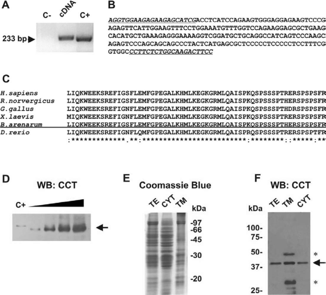
By performing reverse transcription (RT)-PCR using CCT-specific primers and RNA obtained from B. arenarum unfertilized eggs, a band of the expected size was detected (233 base pairs (bp)) (Fig. 1A). The PCR band was excised, cloned and sequenced as described in Materials and methods. The sequence of the amplified fragment is shown in Fig. 1B. The analysis of this sequence fragment revealed a high identity with CCT mRNA sequences of several vertebrates (data not shown) and the amino acidic sequence comparison from different species shows a high degree of identity (Fig. 1C). This high identity score can be explained considering that the amplified region encoded the CCT membrane binding domain (Kent, Reference Kent1997; Cornell & Taneva, Reference Cornell and Taneva2006). In order to analyse CCTα expression in B. arenarum eggs, an antiserum raised against the rat CCTα was used. Figure 1D shows that the antiserum recognized a unique band in egg cytosolic samples that co-migrated with the positive control (rat liver extract). There was a slight difference in mobility that could be due to differences in primary sequence or different post-translational modifications (e.g. phosphorylation) (Arnold et al., Reference Arnold, DePaoli-Roach and Cornell1997). CTP:phosphocholine cytidylyltransferase is an amphitropic enzyme that binds to cellular membranes (Cornell & Northwood, Reference Cornell and Northwood2000). The presence of the enzyme in total extracts, cytosol, and total membrane fraction was evaluated by SDS-PAGE and western blots using the anti-CCT antibody (Fig. 1F). In all the tested samples, a band immunoreactive to the antibody was found. It is important to note that cytosolic samples rendered cleaner blots than total membrane samples and so they were preferentially selected in the experiments that dealt with the estimation of CCT activity using membrane association as an indicator (see below).
Cytidylyltransferase (CCT) expression by B. arenarum eggs. (A) Detection of CCT expression by reverse transcription polymerase chain reaction (RT-PCR). C–: negative control (skipped reverse transcription); cDNA: egg cDNA; C+: PCR performed using a plasmid harbouring rat CCT sequence. (B) Nucleotide sequence of the CCT fragment amplified using cDNA obtained from B. arenarum unfertilized eggs. (C) Amino acid sequence alignment of CCT from different species. Asterisks indicate amino acid identity. (D) Increasing protein amounts of cytosolic samples from B. arenarum eggs were electrophoresed, western blotted (WB) and revealed with anti-CCT antibody (3–12 μg of protein). C+: rat liver extract as positive control (10 μg of protein). The arrow indicates the band corresponding to CCT (42 kDa). (E) Coomassie blue-stained gel of total extracts (TE), cytosol (CYT) and total membranes (TM) obtained from eggs. Each lane was loaded with 10 μg of protein. Molecular standards are indicated to the right of the panel. (F) A similar gel (20 μg of protein) was blotted and revealed with anti-CCT antibody. The arrow indicates the band corresponding to CCT and the asterisks show non-specific bands in the TM fraction.

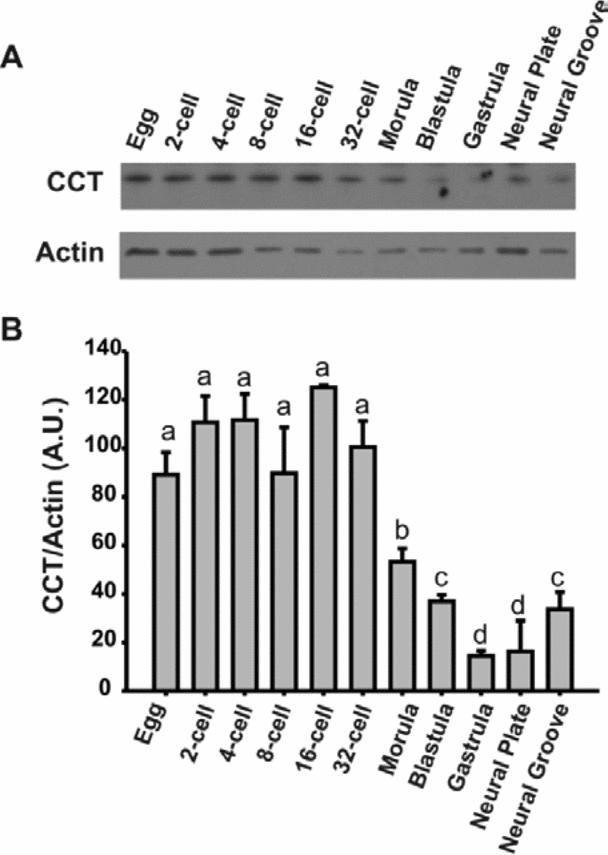
CTP:phosphocholine cytidylyltransferase expression during different developmental stages (unfertilized egg to neurula) was analysed by western blot (Fig. 2A). The levels of CCT were quantified using actin as its corresponding loading control. CTP:phosphocholine cytidylyltransferase protein levels remained high and constant until the morula stage, at which they showed a statistically significant decrease reaching a minimum at the gastrula stage, and after which they started a slow recovery (Fig. 2B). This observed decrease in the gastrula stage followed by a slow recovery coincides with events characteristic of MBT, such as the beginning of zygotic gene transcription (Newport & Kirschner, Reference Newport and Kirschner1982).
Cytidylyltransferase (CCT) protein expression during development. (A) Representative CCT western blot of total extracts from egg and embryos at different stages (10 μg protein per lane). Actin detection was performed to ensure equal loading. (B) Densitometric analysis (CCT/actin) of three independent experiments similar to the one in (A). A.U.: arbitrary units.
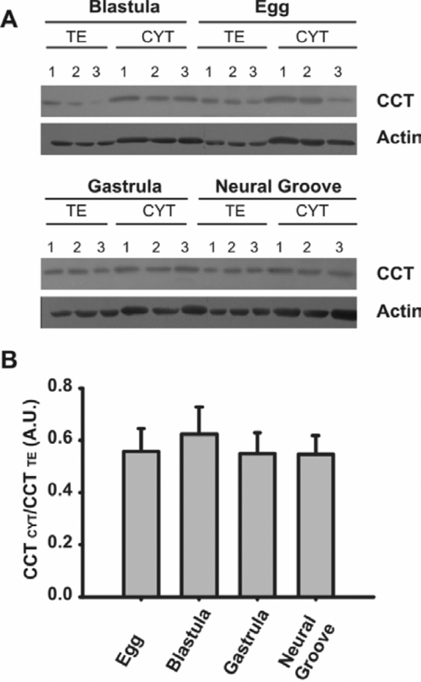
CTP:phosphocholine cytidylyltransferase activity can be regulated by membrane association; a soluble homodimer is an inactive reservoir that is activated after binding to the lipid bilayer (Vance & Vance, Reference Vance and Vance2004). As membrane biosynthesis is essential during development and CCT protein levels decrease in the morula/gastrula stage (Figure 2), it was hypothesized that CCT could be regulated by its translocation to the membrane in order to increase its activity for supplying PC adequately. To test this hypothesis, CCT levels were measured in cytosol samples and in total extracts and the cytosol:total CCT ratio calculated in selected stages (egg, blastula, gastrula and neurula). Cytosolic and total extract samples of staged embryos (n = 3) were run together to ease quantification (Fig. 3A). Figure 3B shows the result of densitometry and calculations of the ratios (see Materials and methods). It was observed that in all the stages analysed, approximately 50% of CCT is cytosolic and there is no statistically significant difference among them. Considering that total CCT diminished in blastula, gastrula and early neurula (Fig. 2), and the ratio of cytosolic: total CCT protein does not change, it can be assumed that the membrane-associated (active) form of CCT per embryo is lower in these stages compared with eggs and embryos in early cleavage. However, it should be remembered that, according to Alonso et al. (Reference Alonso, Bonini de Romanelli and Bazan1982a), there is PC biosynthesis during the first stages of B. arenarum development, which continues until at least late gastrula.
Cytosolic versus total extract cytidylyltransferase (CCT) protein in staged embryos. (A) CCT detected in egg and embryonic total extracts (TE) and cytosol (CYT) samples (from three independent fertilization events) by western blot (egg, blastula, gastrula and neural groove, 10 μg TE protein and 15 μg CYT protein per lane). Actin detection was performed to ensure equal loading. (B) Densitometry analysis of the blot shown in (A). A.U.: arbitrary units.
CK expression during development
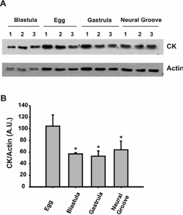
Mouse embryos homozygous for an insertional mutation in Chka allele were only recovered at the blastocyst stage but not at embryonic day 7.5 (Wu et al., Reference Wu, Aoyama, Young and Vance2008), indicating that CKα, as well as CCTα, is crucial for the early development of mouse embryos. Choline kinase protein abundance was assayed in total extracts from B. arenarum egg, blastula, gastrula and neurula stages by western blot (Fig. 4A) and the correspondent densitometry analysis is shown in Fig. 4B. The results demonstrated that CK expression pattern in development is analogous to CCT behaviour. Also, CK protein levels at the neurula stage are similar to the gastrula levels, probably indicating a slower recovery than the one observed for CCT.
CK protein expression during development. (A) CK detected in egg and embryonic total extracts (from three independent fertilization events) by western blot (egg, blastula, gastrula and neural groove, 10 μg protein per lane). Actin detection was performed to ensure equal loading. (B) Densitometric analysis (CK/actin) of the blot shown in (A). A.U.: arbitrary units.
PC and PE in eggs and embryos
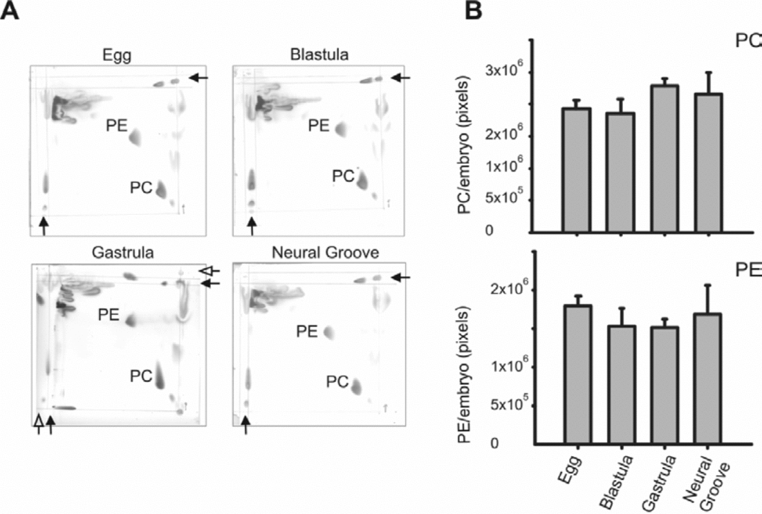
Considering that PC and PE are the major phospholipids of cell membranes (Jackowski & Fagone, Reference Jackowski and Fagone2005), and as the current results suggest that the regulatory enzymes of the Kennedy pathway (CCT and CK) undergo a decrease in their expression (protein abundance) along development, the content of PC and PE were investigated, and the PC:PE ratio per embryo calculated. Phosphatidylcholine and PE were analysed and estimated by bi-dimensional thin-layer chromotography (TLC) and copper/phosphoric stain (Marcucci et al., Reference Marcucci, Paoletti, Jackowski and Banchio2010) (Fig. 5A, B). In agreement with previous studies (Barassi & Bazan, Reference Barassi and Bazan1974; Alonso et al., Reference Alonso, Bonini de Romanelli and Bazan1982a), the current analysis demonstrated that the mass of PC and PE, and the ratio PC:PE is maintained in all the embryonic stages studied.
Phosphatidylcholine (PC) and phosphatidylethanolamine (PE) in Bufo arenarum eggs and embryos. Total phospholipids were isolated from eggs and staged embryos (25 in each case) and analysed by bi-dimensional thin-layer chromatography (TLC). (A) Typical bi-dimensional TLC revealed with copper/phosphoric reactive (one for each stage: egg, blastula, gastrula and neural groove). Black arrows indicate the point of loading of PC standard and the white arrow the point of loading of the PE standard. (B) Semi-quantification of PC and PE in eggs and embryos (see Material and methods for details).
Discussion
Although several works have dealt with lipid composition and synthesis in amphibian eggs and embryos (Mes-Hartree & Armstrong, Reference Mes-Hartree and Armstrong1976, Reference Mes-Hartree and Armstrong1980; Alonso et al., Reference Alonso, Bonini de Romanelli and Bazan1982a; Petcoff et al., Reference Petcoff, Holland and Stith2008), no study has focused on the expression of the PC key biosynthetic enzymes during normal development in any vertebrate. The expression of CCT and CK at the protein level in eggs/early embryos of B. arenarum was examined and it was found that both enzymes are expressed highly at the very beginning of embryonic development (e.g. until the 32-cell stage), after which they decrease through the morula, blastula and gastrula stages. Following this transient decrease, CCT and CK levels rise again at the early neurula stage. This last behaviour could be explained by the fact that during MBT, zygotic gene transcription is activated and more mRNA is available for translation.
Data from different cell types endorse the view that when prompt PC synthesis is required, cells respond either by gene induction (transcription and translation) or CCT activity is up-regulated based on its interaction with membrane lipid components (Jackowski, Reference Jackowski1994; Sugimoto et al., Reference Sugimoto, Banchio and Vance2008). Nevertheless, in the stages at which CCT expression decreases, there is no indication of a compensatory increment of CCT activity by membrane association. Apparently, in the current model, none of these mechanisms seems to be activated, as the CCT protein level decreases and the cytosol:total extract ratio remains constant, although cell proliferation and membrane generation are undoubtedly going on.
At first glance, the decrease in CK and CCT content observed between the 32-cell stage to gastrula may seem unexpected in the context of membrane biogenesis requirements and the previous findings obtained from KO mice (Wang et al., Reference Wang, Magdaleno, Tabas and Jackowski2005; Wu et al., Reference Wu, Aoyama, Young and Vance2008). Bufo arenarum is an amphibian that has external development. Externally developing embryos are limited to the resources contained within the egg, therefore these embryos contain large reserves of lipid droplets, glycogen and yolk, which provide the energy and building blocks required for embryogenesis (Jorgensen et al., Reference Jorgensen, Steen, Steen and Kirschner2009). Also, it is interesting to note that in other amphibians (Rana pipiens, Triturus pyrrhogaster, Cynops pyrrhogaster and Xenopus laevis) yolk consumption starts around the blastula–gastrula stages (Karasaki, Reference Karasaki1963; Komazaki et al., Reference Komazaki, Tanaka and Nakamura2002; Jorgensen et al., Reference Jorgensen, Steen, Steen and Kirschner2009), the same time at which CCT and CK enzyme levels are decreasing in B. arenarum. The utilization of these reservoirs, from which nascent membranes may obtain their constituents, could explain how cell division takes place – even though key players of the Kennedy pathway (CCT and CK) show a considerable expression decrease during morula, blastula and gastrula stages. In further support of this view, PC and PE content per egg/embryo were also examined in the current work and there were no statistically significant differences among the evaluated stages. Similar findings in phospholipid content have been described in B. arenarum by other authors (Alonso et al., Reference Alonso, Bonini de Romanelli and Bazan1982a,b) and in Xenopus laevis (Mes-Hartree & Armstrong, Reference Mes-Hartree and Armstrong1976). The fact that PC and PE total content is unchanged throughout early development suggests that these phospholipids are in reservoirs large enough to supply the embryo initial needs. There are at least three mechanisms by which these phospholipids can be mobilized from the yolk platelets to the sites of membrane biogenesis: (1) diffusion through the cytoplasm; (2) vesicular transport; and (3) lipid transfer protein-mediated transport. Nevertheless, Rusiñol et al. (Reference Rusiñol, Salomon and Bloj1987) suggested that B. arenarum fertilization does not activate protein-mediated phospholipid transfer.
As mentioned above, amphibian eggs not only have a reservoir of phospholipids but also a reservoir of maternal proteins and indeed the current work has shown that the amounts of CCT and CK proteins in the first stages analysed are high. In this regard, it is important to note that, in a mammalian model, these egg enzyme reservoirs also seemed to be high, as heterozygous mice for CK and CCT have no or very mild phenotype, further supporting the idea that enzyme levels are more than sufficient to maintain PC-associated functions (Wang et al., Reference Wang, Magdaleno, Tabas and Jackowski2005; Wu et al., Reference Wu, Aoyama, Young and Vance2008).
In B. arenarum, Alonso et al. (Reference Alonso, Bonini de Romanelli and Bazan1982a) have shown that 32P was actively incorporated into individual phospholipids in the absence of measurable net synthesis of phospholipids during the first stages of development (fertilized egg, late blastula, late gastrula). As phospholipid homeostasis is regulated by the expression and activity of biosynthetic enzymes and also by turnover through phospholipases activity (Jackowski, Reference Jackowski1994), the current results suggest that PC turnover plays an important role during early embryogenesis. Moreover, given that the net synthesis of PC in early development does not seem essential, it can be hypothesized that perhaps what matters during early development is not the end product of the pathway (e.g. PC) but the enzyme activities themselves, the intermediate metabolites of the route or the sub-cellular location where they are generated.
Altogether, previous published findings, and the results presented in the current manuscript, suggest the existence in the embryo of mechanisms that are in charge of maintaining the balance of phospholipids. This balance is maintained by regulating the activity of phospholipases that coordinate with de-novo synthesis of phospholipids and yolk platelet reservoir utilization to fulfil the needs of early developmental phospholipids.
Financial support
This work was supported by Universidad Nacional de Rosario (grant no. BIO262).