Introduction
Marijuana (Cannabis sativa) is the most widely cultivated, trafficked, and abused illicit drug in the world. 1 In recent years, the use of cannabis during pregnancy in North America has increased considerably, particularly among younger women. Reference Singh, Filion, Abenhaim and Eisenberg2 Indeed, recent estimates suggest that approximately 3%–7% of pregnant women in North America use cannabis. Reference Singh, Filion, Abenhaim and Eisenberg2 An integrative review concluded that women who used cannabis during pregnancy believed that there were no significant risks to the mother or fetus. Reference Carlier, Huestis, Zaami, Pichini and Busardò3 In fact, it is not uncommon for women to use cannabis to treat pregnancy-related nausea and vomiting. Reference Carlier, Huestis, Zaami, Pichini and Busardò3
While it has been shown that cannabis negatively affects female fertility in adults, Reference Brents4 little is known regarding the long-term influence of maternal exposure to cannabis on ovarian development in the offspring. Given that Δ-9-tetrahydrocannabinol (Δ9-THC) efficiently crosses the placental barrier and can be detected in cord blood and fetal tissue, Reference Carlier, Huestis, Zaami, Pichini and Busardò3 this compound could have direct effects on ovarian development. Since ovarian primordial follicle assembly takes place during this time and largely determines the long-term reproductive capacity of female mammals, exposure to exogenous chemicals during this sensitive window may have persistent effects throughout the lifespan. Reference Allais, Albert and Lefèvre5 In addition, cannabinoid receptors 1 and 2 (CB1 and CB2), which bind to cannabinoids such as Δ9-THC, are expressed in the human and rodent embryo from very early stages of development. Reference Buckley, Hansson, Harta and Mezey6,Reference Biegon and Kerman7 The preimplantation mouse embryo has been shown to express both receptors from the two-cell stage, and CB1 and CB2 have been detected in the ectoplacental cone from gestational day (GD) 8 in the rat. Reference Buckley, Hansson, Harta and Mezey6 A recent study reported that in utero treatment with a selective CB2 agonist influenced oocyte reserve and reduced the reproductive capacity of mice. Reference De Domenico, Todaro and Rossi8
We have previously shown that Δ9-THC exposure during pregnancy leads to placental insufficiency and symmetrical fetal growth restriction. Reference Natale, Gustin and Lee9 It is known that placental insufficiency can cause severe malnutrition of the fetus, and that the preferential blood flow to vital organs may impact the development of other organs. Indeed, studies have suggested that fetal growth restriction due to placental insufficiency negatively affects ovarian development both in humans and rats. Reference Pampanini, Jahnukainen and Sahlin10,Reference De Bruin, Dorland and Bruinse11 In one of these studies, a significantly lower number of primordial follicles were observed in intrauterine growth restricted animals at postnatal day (PND) 20. Interestingly, no differences were observed in follicle density at PND40, suggesting that compensatory mechanisms occurred during the prepubertal window. In addition, the expression of genes involved in cellular processes such as proliferation, apoptosis, and metabolism was affected at all ages evaluated, potentially implying long-term genetic alterations. Reference Pampanini, Jahnukainen and Sahlin10 Taking all of these studies into account, we hypothesized that prenatal exposure to Δ9-THC would adversely affect ovarian function in adult rats.
Methods
All procedures were performed in accordance with the guidelines of the Canadian Council of Animal Care. Pregnant Wistar rats were purchased from Charles River (La Salle, St. Constant, QC, Canada) and throughout the experimental procedure were maintained at 22 °C on a 12:12-h light–dark cycle with access to food and water ad libitum. Dams were randomly assigned to receive a daily intraperitoneal (IP) injection of either vehicle (1:18 cremophor:saline; n = 5) or 3 mg/kg Δ9-THC (Sigma-Aldrich, St Louis, MO, USA; n = 6) from GD6 to GD22 as previously described. Reference Natale, Gustin and Lee9 This dose results in maternal blood concentrations (8.6–12.4 ng/ml) comparable to those detected after moderate marijuana smoking in adults (13–63 ng/ml), as well as in aborted fetal tissue (4–287 ng/ml) after maternal cannabis use. Reference Klein, Karanges and Spiro12–Reference Falcon, Pichini and Joya14 Moreover, it has been previously demonstrated that 3 mg/kg Δ9-THC per day does not cause fetal demise, alterations in litter size, gestational length, or maternal weight gain. Reference Natale, Gustin and Lee9 Dams were allowed to deliver normally and litters were randomly culled to four female pups and four male pups each. Offspring were then euthanized at 6 months of age by IP pentobarbital overdose. Blood was drawn by cardiac puncture for quantification of estrogen (Biovision, Milpitas, CA, USA), progesterone (Cusabio, Wuhan Huamei Biotech Co., Wuhan, China), and free testosterone (R&D Systems, Minneapolis, MN, USA) by ELISA. The ovaries were collected, fixed in 10% formalin, and embedded in paraffin for histological analysis.
Paraffin-embedded ovaries were serially sectioned (8 μm) and stained with hematoxylin–eosin to carry out follicle quantitation and morphometric analysis. Every 10th section was analyzed and follicles were counted and classified as primordial, transitioning, primary, secondary, antral, and atretic as previously described. Reference Tsoulis, Chang and Moore15 The area of each section was measured using an Olympus IX81 microscope and Infinity Analyze software, and the total number of each type of follicle was normalized to the total area analyzed from each ovary.
Immunohistochemistry (IHC) was performed to assess the expression of the pro-angiogenic factor vascular endothelial growth factor (VEGF) and its receptor (VEGFR-2), as well as the anti-angiogenic factor thrombospondin 1 (TSP-1) in ovarian sections. Immunofluorescence was also performed to determine the levels of the endothelial marker CD31. For IHC, tissues were deparaffinized using reagent-grade xylene and subjected to a series of decreasing ethanol concentrations for rehydration. Endogenous peroxidase activity was quenched through a 10-minute incubation period in 3% hydrogen peroxide followed by antigen retrieval using citrate buffer with Tween 20. To reduce nonspecific binding of antibodies, samples were blocked in 5% bovine serum albumin (with 0.02% sodium azide) for 10 min at room temperature. Sections were exposed to the following primary antibodies overnight at 4 °C: anti-VEGF (Abcam; 1:600); anti-VEGFR-2 (Cell Signaling; 1:400); anti-TSP-1 (Abcam; 1:100). Slides were then incubated in anti-mouse or anti-rabbit (Sigma; 1:100) biotinylated secondary antibody for 2 h at room temperature followed by Extravidin (Sigma, 1:50) for 1 h at room temperature. Antigens were visualized using 3,3′-Diaminobenzidine Tetrahydrochloride (DAB; Sigma) and tissues were counterstained with hematoxylin. Slides were imaged using bright-field microscopy, and the percentage of immunopositive tissue was quantified using morphometry software (ImagePro Plus, Media Cybernetics, Rockville, MD, USA). For immunofluorescence, tissues were deparaffinized and rehydrated as above. Slides were exposed to 0.2% sodium borohydride in PBS for 12 min and 5% bovine serum albumin for 15 min to reduce nonspecific binding. Sections were then stained with anti-CD31 (Abcam; 1:25) overnight at 4 °C. The following day, tissue samples were stained with secondary antibody (Alexa Fluor594nm, red). Slides were counterstained and cured using Prolong Gold anti-fade mounting medium. Representative images (n = 4/ovary) were obtained and image analysis was conducted using Metamorph imaging software (Molecular Devices, San Jose, CA, USA).
For hormone levels, follicle quantitation, IHC, and immunofluorescence, one female offspring per dam were included in the statistical analysis. A Student’s t-test was performed between vehicle (n = 5) and Δ9-THC-exposed (n = 6) animals for each outcome measure. Data are presented as the mean plus the standard error of the mean and a p-value of less than 0.05 was considered statistically significant.
Results
Since sex steroids are known to play a crucial role in the regulation of ovarian function and follicular development, at 6 months of age, blood was collected from Δ9-THC-exposed offspring for hormone quantification. No differences were found in the levels of circulating estrogen (vehicle: 57.92 ± 2.53 pg/ml; Δ9-THC: 61.11 ± 2.86 pg/ml; intra-assay CV: 4.99%), progesterone (vehicle: 94.12 ± 15.48 ng/ml; Δ9-THC: 87.76 ± 7.52 ng/ml; intra-assay CV: 10.6%), or testosterone (vehicle: 3.26 ± 0.65 ng/ml; Δ9-THC: 3.46 ± 0.74 ng/ml; intra-assay CV: 7.25%).
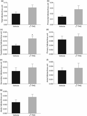
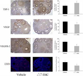
When the ovaries from adult offspring were examined, Δ9-THC-exposed animals had significantly more transitioning follicles/area unit (Fig. 1c). However, this difference did not persist in later stages of follicle development (Fig. 1d–g). Additionally, there was a significant increase in the percentage of TSP-1-positive granulosa cells, as well as a significant decrease in the percentage of VEGF and VEGFR-2-positive granulosa cells in ovaries from Δ9-THC-exposed offspring. Consistent with this shift to an anti-angiogenic phenotype, a significant decrease in ovarian blood vessel density, assessed by CD31 levels, was also detected (Fig. 2).
Number of (a) total, (b) primordial, (c) transitioning, (d) primary, (e) secondary, (f) antral, and (g) atretic follicles per area unit in vehicle (n= 5) and prenatally Δ9-THC-exposed (n = 6) rat ovaries. Mean ± SEM. Student’s t-test; *p < 0.05.

Expression of thrombospondin 1 (TSP-1), vascular endothelial growth factor (VEGF), vascular endothelial growth factor receptor 2 (VEGFR-2), and CD31 in vehicle (n = 5) and prenatally Δ9-THC-exposed (n = 6) rat ovaries. Mean ± SEM. Student’s t-test; *p < 0.05.

Discussion
Given that the rodent embryo expresses the cannabinoid receptors from very early stages of development, and that gestational Δ9-THC exposure has been shown to result in intrauterine growth restriction, we suspected that prenatal exposure to this compound may have direct and/or indirect effects on fetal ovarian development that could manifest during adulthood.
We have previously reported that fetal exposure to Δ9-THC resulted in placental insufficiency culminating in symmetrical fetal growth restriction in the animals used in this study; Reference Natale, Gustin and Lee9 this growth restriction is similar to what has been reported in humans following maternal cannabis use. Reference Carlier, Huestis, Zaami, Pichini and Busardò3 Interestingly, studies indicate that low birth weight may be a risk factor for earlier age at menopause in women. Reference Steiner, D’Aloisio, DeRoo, Sandler and Baird16 Since the age at which menopause occurs is thought to be a reflection of both the size of the initial follicle pool, as well as the rate of oocyte loss, Reference Gougeon and Chainy17 factors that affect either or both of these parameters may lead to premature reproductive aging.
With this in mind, we assessed ovarian follicle dynamics of prenatally Δ9-THC-exposed rats at 6 months of age. Female rats that were exposed to Δ9-THC during fetal development had a significant increase in the number of transitioning follicles/area unit. This could be a result of an increase in the activation of dormant primordial follicles, similar to what has been reported in other models of in utero insults. Reference Tsoulis, Chang and Moore15 Since this difference was not observed in later stages of follicle development (i.e. primary, secondary, and antral), it is possible that a significant portion of the transitioning follicles did not continue to develop. It has previously been suggested that primordial follicles that are prematurely recruited into the growing pool are subsequently lost, resulting in no net loss or gain of developed follicles. Reference Tsoulis, Chang and Moore15 In support of this hypothesis, we did not observe any differences in the levels of circulating estradiol, progesterone or testosterone, or in the number of developed follicles, which are largely responsible for the synthesis of steroid hormones. However, the increase in transitioning follicles may lead to a premature loss of the primordial follicle pool and therefore early reproductive senescence, Reference Nelson18 although this remains to be confirmed.
Since ovarian follicle development is largely dependent on angiogenesis, Reference Robinson, Woad and Hammond19 ovarian sections that were not used for follicle dynamics evaluation were used to assess levels of pro and anti-angiogenic proteins. It has previously been reported that cannabis and Δ9-THC exposure during pregnancy contributed to reduced blood vessel formation in human and rodent placenta, suggesting that this compound has anti-angiogenic effects. Reference Natale, Gustin and Lee9,Reference Chang, Li and Li20 Moreover, early life insults, which result in reduced birth weight have been reported to reduce ovarian vascularization. Reference Chan, Bernal and Vickers21 Similarly, in the current study, ovaries from Δ9-THC-exposed animals had reduced blood vessel density in association with decreased expression of the pro-angiogenic factor VEGF and its receptor VEGFR-2, and increased expression of the anti-angiogenic factor TSP-1. TSP-1 is a potent inhibitor of VEGF signaling, and VEGF and TSP-1 are inversely expressed throughout folliculogenesis, with VEGF levels increasing and TSP-1 decreasing as follicle progression occurs. Reference Greenaway, Lawler and Moorehead22 While primordial follicles rely on the stroma for nutrient delivery, after activation and development of a primary follicle, endothelial cells are recruited from neighboring blood vessels so that follicular development can proceed. Reference Fraser and Wulff23 Therefore, the increase in TSP-1 and decrease in VEGF and VEGFR-2 could, at least in part, underlie the follicle dynamics observed in Δ9-THC-exposed animals, since a significant decrease in ovarian blood vessel formation could result in follicular development arrest after stages that rely solely on the stroma.
On the other hand, in vitro treatment of immortalized rat granulosa cells with TSP-1 has been demonstrated to increase the expression of pro-apoptotic factors, Reference Greenaway, Lawler and Moorehead22 while VEGF-VEGFR-2 signaling appeared to have opposing effects. Reference Greenaway, Connor and Pedersen24 The increase in TSP-1 and decrease in VEGF and VEGFR-2 observed in this study could therefore manifest as increased apoptosis (atresia), and ultimately a loss of developing follicles. Although we did not observe a statistically significant increase in the number of atretic follicles in the Δ9-THC-exposed animals at 6 months of age (p = 0.197), this may be a reflection of the small sample size (n = 6). Alternatively, it is possible that the deficits in angiogenesis may manifest more clearly as increased follicle atresia and depletion of the follicle reserve as the animal ages. Thus, to further investigate the effects of gestational exposure to Δ9-THC on adult reproductive health, it would be prudent to monitor the ovarian follicle reserve at a more advanced age, closer to that at which rats typically experience reproductive senescence (9–12 months of age). Additionally, it would be of great interest to assess the fertility of these animals, since it is possible that not only the number but also the quality of the oocytes could be affected.
In conclusion, at 6 months of age, animals that were exposed to Δ9-THC in utero had accelerated folliculogenesis with apparent follicular development arrest, as well as decreased blood vessel density. These results suggest for the first time that the popular use of cannabis during pregnancy may have long-term effects on ovarian development in the offspring, which may negatively impact postnatal reproductive health.
Acknowledgments
None.
Financial support
This study was supported by a CIHR Project Grant to ACH (PJT-155981) and an HSFC Grant-in-Aid to DBH (G-19-0,026,343). AAMP was sponsored by the Mexican National Council of Science and Technology (CONACYT).
Conflicts of interest
None.
Ethical standards
The authors assert that all procedures contributing to this work comply with the ethical standards of the relevant national guides on the care and use of laboratory animals (Canadian Council of Animal Care) and has been approved by the institutional animal care committee at Western University.