Introduction
Refractive errors represent conditions in which distant objects are not focused on the retina and as a result are seen as blurred. They reflect mismatches between the optical power and axial length of the eye (Flitcroft, Reference Flitcroft2013). Myopia, one of the most common types of refractive errors, typically results from excessive axial elongation of the eye. Its prevalence has risen rapidly over the past few decades, to epidemic levels in some countries although the etiology of this “epidemic” is still poorly understood (Wallman & Winawer, Reference Wallman and Winawer2004; Vitale et al., Reference Vitale, Sperduto and Ferris2009; Wojciechowski, Reference Wojciechowski2011; Dolgin, Reference Dolgin2015).
In terms of understanding how myopia develops, significant advances have been made through studies using animal models, with form-deprivation and hyperopic optical defocus (imposed with negative lenses) manipulations proving to be robust methods for stimulating eye elongation, and so inducing myopia. The optical defocus effect is also sign-dependent, with myopic defocus (imposed with positive lenses), slowing rather than accelerating eye elongation in most models (Wiesel & Raviola, Reference Wiesel and Raviola1977; Wildsoet & Wallman, Reference Wildsoet and Wallman1995; Wallman & Winawer, Reference Wallman and Winawer2004). Investigations into ocular molecular and cellular mechanisms underlying the regulation of eye growth represent one of the major efforts in current myopia research, with the hope of uncovering novel antimyopia treatment strategies for the current epidemic. This approach is supported by accumulating evidence from animal model studies, both direct and indirect, for local control of eye elongation (Troilo et al., Reference Troilo, Gottlieb and Wallman1987; Wildsoet, Reference Wildsoet2003; Wallman & Winawer, Reference Wallman and Winawer2004). Of particular interest is how growth modulatory signals generated in the retina, the presumed origin of such signals, are relayed to the choroid and sclera, which together define the position of the retina and eye shape more generally (Crewther, Reference Crewther2000; Wildsoet, Reference Wildsoet2003; Wallman & Winawer, Reference Wallman and Winawer2004; Rymer & Wildsoet, Reference Rymer and Wildsoet2005; Rada et al., Reference Rada, Shelton and Norton2006; Nickla & Wallman, Reference Nickla and Wallman2010).
Our previous studies have focused on the role in eye growth regulation of the retinal pigment epithelium (RPE), which is uniquely located to participate as a relay in a retina-scleral signal cascade, and has multiple receptors whose functions are only poorly understood, if at all (Zhang et al., Reference Zhang, Liu and Wildsoet2012; Reference Zhang, Liu, Ho and Wildsoet2013; Zhang and Wildsoet, Reference Zhang and Wildsoet2015). In brief, RPE is a monolayer of polarized, pigmented epithelial cells lying between the neural retina and vascular choroid, interconnected by tight junctions to form a critical component of the blood/retina barrier (Rymer & Wildsoet, Reference Rymer and Wildsoet2005; Maminishkis et al., Reference Maminishkis, Chen, Jalickee, Banzon, Shi, Wang, Ehalt, Hammer and Miller2006; Zhang and Wildsoet, Reference Zhang and Wildsoet2015). The RPE is also a major source of cytokines and growth factors, at least some of which play important roles in establishing the immune privilege of the eye and maintaining the structural integrity of the retina and choroid. In the context of eye growth regulation, we have identified three members of the Bone Morphogenetic Protein family (BMP2, BMP4, and BMP7), as candidate signal molecules. Specifically in young chicks, all show sign-dependent differential gene expression in RPE in response to short term, imposed optical defocus (2 or 48 h) (Zhang et al., Reference Zhang, Liu and Wildsoet2012; Reference Zhang, Liu, Ho and Wildsoet2013). Differential gene expression of BMPs has also been reported in combined retina/RPE samples in another study using the form-deprivation myopia chick model (McGlinn et al., Reference McGlinn, Baldwin, Tobias, Budak, Khurana and Stone2007). These results together open the possibility that the RPE acts as a conduit for relaying growth modulatory retinal signals to the choroid and/or sclera.
In the study reported here, we investigated the gene expression of BMP2, 4, 7 and BMP receptors (BMPR1A, BMPR1B, BMPR2) in chick retina and choroid after short-term optical defocus treatment (+10 and −10 D lenses, worn for either 2 or 48 h). The aims of this study were to: (1) determine if retinal and choroidal tissues express BMP receptors, with sensitivity to optical defocus consistent with roles of BMP2, 4, and 7 in eye growth regulation, and to (2) compare the significant gene expression changes of BMP2, 4, and 7 reported previously in chick RPE with the changes in retina and/or choroid.
Materials and methods
Chicks and lens treatment
White-Leghorn chickens were hatched in the Animal Facilities at University of California, Berkeley, from eggs obtained from University of California, Davis (CA). Chicks were raised under a 12 h light/12 h dark cycle, with free access to food and water. Accelerated or slowed eye growth patterns were induced with monocular −10 or +10 D lens treatments, applied for 2 or 48 h from 19 days of age. The contralateral (fellow) eyes of treated birds served as controls. Age-matched untreated chicks also were included as additional controls. Each treatment group consisted of two or three independent repetitions, with tissue from 3–4 chicks included in each repeat. Experiments were conducted according to the ARVO Statement for the Use of Animals in Ophthalmic and Vision Research and approved by the Animal Care and Use Committee (ACUC) at University of California, Berkeley (Berkeley, CA).
Measurement of refractive errors & ocular dimensions in vivo
Ocular dimensional effects of the positive and negative lens treatments were characterized in vivo as previously described (Zhang et al., Reference Zhang, Liu and Wildsoet2012; Reference Zhang, Liu, Ho and Wildsoet2013). In brief, for both eyes of each bird, refractive errors (RE) were measured using retinoscopy and axial ocular dimensions measured using high-frequency A-scan ultrasonography, both at the beginning and end of the lens treatment period. All measurements were performed under isoflurane anesthesia (1% in oxygen). Both RE and ocular dimensions were expressed as interocular differences between treated and fellow eyes or right and left eyes, normalized to pre-treatment values.
Ocular tissue collection
Retinal and choroidal samples were collected from lens-treated and fellow control eyes as well as eyes from untreated birds, at approximately the same time of day in all cases, as described previously (Zhang et al., Reference Zhang, Liu and Wildsoet2012; Reference Zhang, Liu, Ho and Wildsoet2013). In brief, chicks were sacrificed and eyes immediately enucleated. Retinas were first isolated, by gently peeling them from RPE; pieces of retina contaminated with RPE were discarded. Next, the RPE was gently rinsed off choroid with cold 1× PBS and choroidal tissue isolated from the underlying sclera. Both retinal and choroidal tissues were homogenized in cell lysis buffer (RLT buffer from RNeasy Mini kits, Qiagen, Valencia, CA) over ice and lysed samples then stored at −80°C for later use.
RNA purification and reverse transcription
Total RNA from tissue samples of retina and choroid was purified using RNeasy Mini Kits and RNeasy Fibrous Tissue Mini Kits, respectively (Qiagen). On-column DNase digestion was performed according to the manufacturer's protocol. RNA quantification and A260/A280 optical density ratio were measured with a spectrophotometer (NanoDrop 2000; NanoDrop Technologies, Inc., Wilmington, DE). That genomic DNA contamination was minimal, was confirmed by examining RNA samples without RT enzymes. Total RNA was reverse transcribed to cDNA (SuperScript III First-Strand Synthesis System for RT-PCR, Invitrogen, Carlsbad, CA), and then subjected to real-time PCR for gene expression studies.
Real-time PCR and gene expression levels
In total, the expressions of six genes, comprising three growth factors, BMP2, BMP4, BMP7, and three receptors, BMPR1A, BMPR1B, and BMPR2, were examined in both chick retina and choroid. Chick glyceraldehyde 3-phosphate dehydrogenase (GAPDH) was used as the reference gene. The validation of GAPDH as a reference gene and the efficiency of GAPDH primers have been reported previously (Zhang et al., Reference Zhang, Liu and Wildsoet2012; Reference Zhang, Raychaudhuri and Wildsoet2016). The study made use of the same primers as characterized and used in previous related studies (Zhang et al., Reference Zhang, Liu and Wildsoet2012; Reference Zhang, Liu, Ho and Wildsoet2013). The amount of cDNA template used in each real-time PCR reaction varied across tissues and between genes, according to expression levels. QuantiTect SYBR Green PCR Kits (Qiagen) and a StepOnePlus Real-Time PCR System (Life Technologies, Grand Island, NY) were used for gene expression quantification.
Gene expression levels were calculated as described previously (Zhang et al., Reference Zhang, Liu and Wildsoet2012; Reference Zhang, Liu, Ho and Wildsoet2013). First, the efficiency (E) of primers was calculated using 10-fold serial dilutions of cDNA. Second, mean normalized expression (MNE) values were calculated for technical repeats of target genes. Next, mean mRNA expression levels for all biological repeats were calculated. Finally, differential gene expression values were derived; expression levels representing lens-treated eyes are expressed as a percentage of equivalent values for fellow eyes, with a similar calculation undertaken using the data for the two eyes of untreated chicks.
Statistical analysis
Paired Student’s t-tests were used to compare lens-treated eyes with their fellow control eyes, while one-way ANOVAs combined with post-hoc analysis (Fisher’s least significant difference) were used for comparisons involving more than two groups.
Results
Refractive errors and ocular dimension changes
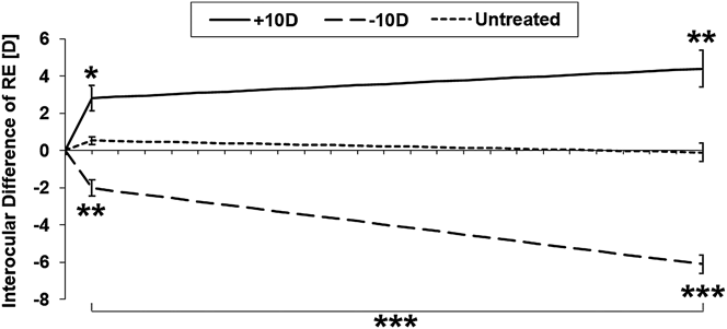
Baseline refractive errors measurements made immediately prior to the initiation of lens treatments, consistently revealed low hyperopia, with minimal difference between the two eyes of individual chicks or between treatment groups (Table 1). Both +10 and −10 D lenses worn for 2 and 48 h induced significant changes in refractive errors (Fig. 1). With the +10 D lens, the interocular differences of refractive errors between treated and fellow eyes changed in the direction of increased hyperopia, i.e., to +2.81 ± 0.65 D after 2 h (P < 0.05, n = 6), and to +4.40 ± 0.9 D after 48 h (P < 0.01, n = 6). In contrast, with the −10 D lens, refractive errors became relative myopic and interocular differences, increasingly negative, i.e., to −2.02 ± 0.43 D after 2 h (P < 0.01, n = 6), and −6.10 ± 0.50 D after 48 h (P < 0.001, n = 6). Neither the fellow control eyes of treated chicks, nor the eyes of untreated chicks, showed any significant change in refractive errors over the same 48 h time frame (P > 0.05).
Interocular differences (means & SEMs in brackets) in axial length (AL), vitreous chamber depth (VCD), retinal thickness (RT) and choroidal thickness (CT), after 2 and 48 h of monocular +10 and −10 D lens treatments, normalized to baseline values.

Baseline measurements for RE were +2.25 ± 0.07 D, AL 10.30 ± 0.02 mm, VCD 5.72 ± 0.02 mm, RT 0.24 ± 0.0008 mm, and CT 0.22 ± 0.004 mm.
Interocular difference of refractive errors, normalized to baseline values, of eyes treated with +10 or −10 D lenses plotted as a function of treatment duration (2 or 48 h), as well as of fellow eyes of same birds and eyes of untreated chicks over same time frame. *P < 0.05, **P < 0.01, ***P < 0.001.

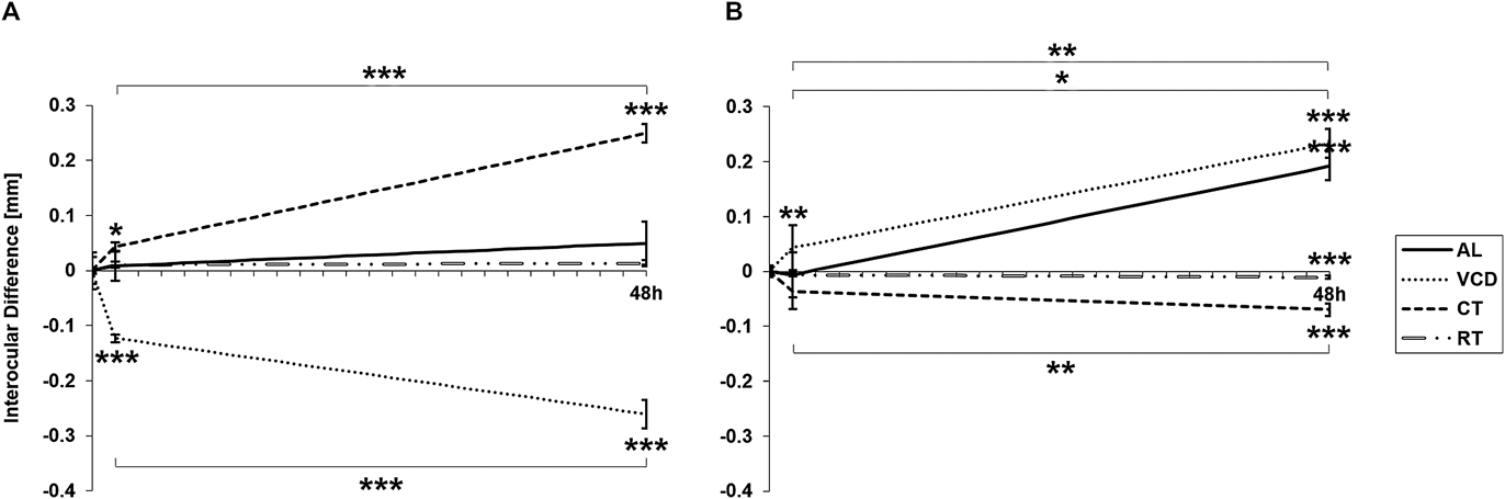
Ocular dimensional changes, also expressed as interocular differences (treated—fellow eyes), are summarized for axial length (AL), vitreous chamber depth (VCD), retinal thickness (RT), and choroidal thickness (CT) along with sample sizes in Table 1 and illustrated in Fig. 2. With the +10 D lens (Fig. 2A), the VCDs of treated eyes had decreased relative to their fellows, by 0.12 ± 0.007 and 0.26 ± 0.026 mm after 2 and 48 h of lens wear respectively (P < 0.001). The effect of 48 h of treatment was also significantly larger than the effect at 2 h (P < 0.001). These VCD changes reflect, at least in part for 2 h, and almost entirely for 48 h, parallel, significant increases in CT, i.e., 0.04 ± 0.008 and 0.25 ± 0.017 mm, respectively (P < 0.05 for 2 h and P < 0.001 for 48 h). No significant changes in AL and RT were detected with the +10 D lens treatment, even after 48 h of treatment. With the −10 D lens treatment (Table 1, Fig. 2B), VCDs of treated eyes was enlarged compared to their fellow eyes, by 0.04 ± 0.017 mm (P < 0.01) and 0.23 ± 0.025 mm (P < 0.001) after 2 and 48 h, respectively. Accordingly, CT was reduced in treated eyes relative to their fellows, although interocular CT differences reached significance only after 48 h of −10 D lens treatment (−0.070 ± 0.011 mm; P < 0.001). Similarly, RT was also reduced in treated eyes, with interocular RT differences reaching significance after 48 h of −10 D lens treatment (−0.01 ± 0.003 mm; P < 0.01). These RT and CT changes contributed to, but did not fully account for the observed VCD changes at 48 h, which largely reflect increased axial elongation over the same period. Thus interocular AL differences were also significantly increased after 48 h (0.19 ± 0.026 mm; P < 0.001).
Interocular differences, normalized to baseline values, in axial length (AL), vitreous chamber depth (VCD), choroidal thickness (CT), and retinal thickness (RT) induced by monocular (A) +10 D and (B) −10 D lens treatments after for 2 and 48 h. *P < 0.05, **P < 0.01, ***P < 0.001.

BMP & BMP receptor gene expression in retina & effects of lens treatment
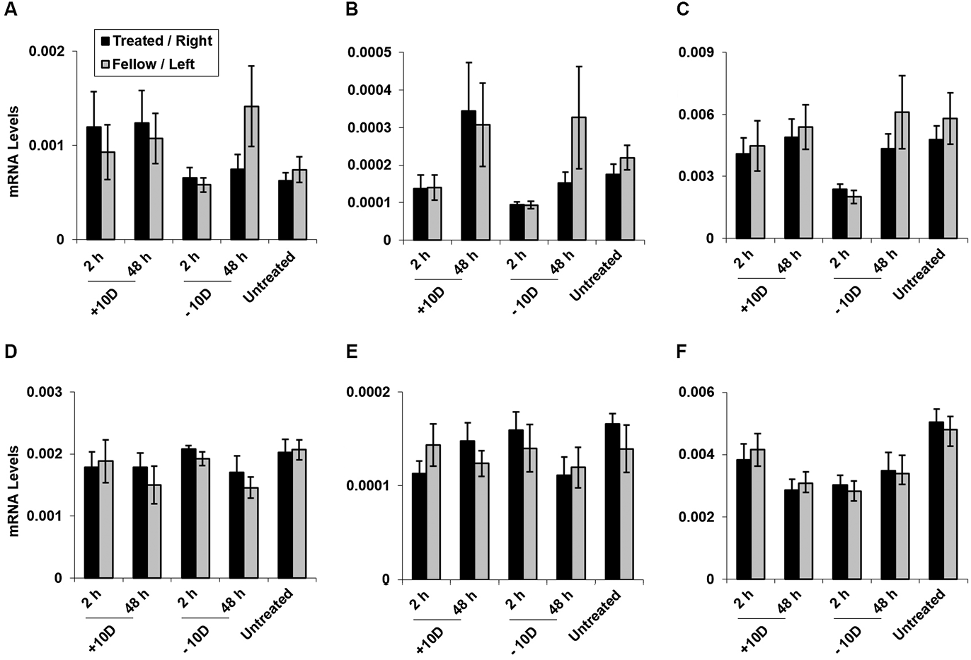
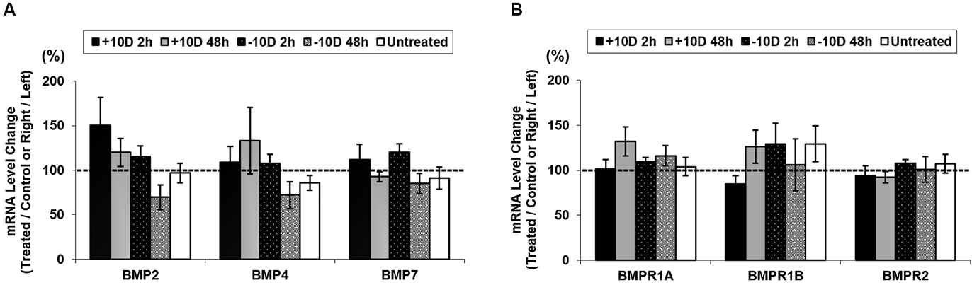
For all of six genes tested, BMP2, BMP4, BMP7, BMPR1A, BMPR1B, and BMPR2, expression reached detectable levels in retinal tissues from both treated and untreated eyes (fellows to treated eyes and eyes of untreated birds) (Table 2). However, none of the six genes showed treatment-related differential gene expression (Figs. 3 & 4).
BMP and BMP receptor gene expression levels (means & SEMs in brackets), for retinal and choroidal tissues from untreated chicks; GAPDH used as reference gene.

mRNA levels of BMP2 (A), BMP4 (B), BMP7 (C), BMPR1A (D), BMPR1B (E), and BMPR2 (F) in retina after monocular +10 or −10 D treatment applied for 2 or 48 h. Comparison made between treated (dark bars) vs. fellow control (light bars) eyes of treated birds or right (dark bars) vs. left (light bars) eyes of untreated birds. None of the genes showed significant treatment-induced differential expression. Note differences in Y-axis scales for individual panels, used to offset gene-dependent differences in expression level.

Lens treatment-induced changes of retinal mRNA levels for BMP2, BMP4, BMP7, BMPR1A, BMPR1B, and BMPR2. Data are expressed as percentages (treated/fellow control eyes for lens treated birds and right/left eyes for untreated birds). None of the changes reached statistical significance.

BMP & BMP receptor gene expression in choroid & effects of lens treatment
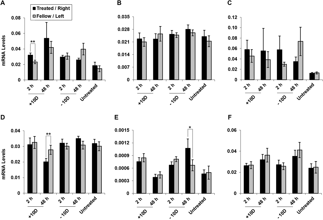
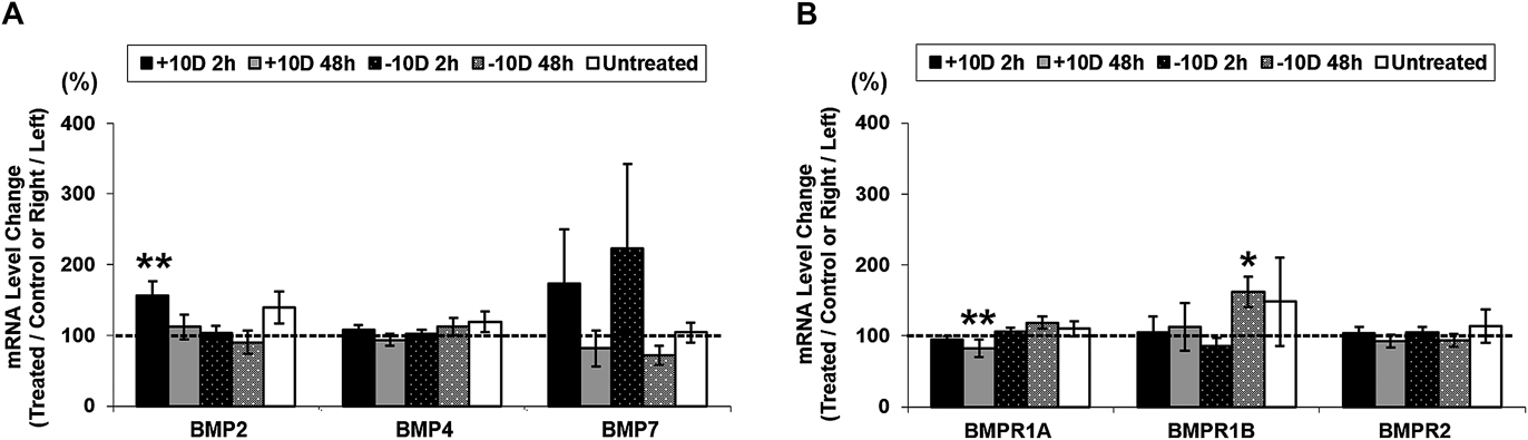
The mRNA expression levels in choroid for the same three BMPs and three BMP receptors in untreated birds are summarized in Table 2 and these data are also illustrated in Fig. 5. Equivalent gene expression data for treated and fellow eyes of lens wearing birds are illustrated in Figs. 5 and 6. Here, and in contrast to the results from retina, significant treatment effects were recorded for three genes, BMP2, BMPR1A, and BMPR1B. Specifically, with the +10 D lens, the expression of both BMP2 and BMPR1A genes were significantly altered; BMP2 gene expression was up-regulated by 156.7 ± 19.7% after 2 h of treatment (P < 0.01, n = 15), and BMPR1A gene expression was down-regulated to 82.3 ± 12.5% after 48 h of treatment (P < 0.01, n = 12). Significant effects of the −10 D lens treatment were limited to one receptor gene, BMPR1B, which was up-regulated by 162.3 ± 21.2% after 48 h of treatment (P < 0.05, n = 13).
mRNA levels of BMP2 (A), BMP4 (B), BMP7 (C), BMPR1A (D), BMPR1B (E), and BMPR2 (F) in choroid after +10 or −10 D lens treatment for 2 or 48 h. Comparison is made between treated vs. contralateral control eyes of monocularly treated birds or right vs. left eyes of untreated birds. Note differences in Y-axis scales for individual panels, used to offset gene-dependent differences in expression level. BMP2, BMPR1A, and BMPR1B all showed significant treatment-induced differential gene expression. *P < 0.05; **P < 0.01.

Lens treatment-induced gene expression changes in choroid for BMP2, BMP4, BMP7, BMPR1A, BMPR1B, and BMPR2. Data are expressed as percentages (treated/fellow control eyes or right/left eyes). BMP2, BMPR1A, and BMPR1B all showed significant treatment-induced differential gene expression.

Discussion
In this study, we have been able to confirm that in normal chickens, all three BMPs, BMP2, BMP4, and BMP7, previously shown to be expressed in RPE, as well as three BMP receptors, BMPR1A, BMPR1B and BMPR2, are expressed in both retina and choroid (Zhang et al., Reference Zhang, Liu and Wildsoet2012; Reference Zhang, Liu, Ho and Wildsoet2013). With imposed optical defocus (+10 or −10 D lenses), BMP2, BMPR1A, and BMPR1B all showed differential gene expression in chick choroid, while none of the six genes investigated showed differential expression in retina. In choroid, BMP2 was up-regulated after 2 h of +10 D lens treatment, while BMPR1A was down-regulated after 48 h, and in contrast, BMPR1B was up-regulated after 48 h of −10 D lens treatment.
In comparison to the defocus-induced patterns of expression for the same genes in RPE that we have previously reported, the gene expression changes in the choroid were smaller in magnitude and also showed different patterns (Zhang et al., Reference Zhang, Liu and Wildsoet2012; Reference Zhang, Liu, Ho and Wildsoet2013). Nonetheless, these new results for retina and choroid potentially offer new insights into the roles of BMPs in both the initiation and maintenance of defocus-driven eye growth responses (as evidenced by biometric changes after 2 & 48 h of treatments, Table 1 & Fig. 2). As reported previously, short-term imposed defocus induced rapid and sustained gene expression changes in chick RPE, and also statistically significant for both +10 or −10 D lenses after 2 or 48 h (Zhang et al., Reference Zhang, Liu and Wildsoet2012; Reference Zhang, Liu, Ho and Wildsoet2013). For example, expression of the BMP2 gene, which showed the fastest and most robust regulation among the three BMPs investigated (BMP2, 4, and 7), was up-regulated 7.2-fold and down-regulated 13.3-fold with 2 h of +10 and −10 D lens treatment respectively. In contrast, choroidal BMP2 gene expression changes are much smaller, 1.57-fold after 2 h of +10 D lens treatment and no change with the −10 D lens treatment. Our working hypothesis is that the RPE is the major source of BMPs, both synthesizing and secreting them, with the choroid being the favored target tissue. Thus assuming observed gene expression changes in RPE are translated to protein secretion, the down-regulation of BMPR1A gene expression in choroid with the +10 D lens treatment and the up-regulation of BMPR1B with the −10 D lens treatment may reflect negative feedback in response to up- or down-regulated BMP secretion in RPE respectively. Either way, our results suggest that the retina and choroid are more likely targets of BMPs rather than major “producers and/or regulators” of BMPs. Nonetheless, more studies of the down-stream BMP signaling pathways in posterior ocular tissues are needed to fully understand the roles of BMPs in the retina-scleral signaling cascades involved in eye growth regulation.
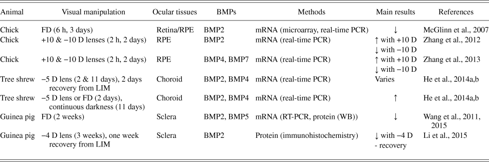
Apart from our past studies involving chick RPE, a number of other recent studies of eye growth regulation have investigated BMPs in different ocular tissues, using chicks, guinea pigs and tree shrews as animal models. The results of these studies are summarized in Table 3. Using the chick form-deprivation myopia model, McGlinn A., et al., reported ∼two fold down-regulation of BMP2 gene expression in retina/RPE after both 6 h and 3 days of treatment (McGlinn et al., Reference McGlinn, Baldwin, Tobias, Budak, Khurana and Stone2007). Interestingly, we did not observe differential gene expression of BMP2 in retina with our −10 D lens treatment, which also accelerates eye growth. It is possible that these different experimental outcomes reflect different retinal mechanisms subserving lens- and form deprivation-induced myopia, as suggested by other unrelated studies (Nickla & Totonelly, Reference Nickla and Totonelly2011; Morgan et al., Reference Morgan, Ashby and Nickla2013). Alternatively, it is possible that observed down-regulation in BMP2 gene expression in McGlinn A., et al. study reflects changes in the RPE, given our previous observation of down-regulation of BMP2 gene expression in chick RPE after 2 h or 2 days of −10 D lens treatment.
Summary of key findings from animal model studies investigating BMP gene and protein expression changes in the context of eye growth regulation.

Form-deprivation, FD; lens-induced myopia, LIM; ↑, increased treated compare to control; ↓, decreased treated compare to control; -, no change treated compare to control; western blot, WB; reverse transcription polymerase chain reaction, RT-PCR.
Choroidal BMP gene expression has been investigated in the tree shrew model using various experimental paradigms, including negative lens treatments, recovery from negative lens-induced myopia, form-deprivation and continuous darkness (He et al., Reference He, Frost, Siegwart and Norton2014a,Reference He, Frost, Siegwart and Nortonb). The expressions of BMP2 and BMP4 genes were up-regulated 1.54 and 1.48 folds respectively in choroid after 2 days of −5 D lens treatment, and in the case of BMP4, up-regulation was still evident after 11 days of −5 D lens treatment. While two days of form-deprivation treatment also induced up-regulation of BMP4 gene expression in tree shrew choroid, differential up-regulation of BMP2 gene expression was attributed in this case to a fellow eye effect, i.e., reduced expression in fellow eyes (He et al., Reference He, Frost, Siegwart and Norton2014a,Reference He, Frost, Siegwart and Nortonb). Curiously, differential gene expression of BMP2 and BMP4 was also documented after 11 days in continuous darkness, when choroids were compared with eyes undergoing form-deprivation, negative lens treatment, and/or normal eyes, (He et al., Reference He, Frost, Siegwart and Norton2014a,Reference He, Frost, Siegwart and Nortonb) and recovery from lens-induced myopia had no effect on BMP2 and BMP4 gene expression (He et al., Reference He, Frost, Siegwart and Norton2014a,Reference He, Frost, Siegwart and Nortonb).
Studies making use of the guinea pig model have focused on the sclera. Wang et al., reported down-regulation of both the BMP2 gene and protein expression in guinea pig sclera after 14 days of form-deprivation, although the results must be questioned based on the methods used for this study. For example, for their gene expression study, they used a reverse transcription polymerase chain reaction and electrophoresed the PCR products on an agarose gel and their protein study, which made use of western blots, was limited to mature BMP; pro-proteins were not examined (Wang et al., Reference Wang, Zhao, Xing, Zhang and Yang2011; Reference Wang, Xue, Zhao, Liu, Ma and Ma2015). Nonetheless, another study investigating guinea pig sclera also found BMP2 protein expression to be decreased with induced myopia after three weeks of −4 D lens treatment (Li et al., Reference Li, Cui, Zhao, Huo, Hu and Zeng2015). No difference in BMP2 protein expression between treated and fellow eyes was detected after one week of recovery from this lens treatment. Only BMP2 protein at 40 kDa was quantified by western blotting in this study, with analysis of overall BMP2 protein expression limited to immunohistochemistry applied to scleral sections (Li et al., Reference Li, Cui, Zhao, Huo, Hu and Zeng2015).
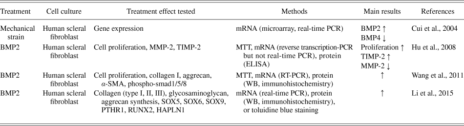
Investigations into the mechanisms by which BMPs may regulate eye growth are limited in number and have focused on the role of BMPs in scleral remodeling using in vitro scleral fibroblast cultures (summary in Table 4) (Cui et al., Reference Cui, Bryant, Sweet and McDonnell2004; Hu et al., Reference Hu, Cui, Yang, Wang, Hu, Li and Zeng2008; Wang et al., Reference Wang, Zhao, Xing, Zhang and Yang2011; Li et al., Reference Li, Cui, Zhao, Huo, Hu and Zeng2015). Synthesis of extracellular matrix (ECM) components, including collagen, glycosaminoglycan, and aggrecan all showed increases with the application of BMP2 protein to cell culture medium (Li et al., Reference Li, Cui, Zhao, Huo, Hu and Zeng2015). Interestingly, both BMP2 and BMP4 genes also show differential expression when mechanical strain is applied to human scleral fibroblast culture (Cui et al., Reference Cui, Bryant, Sweet and McDonnell2004), the in vivo analogy being the effect on the intact sclera of intraocular pressure.
Summary of key findings from in vitro studies investigating BMP gene expression and the effects of exogenous BMPs.

Further provocative data tying BMPs with eye growth regulation and myopia come from human genetic studies. Specifically, two independent studies have pointed to the potential role(s) of BMPs in myopia development, with overlap in two of the three BMP genes, BMP2 and BMP4, targeted in the current and related published RPE studies (Bakrania et al., Reference Bakrania, Efthymiou, Klein, Salt, Bunyan, Wyatt, Ponting, Martin, Williams, Lindley, Gilmore, Restori, Robson, Neveu, Holder, Collin, Robinson, Farndon, Johansen-Berg, Gerrelli and Ragge2008; Verhoeven et al., Reference Verhoeven, Hysi, Wojciechowski, Fan, Guggenheim, Höhn, MacGregor, Hewitt, Nag, Cheng, Yonova-Doing, Zhou, Ikram, Buitendijk, McMahon, Kemp, Pourcain, Simpson, Mäkelä, Lehtimäki, Kähönen, Paterson, Hosseini, Wong, Xu, Jonas, Pärssinen, Wedenoja, Yip, Ho, Pang, Chen, Burdon, Craig, Klein, Klein, Haller, Metspalu, Khor, Tai, Aung, Vithana, Tay, Barathi, Chen, Li, Liao, Zheng, Ong, Döring, Evans, Timpson, Verkerk, Meitinger, Raitakari, Hawthorne, Spector, Karssen, Pirastu, Murgia, Ang, Mishra, Montgomery, Pennell, Cumberland, Cotlarciuc, Mitchell, Wang, Schache, Janmahasatian, Igo, Lass, Chew, Iyengar, Gorgels, Rudan, Hayward, Wright, Polasek, Vatavuk, Wilson, Fleck, Zeller, Mirshahi, Müller, Uitterlinden, Rivadeneira, Vingerling, Hofman, Oostra, Amin, Bergen, Teo, Rahi, Vitart, Williams, Baird, Wong, Oexle, Pfeiffer, Mackey, Young, van Duijn, Saw, Bailey-Wilson, Stambolian, Klaver and Hammond2013). In one study, BMP2 was identified in a genome-wide meta-analyses of multiancestry cohort study encompassing European, American, Australian, and East-Asian populations, aimed at the identification new loci for refractive errors and understanding the mechanisms of refractive error and myopia development (Verhoeven et al., Reference Verhoeven, Hysi, Wojciechowski, Fan, Guggenheim, Höhn, MacGregor, Hewitt, Nag, Cheng, Yonova-Doing, Zhou, Ikram, Buitendijk, McMahon, Kemp, Pourcain, Simpson, Mäkelä, Lehtimäki, Kähönen, Paterson, Hosseini, Wong, Xu, Jonas, Pärssinen, Wedenoja, Yip, Ho, Pang, Chen, Burdon, Craig, Klein, Klein, Haller, Metspalu, Khor, Tai, Aung, Vithana, Tay, Barathi, Chen, Li, Liao, Zheng, Ong, Döring, Evans, Timpson, Verkerk, Meitinger, Raitakari, Hawthorne, Spector, Karssen, Pirastu, Murgia, Ang, Mishra, Montgomery, Pennell, Cumberland, Cotlarciuc, Mitchell, Wang, Schache, Janmahasatian, Igo, Lass, Chew, Iyengar, Gorgels, Rudan, Hayward, Wright, Polasek, Vatavuk, Wilson, Fleck, Zeller, Mirshahi, Müller, Uitterlinden, Rivadeneira, Vingerling, Hofman, Oostra, Amin, Bergen, Teo, Rahi, Vitart, Williams, Baird, Wong, Oexle, Pfeiffer, Mackey, Young, van Duijn, Saw, Bailey-Wilson, Stambolian, Klaver and Hammond2013). The second study of patients with ocular malformations identified BMP4 as a candidate gene for myopia using a positional candidate gene approach (Bakrania et al., Reference Bakrania, Efthymiou, Klein, Salt, Bunyan, Wyatt, Ponting, Martin, Williams, Lindley, Gilmore, Restori, Robson, Neveu, Holder, Collin, Robinson, Farndon, Johansen-Berg, Gerrelli and Ragge2008).
In summary, we examined the expression levels of three BMP genes and three BMP receptors in the chick retina and choroid and showed expression of three of these BMP genes to be differentially regulated by optical defocus in the choroid. These findings add to the growing body of evidence from other animal model and human genetics studies implicating BMPs in eye growth regulation and/or myopia and represent important missing information. Nonetheless, further investigations of this retina–RPE–choroid signaling pathway, both downstream and upstream, are needed to complete this picture and identify key molecules that plausibly could serve as targets for novel ophthalmic anti-myopia therapeutic interventions.
Acknowledgments
The authors thank funding supports from National Eye Institute Grants R01 EY012392 (CFW), K08 EY023609 (YZ), and K12 EY017269 (YZ & YL), and T32 EY007043 (YZ).