Implications
Muscle development in domesticated animals is important for meat production. Previous studies have demonstrated that there are observably different abilities to accumulate muscle postnatally in different breeds of pigs. However, little is known about the breed differences in terms of fetal skeletal muscle growth. Thus, we investigated the Chenghua and Yorkshire pig breeds to clarify the mechanisms of prenatal muscle development in obese and lean pig breeds at two landmark time points before birth. Results of the current study will benefit to better understand the mechanisms of prenatal muscle development and further research on meat production.
Introduction
Skeletal muscle tissue consists of various cell types, such as muscle fibers, adipocytes, endothelial cells and connective tissue cells. Meat originates from skeletal muscle tissue of the slaughtered animal by definition (Te Pas et al., Reference Te Pas, Everts and Haagsman2004). Each cell type of skeletal muscle tissue is supposed to impact the traits of meat quality. However, muscle fibers make up the majority of skeletal muscle mass, and thus meat mass. Therefore, muscle development in domesticated animals is important for meat production.
Prenatal skeletal muscle development plays an important role in postnatal growth. During fetal myogenesis, primary and secondary myofibers undergo both hypertrophy and hyperplasia. The number of myofibers is fixed at birth, and subsequent muscle growth depends entirely on hypertrophy of myofibers (Beermann et al., Reference Beermann, Cassens and Hausman1978; Foxcroft et al., Reference Foxcroft, Dixon, Novak, Putman, Town and Vinsky2006; Xi et al., Reference Xi, Hathaway, Dayton and White2007). Therefore, a change in the number of myofibers during prenatal growth can have a profound effect on the total muscle mass and long-term growth potential of the adult (Fahey et al., Reference Fahey, Brameld, Parr and Buttery2005), and thus livestock meat production. Prenatal myogenesis is regulated by crucial factors such as paired box gene 7 (Pax7) that induces the expression of myogenic regulatory factors including myogenic differentiation 1 (MyoD) and myogenin (MyoG) (Hyatt et al., Reference Hyatt, McCall, Kander, Zhong, Roy and Huey2008). MyoD plays a role in myoblast formation, while MyoG is essential for myoblast fusion and myotube formation (Megeney and Rudnicki, Reference Megeney and Rudnicki1995). Because myogenesis and adipogenesis occur in the same microenvironment and mainly have the same origin (Du et al., Reference Du, Yan, Tong, Zhao and Zhu2010), adipose-related genes appear to be expressed in skeletal muscle. Besides, lean meat itself includes both muscle fibers and fat cells, and fetal muscle development also involves adipogenesis. Promoting intramuscular adipogenesis increases intramuscular fat (IMF) and meat quality is also improved (Du et al., Reference Du, Zhao, Yan, Huang, Nicodemus, Yue, McCormick and Zhu2011). IMF content is an important trait of meat intended for consumption. Peroxisome proliferator-activated receptor γ (PPARG) has been reported to regulate adipogenesis and induce the expression of multiple genes including fatty-acid-binding protein 4 (FABP4) and lipoprotein lipase (LPL), which are involved in gene transcription and regulation of enzymatic activity (Rosen et al., Reference Rosen, Sarraf, Troy, Bradwin, Moore, Milstone, Spiegelman and Mortensen1999; Zimmerman and Veerkamp, Reference Zimmerman and Veerkamp2002).
Among the factors regulating skeletal muscle development, myostatin (Mstn), which is detected in all mammalian species as well as chickens, is expressed in both developing and adult skeletal muscles and is involved in the regulation of fetal and postnatal skeletal muscle mass (McPherron and Lee, Reference McPherron and Lee1997 and Reference McPherron and Lee2002; McPherron et al., Reference McPherron, Lawler and Lee1997). Inhibition of Mstn expression dramatically increases the muscle mass in animals (McPherron and Lee, Reference McPherron and Lee1997; McPherron et al., Reference McPherron, Lawler and Lee1997). In addition, Mstn not only inhibits muscle growth but also affects adipose growth. Mstn-null mice have less fat accumulation than that in wild-type mice (McPherron and Lee, Reference McPherron and Lee2002). Considering that fat content (or IMF content) is a key trait in meat production, Mstn provides a novel opportunity to investigate the molecular mechanisms of muscle development. In addition to its direct effect on muscle and fat development, previous studies have shown that Mstn might negatively regulate muscle growth indirectly through systemic effects on the IGF axis (Williams et al., Reference Williams, Interlichia, Jackson, Hwang, Cohen and Rodgers2011). Furthermore, the growth hormone (GH)/IGF axis plays a critical role in animal growth and development. Recent reports have shown that dysregulation of GH and IGF-1 actions may contribute to the loss of muscle mass (Perrini et al., Reference Perrini, Laviola, Carreira, Cignarelli, Natalicchio and Giorgino2010).
Therefore, it is necessary to investigate the expression of myogenic, adipogenic and growth-related factors to better understand the molecular mechanisms of muscle development. The Chenghua pig breed, a typical southwest-type of Chinese indigenous pig, is known to have a slower growth rate, higher backfat thickness and lower percentage of lean meat than those of commercial pig breeds such as Yorkshire (Yang et al., Reference Yang, Wang, Liu, Zhang, Zhao, Yu, Fan, Li, Xiong and Li2003). There are observably different abilities to accumulate muscle in different breeds of pigs. Thus, it is important to evaluate the breed differences of potential mechanisms in muscle development. However, there are few studies that compare breed differences in terms of skeletal muscle growth before birth, which is important to further elucidate the mechanisms of muscle growth. In this study, we investigated the Chenghua and Yorkshire pig breeds to clarify the mechanisms of muscle development in obese and lean pig breeds by examining the expression of genes related to myogenesis and adipogenesis, and skeletal muscle growth during fetal muscle development.
Material and methods
Animals
All procedures were approved by the Sichuan Agricultural University Animal Care Advisory Committee. Both Chenghua (C, first parity) and Yorkshire (Y, first parity) gilts, mated artificially with the same breed of sire and one sire for four gilts at each developmental stage, were housed individually at the Animal Nutrition Institute of Sichuan Agricultural University. The pregnant pigs were fed a total of 2.0 kg/day of a standard gestation diet with free access to water. At prenatal days 50 (d50) 90 (d90), four dams were sacrificed and six fetuses (n=24) with similar BWs from each dam at each developmental stage within each breed were prepared for sample collection.
Sample collection
Immediately after obtaining the fetuses, blood samples were collected from their umbilical cords. Sera from the centrifuged blood samples were immediately stored at −80°C for further analysis. Then, trunk and limb skeletal muscles were collected from the fetuses. Muscle tissues for histological analysis were stored in 10% neutral buffered formalin. For RNA isolation, muscle samples were snap frozen in liquid nitrogen and stored at −80°C until RNA extraction.
Measurements of serum Mstn, GH, IGF-1 and leptin
Concentrations of Mstn, GH, IGF-1 and leptin in serum were determined using commercial ELISA kits (R&D Systems China Co., Ltd, Shanghai, China).
Histology of skeletal muscles, and immunohistochemical detection and western blotting of Mstn
Muscle tissues were fixed for 48 h in 10% neutral buffered formalin, embedded in paraffin, sectioned using a cryostat (Leica RM2015; Wetzlar, Germany), mounted on slides, and then stained with hematoxylin and eosin. At least three fields of view per section and three sections per fetus were randomly chosen to determine the diameter and number of fetal myofibers.
For immunohistochemistry of Mstn, paraffin-embedded sections were prepared and mounted on slides. Then, A standard protocol was carried out as described (Soslow et al., Reference Soslow, Dannenberg, Rush, Woerner, Khan, Masferrer and Koki2000; Patruno et al., Reference Patruno, Caliaro, Maccatrozzo, Sacchetto, Martinello, Toniolo, Reggiani and Mascarello2008).
For western blotting, a total protein extraction kit was used to extract protein samples from muscle tissues. Then, western blotting was performed with an anti-Mstn antibody (R&D Systems China Co., Ltd) according to a standard protocol as described previously (Patruno et al., Reference Patruno, Caliaro, Maccatrozzo, Sacchetto, Martinello, Toniolo, Reggiani and Mascarello2008) with β-actin as the control protein. The relative expression of Mstn was normalized using β-actin, and then the normalized values were used for comparison of the expression of Mstn protein across breeds within each developmental time point.
Quantitative real-time RT-PCR
Total RNA was extracted using RNAiso reagent (Takara Biotechnology, Dalian, China). RNA samples were reverse transcribed to cDNA for real-time PCR analysis using a PrimeScript® RT reagent Kit with gDNA Eraser Kit (Perfect Real Time; Takara Biotechnology). PCR primer sequences are listed in Supplementary Table S1. Real-time PCR was conducted with iQ™ SYBR® Green Supermix (Bio-Rad, Hercules, CA, USA) reagents in a CFX96™ Real-Time PCR Detection System (Bio-Rad). All procedures were performed according to the manufacturer’s protocols. For normalization, β-actin was used as the internal control. All reactions were performed in triplicate, and the relative gene expression for each gene was calculated using the
 $${\rm 2}^{{{\minus}\Delta \Delta }} ^{{C_{t} }} $$
method (Livak and Schmittgen, Reference Livak and Schmittgen2001).
Cluster analysis of genes detected in fetal skeletal muscles
To compare the expression patterns of genes detected in the fetal skeletal muscles of the two pig breeds, the relative mRNA expression data of each gene were used for hierarchical cluster analysis. Hierarchical clustered genes were based on the expression patterns in Yorkshire and Chenghua pigs during the chosen developmental stages. Data were clustered using the Pearson correlation metric with the average linkage method. A heat map was generated using the heatmap.2 function of the R/gplots package (Warnes et al., Reference Warnes, Bolker, Bonebakker, Gentleman, Liaw, Lumley, Maechler, Magnusson, Moeller, Schwartz and Venables2014).
Statistical analysis
Data are presented as the mean±s.e. Statistical analyses were carried out using SPSS Statistics v20.0 (SPSS Inc., Chicago, IL, USA). Data were analyzed using the t-test within each developmental time point. The model used for analysis was
 $$y_{{ij}} =\mu {\plus}G_{i} {\plus}{\varepsilon}_{{ij}} $$
, where y
ij
is the diameter, number of myofibers, concentration of hormones or normalized gene/protein expression; μ the overall mean; G
i
the i
th breed; and ε
ij
the error for the j
th replicate within the i
th breed. A P-value<0.05 was considered to indicate significance.
Results
Histology of skeletal muscles
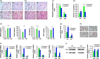
As observed by histological examination, diameter and number of fetal muscles changed in both pig breeds during gestation (Figure 1a to d). Our initial statistical analyses indicated that the traits at each time point and in both breeds were distributed normally. The diameters of myofibers were significantly larger in Chenghua pig fetuses than those in Yorkshire pig fetuses at d50 (11.15 v. 9.89 μm, P<0.001) and d90 (6.99 v. 6.56 μm, P<0.001) (Figure 1c). Moreover, the number of myofibers in Chenghua pig fetuses was higher than that in Yorkshire pig fetuses at d90 (299 v. 267, P=0.001) (Figure 1d).
Analyses of breed differences of Yorkshire and Chenghua. Crosscut (a) and vertical cut (b) muscle sections, and diameter (c) and number (d) of fetal skeletal muscles. Concentration of serum Mstn, Leptin, GH and Ghr (e), and relative mRNA level of related gene expression (f). Quantitative real-time RT-PCR (g), immunohistochemistry (h) and western blotting analysis (i) of Mstn in fetal skeletal muscles of Yorkshire and Chenghua pig fetuses at different stages. Data were shown as mean±s.e. Representative fetal muscle tissue sections were from the trunk and limb skeletal muscles of fetuses (400× magnification, scale bar=50 μm). *P<0.05, **P<0.01. C50=Chenghua fetuses at 50 days of gestation; C90=Chenghua fetuses at 90 days of gestation; Y50=Yorkshire fetuses at 50 days of gestation; Y90=Yorkshire fetuses at 90 days of gestation.

Serum Mstn, leptin, GH and IGF-1 concentrations and relative mRNA expression of genes related to skeletal muscle
Breed differences of serum factors mainly existed at d90 (Figure 1e). The serum Mstn concentration was significantly higher in Yorkshire pig fetuses compared with that in Chenghua pig fetuses at d90 (12.23 v. 9.54 ng/ml, P<0.001) (Figure 1e). In contrast, the GH concentration was significantly higher in Chenghua pig fetuses compared with that in Yorkshire pig fetuses at d90 (0.56 v. 0.46 ng/ml, P=0.006) (Figure 1e). However, there were no statistical differences in serum leptin (449.17 v. 465.53 ng/ml, P=0.445) or IGF-1 (11.59 v. 12.48 ng/ml, P=0.55) of Chenghua and Yorkshire pig fetuses at d90 (Figure 1e). In Yorkshire pig fetuses, the concentrations of serum leptin (309.16 v. 465.53 ng/ml, P<0.001) and IGF-1 (10.01 v. 12.48 ng/ml, P=0.005) had increased significantly during gestation (Figure 1e).
Gene expression in fetal skeletal muscles displays a different expression pattern of serum factors. In contrast to the expression pattern of serum GH, relative mRNA expression of Ghr in fetal skeletal muscles was significantly higher in Yorkshire pig fetuses than that in Chenghua pig fetuses at d90 (3.64 v. 1.30, P<0.001) (Figure 1f). Although there were no significant differences in the concentration of serum leptin (465.53 v. 449.17 ng/ml, P=0.467) or IGF-1 (12.48 v. 11.59 ng/ml, P=0.147) in Yorkshire and Chenghua pig fetuses at d90, the leptin receptor (LEPR) and IGF-1 mRNA levels were higher in Yorkshire pig fetuses at both d50 (LEPR: 27.90 v. 9.17, P<0.001; IGF-1: 12.14 v. 5.28, P<0.001) and d90 (LEPR: 2.17 v. 1.01, P=0.002; IGF-1: 5.05 v. 1.29, P<0.001) (Figure 1f). In accordance with serum Mstn, Yorkshire pig fetuses had a significantly increased mRNA level of Mstn in fetal skeletal muscles compared with that in Chenghua pig fetuses at d90 (2.87 v. 1.21, P<0.001) (Figure 1g).
Different expression patterns of myogenic and adipogenic gene expression in skeletal muscles of Chenghua and Yorkshire pig fetuses
Quantitative real-time RT-PCR was used to detect the relative mRNA levels of myogenic and adipogenic genes in fetal skeletal muscles of Chenghua and Yorkshire pig fetuses at d50 and d90. In general, there were obvious breed differences of the expression patterns of myogenic and adipogenic genes in Chenghua and Yorkshire pig fetuses at d50 and d90.
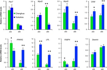
Because Mstn negatively regulates muscle growth, the expression of Mstn was also detected to better understand the mechanisms of muscle development. Quantitative RT-PCR results indicated that the mRNA level of Mstn was higher in Yorkshire pig fetuses compared with that in Chenghua fetuses at d50 (10.78 v. 5.63, P<0.001) and d90 (2.87 v. 1.12, P<0.001) (Figure 1g). Interestingly, the breed difference of myogenic gene expression was mainly detected at d90, whereas only creatine kinase, muscle (CKM) showed a statistical difference of mRNA expression in Chenghua and Yorkshire pig fetuses at d50 (1.76 v. 1.01, P<0.001) (Figure 2). As an important factor for the formation of myoblasts during skeletal muscle development, MyoD had a significantly higher mRNA level in Chenghua pig fetuses compared with that in Yorkshire pig fetuses at d90 (2.78 v. 0.61, P<0.001) (Figure 2). However, during the later stage of skeletal muscle development, as a marker of myoblast fusion and myotube formation, the mRNA expression level of MyoG was obviously higher in Yorkshire pig fetuses than that in Chenghua pig fetuses at d90 (1.69 v. 0.78, P=0.002) (Figure 2). Thus, these myogenic gene expression patterns suggested that the mechanisms of myogenesis during fetal skeletal muscle development might involve different signaling pathways in the two pig breeds, which may be associated with Mstn.
Relative mRNA expressions of myogenic and adipogenic genes examined in skeletal muscles. Quantitative real-time PCR detection was used to quantify mRNA expression level of the target genes in these fetuses at two developmental stages (d50, d90) of breeds. Data were shown as mean±s.e. *P<0.05, **P<0.01.

In addition to negative regulation of muscle development, Mstn plays an important role in adipogenesis. Fat content (or IMF content) is a key point for meat production, which is mainly associated with muscle development. Thus, we investigated the mRNA expression level of adipogenic genes in fetal skeletal muscles of Chenghua and Yorkshire pig fetuses at d50 and d90. Compared with Chenghua pig fetuses, a significant reduction of PPARG (2.57 v. 1.74, P=0.026) and FABP4 (1.23 v. 0.57, P=0.037) mRNA levels was observed in Yorkshire pig fetuses at d50 (Figure 2). Moreover, a significant increase in LPL (11.26 v. 6.19, P<0.001) expression was observed in Yorkshire pig fetuses compared with that in Chenghua pig fetuses at d50. On the other hand, the mRNA levels of PPARG (2.03 v. 1.08, P<0.001), FABP4 (4.42 v. 1.27, P<0.001) and LPL (6.05 v. 1.36, P<0.001) were significantly higher in Yorkshire fetuses than those in Chenghua fetuses at d90 (Figure 2). These results revealed that the adipogenic gene expression profiles of fat, especially IMF, differed in the two pig breeds during development.
Cluster analysis of gene expression in skeletal muscles of Yorkshire and Chenghua pig breeds
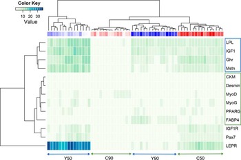
To better understand the mechanisms of muscle development, cluster analysis was conducted for all genes detected in Chenghua and Yorkshire pig fetuses at d50 and d90. A summary heat map and hierarchical clustering of gene expression patterns in the two pig breeds at different developmental stages are shown in Figure 3. The clusters separated fetuses according to their breed (Chenghua v. Yorkshire) and developmental stage (d50 v. d90). As expected by their biological functions in myogenesis and adipogenesis, myogenic genes (MyoG, MyoD, Desmin and CKM) and adipogenic genes (PPARG and FABP4) were clustered into the same group. Because of its regulatory role in both myogenesis and adipogenesis, Mstn was not clustered within this group. Mstn was clustered with the group of Ghr, IGF-1.
Cluster analysis of gene expression in fetal skeletal muscles. Cluster analysis was used to generate a heat map. Green and blue represent lower and higher expression levels of each gene, respectively, with the intensity of the color directly proportional to the degree of gene expression. Vertical columns represent the data for each muscle sample from Yorkshire and Chenghua fetuses, and rows indicate the different genes detected from skeletal muscles. C50=Chenghua fetuses at 50 days of gestation; C90=Chenghua fetuses at 90 days of gestation; Y50=Yorkshire fetuses at 50 days of gestation; Y90=Yorkshire fetuses at 90 days of gestation.

Immunohistochemistry and western blot analysis of Mstn in Yorkshire and Chenghua pig breeds
Because the cluster analysis indicated that Mstn had an obviously different expression pattern from those of myogenic and adipogenic genes, but a similar expression pattern to that of the paracrine factor IGF-1, we analyzed Mstn expression at the protein level. Immunohistochemistry revealed extensive Mstn protein expression in the fetal skeletal muscles of Chenghua and Yorkshire pig fetuses at different developmental stages (Figure 1h). Western blot analysis of Mstn (Figure 1i) indicated that there was a significant difference of Mstn protein expression in the two pig breeds. Yorkshire pig fetuses had an obviously higher level of Mstn protein compared with that in Chenghua pig fetuses at d50 (1.77 v. 1.29, P=0.029) and d90 (1.81 v. 1.41, P=0.037), which was consistent with qRT-PCR results of Mstn gene expression.
Discussion
Histological analysis of skeletal muscle identified differences in the diameters and number of myofibers in Chenghua and Yorkshire pig fetuses. No difference in myofiber number was observed in the two pig breeds at d50. In pig fetal development, d50 (d30 to d55) and d90 (d90 to d105) represent key time points of primary myogenesis and secondary myogenesis during skeletal muscle development, respectively (Beermann et al., Reference Beermann, Cassens and Hausman1978; Foxcroft et al., Reference Foxcroft, Dixon, Novak, Putman, Town and Vinsky2006). The reason for the similar numbers of myofibers at d50 may be equally active primary myogenesis in Chenghua and Yorkshire pig fetuses at this time point. At d90, the number of myofiber in Yorkshire was greater than that in Chenghua. The MyoG gene is essential for myoblast fusion and myotube formation (Megeney and Rudnicki, Reference Megeney and Rudnicki1995). The higher expression level of MyoG was observed in Yorkshire at d90 (Figure 2), and therefore this may be one of the reasons for a great number of myofibers observed in Yorkshire.
We found significant differences in the concentrations of serum Mstn, GH, IGF-1 and leptin in the two pig breeds at d90, and in Yorkshire at d50. Ideally, comparisons would have been made using serum samples of both Yorkshire and Chenghua pig fetuses within each developmental stage. However, obtaining serum from d50 fetuses of Chenghua pigs was difficult because of their small size. Marcell et al. (Reference Marcell, Harman, Urban, Metz, Rodgers and Blackman2001) reported a significant inverse correlation of Mstn and Ghr levels in healthy but elderly humans (>65 years old) and a disassociation existed between GH and autocrine IGF-1 effects on muscle protein synthesis in humans. In the present study, we found that Mstn and genes related to growth (GH and IGF-1) were grouped into the same cluster, suggesting a relationship between Mstn, GH and IGF-1. This result suggested that the regulatory role of Mstn in muscle development might be more relevant than that of IGF-1 but closely related to the GH signaling pathway during primary and secondary myogenesis of fetal skeletal muscles in pigs, which is in accordance with previous studies (Liu et al., Reference Liu, Thomas, Asa, Gonzalez-Cadavid, Bhasin and Ezzat2003; Vijayakumar et al., Reference Vijayakumar, Wu, Sun, Li, Jeddy, Liu, Schwartz, Yakar and LeRoith2012). Liu et al. (Reference Liu, Thomas, Asa, Gonzalez-Cadavid, Bhasin and Ezzat2003) demonstrated upregulation of Mstn in myoblasts during GH receptor antagonism. Moreover, Vijayakumar et al. (Reference Vijayakumar, Wu, Sun, Li, Jeddy, Liu, Schwartz, Yakar and LeRoith2012) demonstrated lower Mstn expression in the muscles of both lean and obese mice with inactivated GH receptors compared with that in normal mice. These results suggested that Mstn might play roles as not only a regulator but also a paracrine factor, which is consistent with a previous study (Guernec et al., Reference Guernec, Berri, Chevalier, Wacrenier-Cere, Le Bihan-Duval and Duclos2003). Furthermore, swine, especially Yorkshire pigs, are typically used as a model for type-1 or type-2 diabetes mellitus in previous studies (Suzuki et al., Reference Suzuki, Poot, Gerrity and Bornfeldt2001; Natarajan et al., Reference Natarajan, Gerrity, Gu, Lanting, Thomas and Nadler2002; Bellinger et al., Reference Bellinger, Merricks and Nichols2006). Guo et al. (Reference Guo, Bond, Jou, Gavrilova, Portas and McPherron2012) found that inhibition of Mstn might be an effective treatment for diabetes. In the present study, significant breed differences of Mstn, GH and Ghr expression were observed in the lean pig breed Yorkshire and obese pig breed Chenghua, indicating that different signaling factors might be involved in the diabetic metabolisms of lean and obese pig breeds via the GH/Ghr signaling pathway.
At d90, MyoD was observed with significantly lower mRNA expression in Yorkshire pig fetuses. Porcine MyoD family play a vital role in myogenesis of skeletal muscle development, and are therefore considered to be candidate genes for traits of meat production. During myogenesis, MyoD and Myf5 are first expressed to specify the myogenic lineage in the myogenic network during embryonic development, while MyoG appeared later to determine the myogenic differentiation (fusion of myoblasts into multi-nucleated muscle fibers) (Te Pas et al., Reference Te Pas, Everts and Haagsman2004). In the current study, both myogenic and adipogenic genes were clustered into the same group owing to the similar expression pattern with higher expression levels in Yorkshire at d90, such as MyoG, CKM, PPARG and FABP4. Gao et al. (Reference Gao, Kishida, Ejima, Gojo and Mazda2013) suggested that Mstn is an autocrine/paracrine negative regulator in myoblast differentiation via inhibition of MyoD in human stem cells. A recent study reported that significant differences in IMF were observed between different genotypes of MyoD in pigs (Verner et al., Reference Verner, Humpolicek and Knoll2007). Interestingly, they also found that there were statistically significant associations between MyoG and fat (Verner et al., Reference Verner, Humpolicek and Knoll2007). Rosen et al. (Reference Rosen, Sarraf, Troy, Bradwin, Moore, Milstone, Spiegelman and Mortensen1999) demonstrated that PPARG was one of the key factors controlling adipogenesis. Lipid deposition in the skeletal muscle was related to adipogenic genes PPARG and FABP4, and thus reinforced the role of FABP4 in IMF development in skeletal muscles (da Costa et al., Reference da Costa, Pires, Fontes and Mestre Prates2013). Therefore, it is plausible that altered myogenic and adipogenic gene expression pattern, which act in myogenesis and adipogenesis, can impact myogenic and adipogenic processes associated with skeletal muscle development, and thus may affect meat production traits.
In conclusion, for skeletal muscle, primary and secondary myogenesis play a vital role in fetal and postnatal muscle development that is important for meat production and health. Our present study indicated significant breed differences in skeletal muscle development of lean and obese pig breeds at two important time points before birth. Moreover, Mstn may serve a negative regulator through the GH/Ghr signaling pathway, which further elucidates the regulatory roles of Mstn in skeletal muscle development before birth and provides novel insights into targeted therapy of diabetes through the Mstn-GH/Ghr signaling pathway.
Acknowledgements
This work was financially supported by National Natural Science Foundation of China (No.30972119) and National Basic Research Program of China (No. 2012CB124701).
Conflicts of Interest
None.
Supplementary material
To view supplementary material for this article, please visit http://dx.doi.org/10.1017/S1751731114002316