Introduction
Internal auditory canal anomalies are uncommon. They are usually unilateral and associated with other inner, middle or external ear anomalies.Reference Demir, Cakmakei, Erdag and Men1 A narrow internal auditory canal is believed to occur as a result of aplasia or hypoplasia of the vestibulocochlear nerve. Narrow duplication of the internal auditory canal is considered a very rare anomaly. It was first described in 1991 by Weissman et al.,Reference Weissman, Arriaga and Curtin2 and only 10 cases have been reported.Reference Lee, Cha, Jeon, Bae, Han and Kim3 Bilateral duplication of the internal auditory canal can be considered extremely rare, being first described in 2007 by Weon et al., followed by a second case reported in 2008 by Bakar et al.Reference Weon, Kim, Choi and Koo4, Reference Bakar, Karadag, Calisir and Adapinar5 A triplicated internal auditory canal case was recently described by Lee et al.Reference Lee, Cha, Jeon, Bae, Han and Kim3
To our knowledge, the presented case represents the third published report of bilateral duplicated internal auditory canal.
Case report
A 14-year-old boy with bilateral congenital dysacusis, who had used sound amplification devices since nine years of age, presented for hearing rehabilitation. He had excellent orofacial reading and normal writing, and attended school regularly.
Physical examination showed no abnormality other than the chief complaint.
Audiological tests demonstrated bilateral, profound, sensorineural dysacusis. Tympanometry produced a type A tympanogram with absent stapedic reflexes. Audiometry revealed thresholds of 80 dB in the right ear and 70 dB in the left ear. Brainstem auditory evoked potential testing produced no response. Videoelectronystagmography testing indicated bilateral peripheral vestibular syndrome.
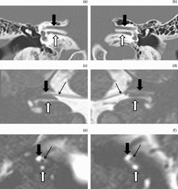
Multidetector computed tomography (CT) (Figure 1) provided high resolution submillimetric slices. Multiplanar and three-dimensional reconstruction of these slices demonstrated bilateral duplication of the internal auditory canal, with narrowing of the lower canals, unilateral cochlear and vestibular dysplasia, bilateral superior semicircular canal malformation, and absence of the posterior semicircular canals.
(a) and (b): coronal multidetector computed tomography, multiplanar reconstruction, demonstrating bilateral duplication of the internal auditory canal. (c) and (d): coronal magnetic resonance imaging (MRI), multiplanar reconstruction, demonstrating the facial nerves entering the upper canals. (e) and (f): sagittal MRI, multiplanar reconstruction, demonstrating the facial nerves in the upper canals and the empty lower canals. Thick black arrow = upper canal; white arrow = lower canal; thin black arrow = facial nerve

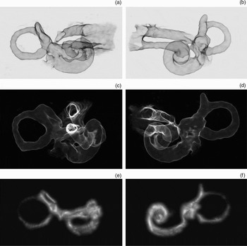
Magnetic resonance imaging information was obtained from a three-dimensional slab of turbo spin-echo sequence (using the ‘Syngo sampling perfection with application optimised contrasts using different flip angle evolution’ system; Siemens, AG, Erlangen, Germany). This demonstrated internal auditory canal and labyrinth malformations; in addition, neither cochlear nerve was able to be demonstrated (Figure 2).
Right (a, c) and left (b, d) three-dimensional, edge-enhanced, post-processed images obtained after temporal bone high-resolution multidetector computed tomography. (e) Right and (f) left three-dimensional, volume rendered, post-processed images obtained after high-resolution, axial, T2-weighted magnetic resonance imaging produced by the ‘Syngo sampling perfection with application optimised contrasts using different flip angle evolution’ system, showing a right dysplastic cochlea, bilateral malformed superior semicircular canals, absent posterior semicircular canals and bilateral duplicated internal auditory canals.

Discussion
The incidence of severe congenital hearing loss is estimated at one in every 1000 live births, with a genetic cause in up to 50 per cent of cases.Reference Bakar, Karadag, Calisir and Adapinar5 Internal auditory canal anomalies constitute 12 per cent of congenital temporal bone anomalies.Reference Lee, Cha, Jeon, Bae, Han and Kim3 The normal diameter of the internal auditory canal is approximately 4 mm, and ranges from 2 to 8 mm. An internal auditory canal of less than 2 mm is considered stenotic.Reference Bakar, Karadag, Calisir and Adapinar5, Reference Ferreira, Shayestehfar and Lufkin6
The inner ear and internal auditory canal have different embryogenesis. The inner ear derives from the otic placode, beginning on the 22nd day of gestation, while the internal auditory canal derives from chondrification and posterior ossification of the mesoderm surrounding the VIIth and VIIIth cranial nerves. The vestibulocochlear nerve begins to develop on the 29th day, and its growth is thought to be induced by the embryonic labyrinth via chemotactic mechanisms.Reference Ferreira, Shayestehfar and Lufkin6
There are two theories which attempt to explain internal auditory canal stenosis.
The first considers narrowing of the internal auditory canal to result from abnormality of the end-organs of the labyrinth, which leads to failure of neural growth induction and consequent vestibulocochlear nerve aplasia or hypoplasia.Reference Ferreira, Shayestehfar and Lufkin6
The other theory considers that such stenosis is the primary defect, leading to aplasia or hypoplasia of the vestibulocochlear nerve by mechanical constriction. However, this is less likely, since most patients with internal auditory canal stenosis have preserved facial nerve function.Reference Ferreira, Shayestehfar and Lufkin6
In our patient, we observed associated inner ear anomalies, with some features similar to those described in previous reports. Narrowing of the lower internal auditory canals was observed, measuring approximately 0.5 mm on the right side and 1.0 mm on the left, and the vestibulocochlear nerves could not be identified. In the superior internal auditory canal, the facial nerves were easily identified. These observations are in accordance with previously reported radiological findings (vestibulocochlear nerves were not identified in 7 out of 8 evaluated IAC and all 8 facial nerves were identified),Reference Lee, Cha, Jeon, Bae, Han and Kim3 and corroborate the hypothesis that vestibulocochlear nerve aplasia or hypoplasia leads to a stenotic internal auditory canal.
It is important to evaluate the internal auditory canal and the cochlear nerve using high resolution CT and magnetic resonance imaging, since absence of the cochlear nerve is an absolute contraindication for cochlear implantation and internal auditory canal stenosis is considered a relative contraindication (with several reports of cochlear implant failure in the presence of such stenosis).Reference Ferreira, Shayestehfar and Lufkin6
• Duplication of the internal auditory canal is rare; bilateral presentation is extremely rare
• The few published case reports describe absence of the vestibulocochlear nerve, corroborating the theory that vestibulocochlear nerve aplasia or hypoplasia leads to a stenotic internal auditory canal
• Absence of the cochlear nerve is an absolute contraindication for cochlear implantation, and internal auditory canal stenosis is considered a relative contraindication, with several reports of cochlear implant failure in the presence of such stenosis