INTRODUCTION
Leishmania major is an obligate intracellular protozoan parasite that causes cutaneous leishmaniasis. The World Health Organization reports that the disease affects over 12 million cases of all forms of leishmaniasis worldwide with 500 000 new cases accruing annually (WHO, 1999). T-cell-mediated immune responses play a central role in the outcome of Leishmania infection (Pearson and Sousa, Reference Pearson and Sousa1996). Infection of mice with L. major leads to highly polarized Th1 and Th2 immune responses in resistant and susceptible inbred mice respectively (Scott, Reference Scott1991). IFN-γ is important for Th1 cell development (Scott, Reference Scott1991) and protection of mice against L. major infection (Scott et al. Reference Scott, Natovitz, Coffman, Pearce and Sher1988). It has been shown that resistant mice, such as C3H/HeN, become susceptible to L. major infection after treatment with anti-IFN-γ antibody (Scott, Reference Scott1991). The essential role of IL-4 in mediating both Th2 cell differentiation and susceptibility to L. major in BALB/c mice has been demonstrated (Sadick et al. Reference Sadick, Heinzel, Holaday, Pu, Dawkins and Locksley1990). IL-4 inhibits macrophage leishmanicidal activity and down-regulates the development of a Th1 response (Oswald et al. Reference Oswald, Gazzinelli, Sher and James1992). Genetically susceptible mice lacking IL-4 are protected from cutaneous infection with L. major (Kopf et al. Reference Kopf, Brombacher, Köhler, Kienzle, Widmann, Lefrang, Humborg, Ledermann and Solbach1996). We reported that an attenuated line of L. major, which was designated L. major H-line, was generated under gentamicin pressure (Daneshvar et al. Reference Daneshvar, Coombs, Hagan and Phillips2003a). We have also used the same attenuating procedure successfully with L. mexicana, L. infantum, and L. donovani (Daneshvar et al. Reference Daneshvar, Coombs, Hagan and Phillips2003a). Attenuation of the parasite is not simply the result of long-term cultivation of promastigotes (Daneshvar et al. Reference Daneshvar, Coombs, Hagan and Phillips2003a). The ability of promastigotes of the attenuated cell line of L. major to infect bone marrow-derived macrophages (BMMs) was similar to that of parasites of L. major WT. In contrast to L. major WT, however, amastigotes of the attenuated L. major H-line survive in only a small proportion of the macrophages (Daneshvar et al. Reference Daneshvar, Coombs, Hagan and Phillips2003a). Recently, we investigated the nature of the immune response of BALB/c mice induced by the attenuated line of L. mexicana (Daneshvar et al. Reference Daneshvar, Hagan and Phillips2003b). In the present study, we show that the L. major H-line failed to induce cutaneous lesions in BALB/c mice over a minimum of 11 weeks observation period, following subcutaneous injection of stationary phase parasites. The attenuated line of L. major potentiates a Th1-like response and controls cutaneous leishmaniasis in BALB/c mice. L. major H-line induced significant protection in mice against challenge with wild-type parasites.
MATERIALS AND METHODS
Parasites
L. major strain (MRHO/IR/75/ER) was provided by Dr A. Khamesipour (Center for Research and Training in Skin Diseases and Leprosy, Tehran, Iran) and the attenuated line was generated by a modification of a previously described method (Daneshvar et al. Reference Daneshvar, Coombs, Hagan and Phillips2003a). All lines were derived from the same original stabilate. Briefly, amastigotes derived from the spleen of an infected BALB/c mouse were transferred into complete haemoflagellate minimal essential medium (HOMEM) (GIBCO) supplemented with 10% (v/v) heat-inactivated foetal calf serum (HI-FCS; Labtech International) and incubated at 25°C. The amastigotes differentiated to promastigotes over 72 h. These were transferred into complete HOMEM containing initially 200 μg/ml and later (see below) 20 μg/ml gentamicin, or medium without gentamicin and incubated again at 25°C. Subsequent subcultures were made with mid- to late-log-phase promastigotes. After 3 subpassages of the promastigotes cultured in gentamicin at 200 μg/ml, these promastigotes were transferred into complete HOMEM medium with 20 μg/ml gentamicin for a further 5 passages. In summary, amastigotes differentiated to promastigotes →3 passages in medium with 200 μg/ml gentamicin →5 passages in medium with 20 μg/ml gentamicin. Attenuation of the parasite using this modified method was evident after a total of 8 passages (referred to as L. major H-line). In parallel to the above, as controls, cultures of the parasites were both subpassaged in medium containing 20 μg/ml gentamicin throughout (our previously published procedure for attenuation) (referred to as L. major H-line20) and in medium without gentamicin (L. major WT). Subpassages after 8 subpassages for both L. major H-line and L. major H-line20 was in complete HOMEM medium with 20 μg/ml gentamicin and WT in medium alone. Attenuation of L. major by this modified process was carried out on 2 separate occasions. The growth rate of promastigotes of L. major H-line, L. major H-line20 and L. major WT was determined in culture. The parasites were grown in HOMEM medium and promastigotes were counted daily.
Mice and infection protocols
Inbred female 12-week-old, BALB/c mice were purchased from Harlan UK. Stationary-phase promastigotes of day 7–9 cultures were collected (Spath and Beverley, Reference Spath and Beverley2001) and washed in phosphate-buffered saline (PBS, pH 7·4). Suspensions of stationary phase promastigotes of the L. major H-line (8 passages) or L. major WT (10 passages) at a concentration of 2·5×107 cells/ml in PBS were prepared, and 200 μl aliquots of these suspensions were injected subcutaneously (s.c.) into the shaven rumps of BALB/c mice. The lesion volume was measured with a capillary micrometer (Royal) at weekly intervals (Mottram et al. Reference Mottram, Souza, Hutchison, Cater, Frame and Coombs1996).
Interaction in vitro between stationary-phase promastigotes and murine peritoneal macrophages
Promastigotes which were serially subpassaged in medium containing gentamicin under the modified schedule described above, or the previously described schedule (designated L. major H-line20) (Daneshvar et al. Reference Daneshvar, Coombs, Hagan and Phillips2003a), and wild type parasites serially passaged in medium without gentamicin were tested for their virulence after 8 passages by assessing their ability to infect and multiply in macrophages in vitro over a 96-h observation period. Stationary phase promastigotes were used. The macrophages were prepared as follows. Peritoneal macrophages were removed from BALB/c mice by peritoneal lavage and transferred into the individual wells of a chamber slide (Nunc/Invitrogen) at 2–3×10⁶ cells/well. The cells were incubated in complete Dulbecco's modified Eagle medium (DMEM; GIBCO) supplemented with 10% (v/v) HI-FCS, 2 mm L-glutamine, 50 U/ml penicillin, and 50 μg/ml streptomycin sulphate (Life Technologies), at 37°C in 5% CO2 95% air overnight. After removal of the non-adherent cells, the adherent cells were exposed to the stationary-phase promastigotes and incubated in 5% CO2 95% air for 3 h at 32°C (in our hands, 32°C is optimal for invasion of macrophages by promastigotes) (Naderer et al. Reference Naderer, Ellis, Fleur Sernee, Curtis, Handman and McConville2005). The free promastigotes were removed by replacing the overlying medium with complete DMEM, and the cells were incubated for a further 96 h at 37°C in 5% CO2 95% air and incubated. The initial infection ratio of parasites to macrophages was 1:1. At the end of the incubation period, the macrophages were fixed and stained with Giemsa's stain, and the infection status of the macrophages was microscopically determined by amastigote detection. The percentage of infected macrophages was determined by examining at least 300 cells for each line.
Detection of L. major parasites in the skin and visceral organs of infected mice
The presence of viable Leishmania parasites in the tissues of challenged and infected mice was detected as described previously (Laskay et al. Reference Laskay, Diefenbach, Rollinghoff and Solbach1995). Briefly, the skin at the site of the infecting inoculum, the proximal draining lymph nodes (PLNs), lungs, spleens, and livers of infected mice were removed aseptically into RPMI medium (GIBCO) at 12 weeks post-infection. Subsequently, the livers, draining PLNs, lungs and spleens of the mice were homogenized in complete HOMEM medium. Bone marrow cells were collected from the femurs and tibias of infected mice. Single-cell suspensions of epidermal cells were prepared from the skin. The skin was cut into 0·75 cm strips, and the strips were floated dermal side down on RPMI medium (GIBCO) supplemented with 1% trypsin (Sigma) for 90 min at 37°C. The sheets then were shaken to release the cells, and the cells were collected by sedimentation and washed. The serial 2-fold dilutions of the cell suspensions from the various organs and tissues were plated in 24-well plates. The cells were then incubated at 25°C for 14 days. The cultures were examined daily for the presence of promastigotes.
Cell proliferation and cytokine production
T-cell proliferation assays were performed as described by Satoskar et al. (Reference Satoskar, Brombacher, Dai, McInnes, Liew, Alexander and Walker1997). Briefly, spleens were aseptically removed from mice at 12 weeks post-infection. Single cell suspensions were prepared from each spleen by gently teasing them in complete DMEM medium, penicillin G (100 U/ml), streptomycin sulphate (100 μg/ml) (Life Technologies, Grand Island, NY), β-mercaptoethanol (50 μm) (Life Technologies, Grand Island, NY), L- glutamine (2 mm), and pyruvate (1 mm) (Sigma). The cell suspension was centrifuged at 200 g for 10 min at 4°C, and the erythrocytes were lysed by re-suspension of the pellet in 3 ml of Boyle's solution (0·17 m Tris-HCl, pH 7·2 and 0·16 m ammonium chloride). After 3 min incubation at 37°C the spleen cells were centrifuged at 200 g for 10 min at 4°C, washed 3 times with PBS and viable cells were enumerated by trypan blue exclusion. Cells were suspended at 5×10⁶ cells/ml in complete DMEM medium and 100 μl aliquots of these suspensions were added in triplicate to the wells of sterile 96-well flat-bottom tissue-culture plates (Falcon). An equal volume of stationary phase promastigotes of L. major WT at a concentration of 5×10⁵ cells per ml was added to each well; the ratio of promastigote/splenocyte was 1:10. One-hundred μl/well aliquots of Concanavalin A (Con A) (5 μg/ml) (Sigma) were used as a positive control for cell proliferation and complete DMEM alone was used as a negative control. The cells were initially incubated at 32°C for 4 h and then incubated at 37°C in 5% CO2 95% air for 56 h. After 56 h [H3] thymidine (1 μCi/well) (Amersham) was added to each well and the plates incubated at 37°C for a further 12 h. The cells were harvested onto filter paper (Tomtec, Hamden, CT) using a cell harvester (Skatron, Lier, Norway), and thymidine uptake was measured by liquid scintillation on a beta-scintillation counter. Supernatants were collected from parallel cultures after 72 h of incubation and stored at −70°C until the cytokine levels in these culture supernatants could be determined. Levels of IFN-γ and IL-4 were measured using ELISA kits purchased from R&D Systems.
Collection of sera and assay for anti-leishmanial IgG1 and IgG2a
Peripheral blood was obtained from the tail veins of the mice. Serum was prepared from the clotted blood of mice at 12 weeks post-infection and stored at −20°C before analysis by enzyme immunoassay. Each well of flat-bottom microtitre plates (Dynatech Laboratories, Billingshurst, UK) was coated with 1 μg of Leishmania lysate (see below) in 0·1 m carbonate buffer pH 9·6 and incubated at 4°C overnight. Following 3 washes in PBS (pH 7·4) containing 0·05% Tween-20, the plates were blocked with 200 μl of blocking buffer (PBS/FCS 10%), and incubated at 37°C for 1 h. After 3 washes, 50 μl of serially diluted serum samples (1/100 starting dilution in PBS/FCS 10%) were added to the wells and incubated for 2 h at 37°C. Bound antibodies were detected by 50 μl/well of biotinylated anti-mouse immunoglobulin G1 (IgG1) or IgG2a (PharMingen) (2 μg/ml), incubated at 37°C for 1 h and subsequently washed 6 times. One-hundred μl of streptavidin-linked peroxidase (1/1000 dilution in PBS/FCS 10%) were added and incubated at 37°C for 1 h. After the final wash with washing buffer, 100 μl of TMB substrate were added to each well. Absorbances were measured at 630 nm with the reference filter at 405 nm on an ELISA reader after 1 h of incubation.
Leishmania lysate preparation
Leishmania lysate was prepared from stationary-phase promastigotes of L. major WT. Promastigotes were washed with ice-cold PBS 3 times and disrupted 6 times by freezing at (−80°C) and thawing at (37°C) at 109 cell/ml in PBS. After each thaw, the suspension was passed through a 25-gauge needle and the suspension centrifuged at 100 00 g and 4°C for 10 min. The protein concentration was determined by the method of Bradford and the supernatant aliquoted and stored at −20°C.
Vaccination studies
Experiments were carried out to determine whether pre-treatment of mice with L. major H-line could protect susceptible BALB/c mice from infection with wild-type L. major. Two groups of BALB/c mice (5 mice/group) were injected s.c. with 5×10⁶ stationary-phase promastigotes of the L. major H-line or PBS into their shaven rumps. After 4 weeks, the immunized mice and control mice were challenged s.c. with 5×10⁶ stationary phase promastigotes of L. major WT into the rump on the opposite side. The lesion volume was measured weekly for 10 weeks as described above. (The 10 week time-point was chosen because at this time some mice infected with WT parasites had large lesions and had to be sacrificed to avoid unnecessary suffering.)
Statistical analysis
Data are expressed as the mean±s.e.m. for each group. Statistical analysis was performed using SPSS t-test (for the final determination of significance of the treatments).
RESULTS
Evaluation of growth rate of promastigotes in vitro
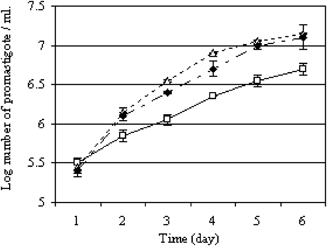
In order to compare the growth rate of promastigotes of L. major H-line with L. major H-line20 and L. major WT in cultures, parasites were grown in HOMEM medium and promastigotes were counted daily. As Fig. 1 shows, the growth rate of promastigotes of L. major H-line was significantly slower than L. major H-line20 or wild type after 11 subpassages. This was evident when cell numbers were compared on day 6 when there were significantly fewer promastigotes of L. major H-line compared with L. major H-line20 and L. major WT (P<0·0001) (Fig. 1).
Growth rates of promastigotes of Leishmania major H-line (□); L. major H-line20 (◆); L. major WT (▵) after 11 subpassages. The growth rate of promastigotes of L. major H-line was significantly slower than L. major H-line20 or wild type on day 6. Bars represent±s.e.m. of at least 3 experiments.

Infection of peritoneal macrophages with L. major WT and L. major H-line20 after 8 subpassages
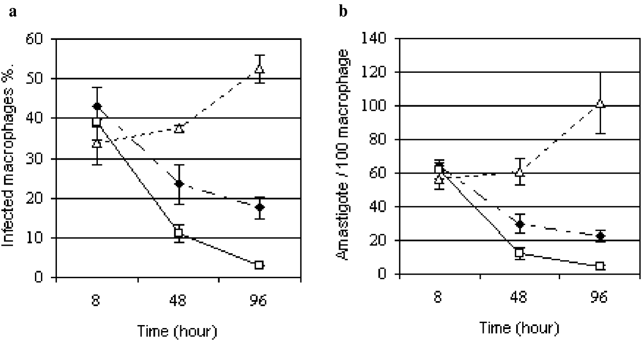
The percentage of macrophages infected with L. major H-line was similar to that of L. major H-line20 at 8 h post-infection. In contrast to L. major H-line20, amastigotes of L. major H-line survived in only a small proportion of the macrophages. The percentage of macrophages infected with L. major H-line20 at 8 h after infection was 43% decreasing to 17·5% at 96 h after infection (Fig. 2A). The percentage of macrophages infected with L. major H-line decreased over time, from 39% at 8 h to 3% at 96 h after infection. For L. major H-line the number of amastigotes within the macrophage at 8 h was 61·5 amastigotes/100 macrophages but declined to 4 amastigotes/100 macrophages at 96 h after infection, whereas, the number of amastigotes of L. major H-line20 within the infected macrophage at 8 h was 64 amastigotes/100 macrophages decreasing to 22·5 amastigotes/100 macrophages at 96 h (Fig. 2B). The percentage of macrophages infected with wild-type L. major at 8 h after infection was 34% and increased to 52·5% at 96 h after infection (Fig. 2A). For L. major WT, the number of amastigotes within the macrophage at 8 h was 59·5 amastigotes/100 macrophages and increased 102 amastigotes/100 macrophages at 96 h after infection (Fig. 2B).
Peritoneal macrophages were exposed to stationary-phase promastigotes of Leishmania major. Percentage infected macrophages (a) and number of amastigotes per 100 macrophages (b). L. major H-line, attenuated initially in the presence of 200 μg/ml of gentamicin, after 8 passages (□); L. major H-line20 attenuated in the presence of 20 μg/ml gentamicin after 8 passages (◆); L. major WT after 8 passages in gentamicin-free medium (▵). Data are mean +/−s.e.m. of 3 experiments.

Infectivity and dissemination of the attenuated line of L. major in mice
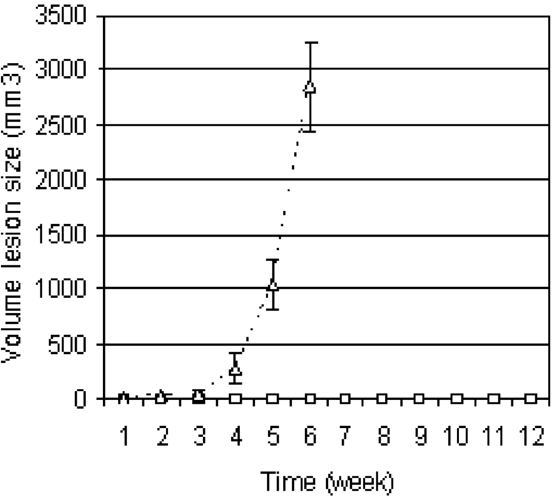
To examine the infectivity of the attenuated line, BALB/c mice were injected subcutaneously with L. major H-line or L. major WT promastigotes. The in vitro results suggested that L. major H-line is unlikely to survive within murine macrophages in significant numbers beyond 96 h. To examine the infectivity of the attenuated line, groups of 5 BALB/c mice were injected subcutaneously with 5×10⁶ stationary-phase L. major H-line (8 passages) or L. major WT (10 passages) promastigotes. In contrast to the progressive non-healing lesion resulting from infection with wild-type parasites, 10 mice infected with L. major H-line did not develop lesions during the 12-week period of the study (Fig. 3). Dissemination and survival of the 2 lines in the skin and visceral organs of BALB/c mice were investigated. At 12 weeks, parasites were found at the site of injection of the attenuated parasites, but their dissemination was limited to the draining popliteal lymph nodes, whereas wild-type parasites could be found in the spleen, liver, lung, draining popliteal lymph nodes, and the skin at the site where the parasites were injected.
Lesion size of BALB/c mice infected with 5×10⁶ stationary-phase promastigotes of Leishmania major H-line (□) or L. major WT (▵). Lesion size was measured weekly. Data are mean +/−s.e.m. of 3 experiments.

Cytokine production by splenocytes from L. major H-line and L. major WT-infected mice
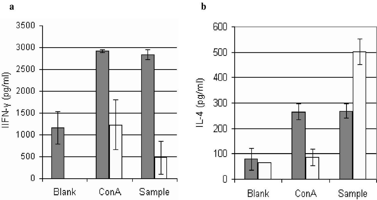
Splenocytes from mice infected with either the L. major H-line or L. major WT were harvested at 12 weeks post-infection and stimulated in vitro with stationary phase promastigotes of L. major WT. Spleen cells taken from mice infected with L. major WT secreted a relatively low level of IFN-γ when stimulated with stationary phase promastigotes. In comparison, cells taken from mice infected with the L. major H-line secreted a higher level of IFN-γ (P<0·0001) (Fig. 4). IL-4 in the supernatant of promastigote-stimulated splenocytes from the mice infected with L. major WT was significantly higher than that in the supernatant of splenocytes of mice infected with the L. major H-line (P<0·002) (Fig. 4).
Comparison of cytokine production in supernatant of cultured splenocytes from mice infected with stationary-phase promastigotes of the Leishmania major H-line (closed bars) or L. major WT (open bar) at 12 weeks post-infection. Supernatants were collected from parallel cultures of splenocytes stimulated with stationary-phase promastigotes, Con A, or non-stimulated, after 72 h and analysed for IFN-γ (a) and IL-4 (b). Bars represent±s.e.m. of triplicate wells.

Serum antibody generated in mice following infection with L. major WT or the L. major H-line
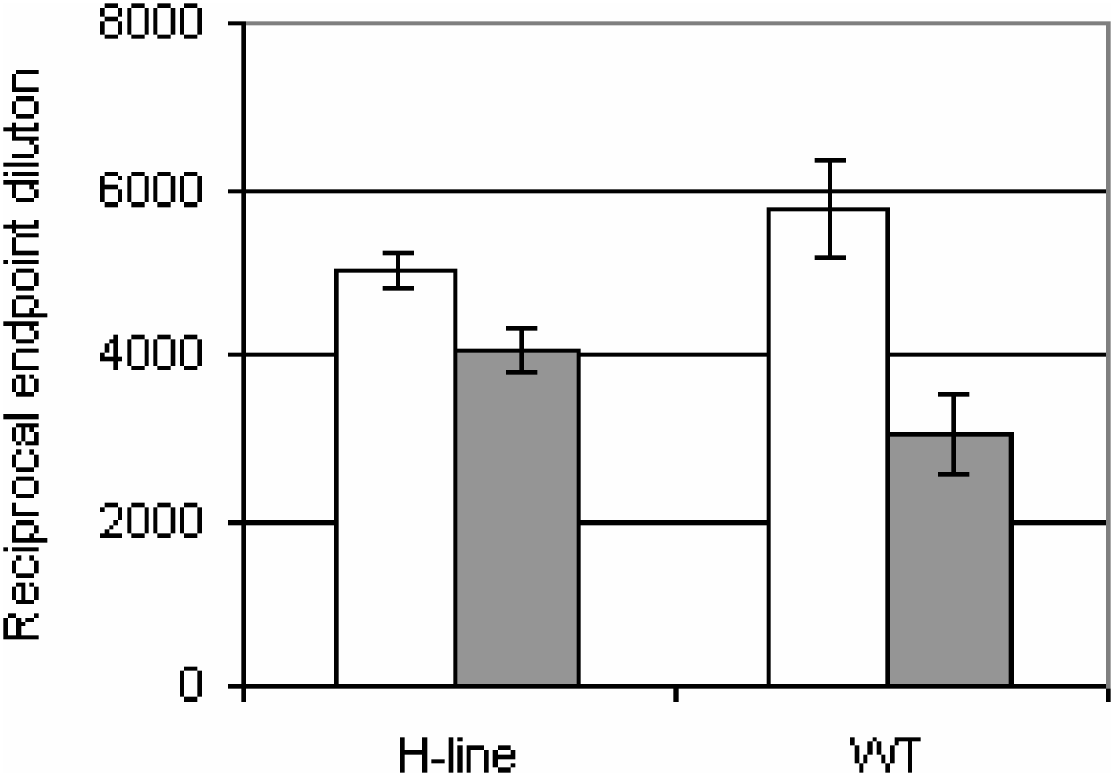
Serum levels of anti-Leishmania-specific IgG1 and IgG2a were determined at 12 weeks post-infection. Mice infected with L. major WT had pronounced Leishmania-specific antibody levels, predominantly of the IgG1 subclass. Mice infected with L. major H-line also had high antibody titres, principally of the IgG1 subclass with higher IgG2a titres than those recorded after infection with L. major WT but there was no significant difference between them (P<0·09). The level of IgG1 antibody in the serum of mice infected with the L. major H-line was, however, significantly lower than that in the mice infected with L. major WT (P<0·009) (Fig. 5).
Comparison of specific antibodies in the serum of BALB/c mice 12 weeks after infection with stationary phase promastigotes of the Leishmania major H-line or L. major WT. The serum levels of IgG1 (open bars) and IgG2a (closed bars) were measured. Bars represent Mean±s.e.m. of 7 identified sera.

Vaccine potential of L. major H-line
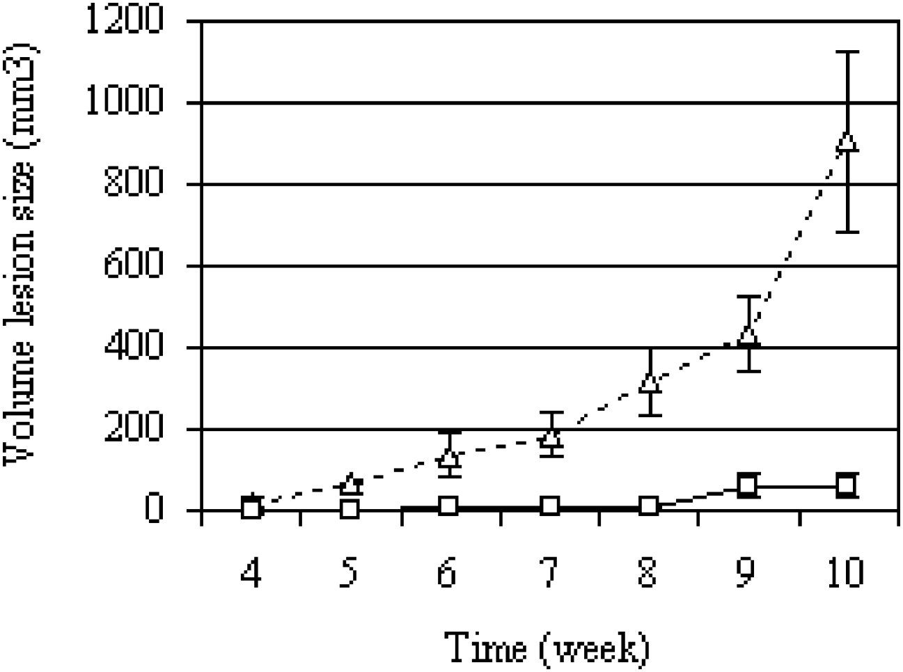
Experiments were carried out to determine whether pre-treatment of mice with L. major H-line could protect susceptible BALB/c mice from infection with wild-type L. major. All non-inoculated mice infected with wild-type L. major developed progressive non-healing lesions, and the mean lesion volume at 10 weeks after infection was about 900 mm3 (Fig. 6). In contrast, the lesion developed slowly in inoculated mice, and the mean lesion volume at 10 weeks post-infection was 60 mm3 (Fig. 6). Another important effect of vaccination with the attenuated line was that the wild-type challenge parasites were confined to the site of injection, at least up to 11 weeks after challenge. Wild-type parasites given alone rapidly disseminated to the internal organs, as noted above.
The mean lesion growth of Leishmania major in BALB/c mice not vaccinated (▵) or vaccinated with stationary-phase promastigotes L. major H-line (□) 4 weeks before challenge with 5×10⁶ wild-type parasites. Bars represent Mean±s.e.m. of 15 mice.

DISCUSSION
We previously reported that culture of Leishmania promastigotes in vitro under pressure of gentamicin resulted in the selection of attenuated lines that invaded but were unable to survive within bone marrow-derived BALB/c mice macrophages in vitro, whereas wild-type parasites survived and multiplied within infected macrophages (Daneshvar et al. Reference Daneshvar, Coombs, Hagan and Phillips2003a). Attenuated lines of L. mexicana and L. major failed to induce cutaneous lesions but induced significant protection in BALB/c mice (Daneshvar et al. Reference Daneshvar, Coombs, Hagan and Phillips2003a).
Here we show that L. major WT can be attenuated rapidly and reproducibly by culturing promastigotes initially in the presence of 200 μg/ml and then in 20 μg/ml gentamicin after 8 passages (as described in the Materials and Methods section). The percentage of macrophages infected with L. major H-line20 at 8 h after infection was 43% and decreased to 17·5% at 96 h after infection. The percentage of macrophages infected with L. major H-line decreased over time, from 39% at 8 h to 3% at 96 h after infection. Significantly, we have found that the growth rate of promastigotes of L. major H-line is slower than those of L. major WT and L. major H-line20 all after 11 subpassages. There is a significant difference (P<0·0001) between the growth rate of promastigotes of L. major H-line and L. major WT on day 6. These findings suggest that the modified method attenuates L. major WT in fewer passages than the previously described method and L. major H-line is unlikely to survive within murine macrophages in significant numbers beyond 96 h. In addition, the attenuated line parasites that looked rounded up within macrophages are not necessarily amastigotes. BALB/c mice, which are susceptible to L. major (Kropf et al. Reference Kopf, Brombacher, Köhler, Kienzle, Widmann, Lefrang, Humborg, Ledermann and Solbach1996) when infected with L. major H-line failed to develop cutaneous lesions, whereas all mice infected with L. major WT developed non-healing lesions. The mechanisms underlying the development of non-healing lesions, in genetically susceptible mice, are under control of specific cytokines, including IL-12, IFN-γ, and IL-4 (Heinzel et al. Reference Heinzel, Rerko, Ahmed and Pearlman1995; Scharton-Kersten and Scott, Reference Scharton-Kersten and Scott1995; Kropf et al. Reference Kropf, Etges, Schopf, Chung, Sypek and Muller1997). Genetically susceptible mice lacking IL-4 are protected from cutaneous infection with L. major WT (Kropf et al. Reference Kopf, Brombacher, Köhler, Kienzle, Widmann, Lefrang, Humborg, Ledermann and Solbach1996), whereas in the presence of IFN-γ and IL-12 cutaneous lesions fail to develop or lesions heal (Scharton-Kersten and Scott, Reference Scharton-Kersten and Scott1995). In the present study we found that the resistance of BALB/c mice to cutaneous lesion growth following infection with L. major H-line was associated with the ability of their splenocytes to produce IFN-γ following specific antigen stimulation. In the supernatant of cultured promastigote-stimulated splenocytes of mice infected with L. major H-line the level of IFN-γ was significantly higher (P<0·0001) and the level of IL-4 was lower (P<0·002) than in the supernatants of splenocytes of mice infected with L. major WT. These results are consistent with reports that survival of L. major amastigotes within the macrophage is prevented when the macrophages are stimulated by IFN-γ (Nacy et al. Reference Nacy, Fortier, Meltzer, Buchmeier and Schreiber1985), and IL-4 inhibits macrophage leishmanicidal activity and down-regulates the development of a Th1-like response (Satoskar et al. Reference Satoskar, Brombacher, Dai, McInnes, Liew, Alexander and Walker1997). These results also conform with reports that a Th1 response protects against cutaneous leishmaniasis, but the induction of a simultaneous Th2 response abrogates the protective Th1 effector function (Reiner and Locksley, Reference Reiner and Locksley1995; Sjölander et al. Reference Sjölander, Baldwin, Curtis and Handman1998). We assayed for a Th1 and Th2 response indirectly by examining immunoglobulin isotypes compartment. The level of Leishmania-specific IgG1 in the serum of mice infected with L. major WT was higher than the level of IgG2a whereas, the amount of specific IgG1 in the serum of mice infected with the L. major H-line was lower than IgG1 in the mice infected with L. major WT. The production of IgG1 antibody, which is promoted by Th2 cells, was markedly less in the mice infected by the L. major H-line, compared with that in the L. major WT infected mice, indicating possible inhibition of Th2 function by the L. major H-line. These data are in agreement with reports that IgG1 regulation is not completely dependent upon IL-4 signalling (Snapper and Paul, Reference Snapper and Paul1987).
In the present study we report that L. major H-line was localized in the skin at the site where the promastigotes were injected and in the draining PLN after 12 weeks post-infection, without any dissemination to the visceral organs. In contrast, infection with L. major WT led to rapid parasite spread to the bone marrows, spleens, draining PLNs, and lungs. Other studies have shown that dissemination of the parasite in the visceral organs of susceptible mice is the result of the development of a non-protective Th2 response (Liew and Donnell, Reference Liew and O'Donnell1993). The local restriction of the parasite prior to the development of T cell responses appears to be mediated by innate immunity and this activity is considered to play an important role in the subsequent development of a protective T cell response (Laskay et al. Reference Laskay, Diefenbach, Rollinghoff and Solbach1995). We previously reported that BALB/c mice infected with the L. mexicana H-line developed a CD4+Th1-like response, indicated by the cytokine profile of their splenocytes stimulated by L. mexicana WT promastigotes (Daneshvar et al. Reference Daneshvar, Hagan and Phillips2003b). This profile was sustained after these mice were challenged with L. mexicana WT, whereas mice infected with L. mexicana WT alone developed a CD4+Th2-like cytokine profile (Daneshvar et al. Reference Daneshvar, Hagan and Phillips2003b). These findings suggest that attenuated lines of both lines of L. mexicana and L. major hold considerable promise for vaccination against leishmaniasis.
The advantages of using attenuated lines as vaccines are that they most closely resemble natural infections; they are likely to generate an immune profile associated with protection; they lead to a relatively small antigen exposure, which might preferentially generate a protective Th1 response; and the host is beneficially exposed to a wide range of antigens (Constant et al. Reference Constant, Pfeiffer, Woodard, Pasqualini and Bottomly1995). The principal disadvantage of an attenuated vaccine is the possibility that pathogenic revertants may spontaneously arise. Furthermore, even attenuated organisms might cause disease in immunocompromised individuals. Such problems of course apply to all attenuated vaccines, including those currently in use in humans, both viral such as the polio vaccine and bacterial vaccines such as BCG. Having established that a Leishmania vaccine line shows no evidence of revertants over multiple passages in vitro and is safe by testing initially in mice and later in human volunteers, a judgment has to be taken on the balance of the benefit of protection from the vaccine against the use of drug treatment which is often ineffective and which itself may cause serious side-effects. It is therefore extremely encouraging that the L. major H-line successfully limited lesion development and parasite growth in BALB/c mice following s.c. vaccination. These findings support further investigation of the proposal that L. major H-line is a potential vaccine candidate. We are currently investigating the molecular basis of the observed attenuation, which would allow us to assess the likelihood of spontaneous reversion.