INTRODUCTION
While the occurrence of allergens in mites (phylum Arthropoda, subphylum Chelicerata, class Arachnida, subclass Acari) is quite often well documented, the knowledge of similar proteins in ticks is rather limited. Allergens of house dust mites can be divided into several groups (1–19). It has been reported that their homology within single groups is quite high (70–80%) but they differ in molecular weight, level of glycosylation, content of lipids or in biochemical function (Thomas et al. Reference Thomas, Smith, Hales, Mills and O'Brien2002).
Hard ticks belong to the phylum Arthropoda, subphyllum Chelicerata, class Arachnida, subclass Acari, order Ixodida, and family Ixodidae. They are well-known blood-feeders, vectors of many pathogenic microorganisms, causing diseases both in animals and humans. While taking a bloodmeal ticks encounter a large diversity of pathogens from hosts, their organisms undergo dramatic changes on morphological, physiological and molecular levels. In the complex response of the gene expression towards the presence of the pathogenic agent, it is possible to expect the production of proteins that represent the molecular factors of innate immunity either involved in defence reactions or pathogen pattern recognition (Ribeiro, Reference Ribeiro1995; Mans and Neitz, Reference Mans and Neitz2004; Sonenshine and Hynes, Reference Sonenshine and Hynes2008). During blood feeding, ticks secrete different bioactive molecules into their host. The set of secreted molecules depends on the length of blood feeding or on the developmental stage of the tick, as well as on the immune response of the host (Lawrie and Nuttall, Reference Lawrie and Nuttall2001). The majority of molecules are secreted by salivary glands and their description is an object of many recent studies, for example on Argas monolakensis (Mans et al. Reference Mans, Andersen, Francischetti, Valenzuela, Schwan, Pham, Garfield, Hammer and Ribeiro2008), Dermacentor andersoni (Alarcon-Chaidez et al. Reference Alarcon-Chaidez, Sun and Wikel2007) or Ixodes ricinus (Leboulle et al. Reference Leboulle, Rochez, Louahed, Rutti, Bollen and Godfroid2002; Rudenko et al. Reference Rudenko, Golovchenko, Edwards and Grubhoffer2005; Chmelař et al. Reference Chmelař, Anderson, Mu, Jochim, Valenzuela and Kopecký2008).
Allergens or allergen-like proteins were identified and characterized in the hard tick I. ricinus and in the soft tick Argas reflexus (Jacot, personal communication; Hilger et al. Reference Hilger, Bessot, Hutt, Grigioni, de Blay, Pauli and Hentges2005). An alerg 1 gene of the castor bean tick encodes an allergen-like protein that is a member of the ML (MD-2-related lipid-recognition) domain protein family as well as group 2 of mite allergens. The ML protein family is comprised of 4 groups and is common to animals, plants and fungi. Its members contain an N-terminal signal peptide and conserved cysteine residues that form disulfide bridges (Wright et al. Reference Wright, Li and Rastinejad2000). Often, their function is connected with innate immunity or with lipid binding and transport (Inohara and Nuñez, Reference Inohara and Nuñez2002). I. ricinus allergen-like protein shows an homology of about 30% to group 2 mite allergens and contains a domain with putative lipid-binding function (Jacot, personal communication). An allergen Arg r 1 from the pigeon tick A. reflexus has 4 conserved cysteine residues that form disulfide bridges, an N-terminal signal peptide, and other characteristics typical for a protein family of lipocalins including the G-X-W pattern. Based on the homology with other histamine-binding proteins of different tick species and due to possible antibody cross-reactivity with similar proteins from Rhipicephalus appendiculatus and Haemaphysalis punctata allergen Arg r 1 was grouped with histamine-binding proteins (Rolla et al. Reference Rolla, Nebiolo, Marsico, Guida, Bigo, Riva and Zanotta2004; Hilger et al. Reference Hilger, Bessot, Hutt, Grigioni, de Blay, Pauli and Hentges2005).
This is a report on the identification and characterization of a new tick gene encoding Der-p2 allergen-like protein from I. ricinus, analysis of protein structure, relation to other members of ML domain protein family and similar proteins from the other tick species. We defined the tissue and stage-specific expression pattern of the gene and analysed the Der-p2 allergen-like protein interaction with immunoglobulin E (IgE).
MATERIALS AND METHODS
Ticks
Hard ticks I. ricinus were provided by the Biology Centre, Institute of Parasitology, Academy of Sciences of the Czech Republic. Uninfected ticks were reared for several generations in the animal facilities of the institute and fed on guinea pigs that were raised to be free of infection under the strict hygiene regulations of the Central Commission for the Protection of Animals (§21, section 3e, law 246/1992). Blood-fed larvae, nymphs and females were obtained by attaching a container (cell) with ticks to a shaved area on the back of an adult uninfected guinea pig and left on the animal back until the specified engorgement stage. After the separation of the fed ticks from the guinea pig, RNA was isolated from them using TRI Reagent (Sigma, USA). The fully engorged females were kept at 4°C for a further 3 days for blood digestion prior to RNA isolation.
First strand cDNA synthesis and polymerase chain reaction (PCR)
First strand cDNA was synthesized by use of RevertAid H Minus First Strand cDNA Synthesis Kit (MBI Fermentas, Lithuania) according to the protocol provided by the manufacturer. Primers AL1F and AL1R were designed according to the known partial nucleotide sequence of the gene encoding I. ricinus allergen-like protein (AJ547805) and used for amplification of I. ricinus gene for the Der-p2 allergen-like protein (AL1F 5′-ATGTTCCGTTACGTTGTGTTCC-3′/AL1R 5-CATCCCCACCGCTGTAAC-3′; Generi Biotech, Czech Republic).
RT-PCR analysis of stage specific and tissue specific patterns and time expression of the Der-p2 allergen-like gene
RT-PCR was performed using Enhanced Avian HS RT-PCR kit (Sigma, USA). An equal amount of total RNA isolated from unfed and engorged larvae, nymphs and females of I. ricinus and different tissues (salivary glands, gut, ovarium, Malphigian tubes, and haemolymph) of partially engorged females was used as a template in RT-PCR in order to reveal the Der-p2 allergen-like gene expression in different developmental and feeding stages. To determine time expression of the gene, an equal amount of total RNA from unfed, 1, 3, 5, 7 days fed, and totally engorged adult females were used as a template in RT-PCR. The primers, AL2F 5′-CACCATGTTCCGTTACGTTGTGTTCC-3′, AL2R 5′TCACTCGATGACGATGTCCGA-3′ were used for the amplification with an annealing temperature of 55°C. A constitutively expressed actin gene (AY333957) was chosen as a control. The control amplification was performed at the annealing temperature of 55°C with the primers Act-F1 5′-CGTCTGGATCGGCGGCTCTAT-3′ and Act-R1 5′-ACGCGCACTCTTTTCCACAATCTC-3′. All used RNAs were first checked for DNA contamination by adding them directly into the PCR reaction with specific primers.
Bioinformatics/sequence analysis
Sequences were compared against the GenBank non-redundant (NR) protein database using the BlastX program from the executable package at ftp://ftp.ncbi.nlm.nih.gov/blast/executables (Altschul et al. Reference Altschul, Madden, Schäffer, Zhang, Zhang, Miller and Lipman1997) and searched against the Conserved Domains Database (CDD) (ftp://ftp.ncbi.nlm.nih.gov/pub/mmdb/cdd), which includes all Pfam (Bateman et al. Reference Bateman, Birney, Durbin, Eddy, Howe and Sonnhammer2000) and Smart (Schultz et al. Reference Schultz, Milpetz, Bork and Ponting1998) protein domains, as well as against the VectorBase (Lawson et al. Reference Lawson, Arensburger, Atkinson, Besansky, Bruggner, Butler, Campbell, Christophides, Christley, Dialynas, Hammond, Hill, Konopinski, Lobo, MacCallum, Madey, Megy, Meyer, Redmond, Severson, Stinson, Topalis, Birney, Gelbart, Kafatos, Louis and Collins2009) (http://www.vectorbase.org) and the Gene Index Project (Quackenbush et al. Reference Quackenbush, Cho, Lee, Liang, Holt, Karamycheva, Parvizi, Pertea, Sultana and White2001) (http://compbio.dfci.harvard.edu/tgi/cgi-bin/tgi/Blast/index.cgi). Protein secondary structure prediction was performed by PSIpred program (http://bioinf.cs.ucl.ac.uk/psipred) (McGuffin et al. Reference McGuffin, Bryson and Jones2000). Putative signal peptide, possible sites of O-glycosylation, N-glycosylation and phosphorylation were determined with the prediction servers Signal P 3.0 (http://www.cbs.dtu.dk/services/SignalP) (Bendtsen et al. Reference Bendtsen, Nielsen, von Heijne and Brunak2004), NetOGlyc 3.1 (http://www.cbs.dtu.dk/services/NetOGlyc) (Julenius et al. Reference Julenius, Mølgaard, Gupta and Brunak2005), NetNGlyc 1.0 (http://www.cbs.dtu.dk/services/NetNGlyc) and NetPhos 2.0 (http://www.cbs.dtu.dk/services/NetPhos) (Blom et al. Reference Blom, Gammeltoft and Brunak1999), respectively.
Tertiary structure prediction
For prediction of the 3-D structure of the Der-p2 allergen-like protein CPHmodel 2.0 Server was used (http://www.cbs.dtu.dk/services/CPHmodels) to find the homologues for the I. ricinus molecule. A pdb file of the molecule (according to the similar sequences of structures from the protein databank) was downloaded and used in Polyview-3D for protein visualization (http://polyview.cchmc.org/polyview3d.html) (Porollo and Meller, Reference Porollo and Meller2007).
Expression and purification of the recombinant protein
The gene encoding the Der-p2 allergen-like protein without signal peptide, prepared by PCR using the primers AL3F 5′CACCAGCGCAGAAAAGCCGT-3′ and AL2R (sequence above), was expressed in pET100/D-TOPO (Invitrogen, USA) expression vector as recombinant fusion proteins with N-terminal 6xHis tag. Rosetta-gami competent cells (Novagen, Germany) were transformed by recombinant construct. All procedures were performed according to the manufacturer's protocol in Champion pET directional TOPO expression kits (Invitrogen, USA). Isopropyl-β-d-thiogalactopyranoside (IPTG) (1 mm final concentration) was used for the induction of recombinant protein expression. Recombinant Der-p2 allergen-like protein was purified by Co2+-affinity chromatography using cartridge Co-MAC (Novagen, Germany). Bacterial lysate was prepared using lysis buffer containing 8 m urea, 0·1 m NaH2PO4, 0·01 m Tris-HCl, pH 8·0. The purification under denaturing conditions was performed according to the manufacturer's protocol. The cartridge was equilibrated with 5 volumes of binding buffer (6 m urea, 0·5 m NaCl, 0·02 m Tris-HCl, 0·005 m imidazole, pH 7·9), and then the sample in binding buffer was applied. The cartridge was washed with 10 volumes of binding buffer, 6 volumes of washing buffer (6 m urea, 0·5 m NaCl, 0·02 m Tris-HCl, 0·06 m imidazole, pH 7·9) and the recombinant protein was eluted with 6 volumes of elution buffer (6 m urea, 0·5 m NaCl, 0·02 m Tris-HCl, 1 m imidazole, pH 7·9). The elution fractions were checked on SDS-PAGE and fractions containing recombinant protein given for further tests were dialysed against PBS (phosphate-buffered saline) buffer, pH 7·4 with 6 m urea, 4 m urea, 2 m urea, 1 m urea, 0·5 m urea and PBS, respectively. The presence of the His-tagged Der-p2 allergen-like protein was verified by immunodetection with Ni-NTA conjugates (Qiagen, Germany).
Antimicrobial activity determination
A double-layer technique was used to estimate antimicrobial activity of the recombinant Der-p2 allergen-like protein (Čeřovský et al. Reference Čeřovský, Hovorka, Cvačka, Voburka, Bednárová, Borovičková, Slaninová and Fučík2008). The plates were incubated at 37°C for 6 h with periodic checking for the growth of bacteria (Escherichia coli, Micrococcus luteus).
IgE binding reactivity
Purified recombinant Der-p2 allergen-like protein was diluted to 10 μg/ml in PBS. Each well of a 96-well microplate (Maxisorp F96 MicroWell Plate, Nunc, Denmark) was coated with 50 μl of the recombinant protein and incubated at 4°C overnight. All further incubations were performed at room temperature with at least 5 washes with PBS containing 0·05% Tween 20 (PBST) between them. Then 50 μl of human biotinylated IgE (BioPorto Diagnostic, Denmark) at different concentrations (0·1, 1, 3, 5, 10 μg/ml) were added into a microplate and incubated for 1 h, after which 50 μl of streptavidin-peroxidase (Sigma, USA) diluted 1:1000 in distilled water was added to each well and incubated for 30 min. Orthophenylenediamine (Sigma, USA) with hydrogen peroxide (50 μl/well) was used as the substrate for the enzymatic colour reaction. The reaction was performed in the dark for 15 min and stopped by the addition of 50 μl of 1 m H2SO4. Optical densities were measured at 490 nm with an ELISA spectrophotometer (Multiskan MCC 340, Labsystems Oy, Finland). Bovine serum albumin (BSA) and a recombinant protein produced in a prokaryotic expression system were used as controls.
Statistical analysis
The significance of any differences obtained between single protein groups and used different antibody concentrations was evaluated by the factorial ANOVA with a subsequent Tukey HSD test (Statistica 6.0, Statsoft, Inc. Tulsa, USA). The presented results represent 3 similar experiments.
RESULTS
A gene encoding Der-p2 allergen-like protein, a protein that contains the ML (MD-2-related lipid-recognition) domain was identified in the hard tick I. ricinus. The isolated cDNA sequence of Der-p2 allergen-like protein is 595 bp long and contains an open reading frame (ORF) encoding a protein of 144 amino acids with a predicted molecular mass of 15·57 kDa and isoelectric point 7·92 for the pre-protein and 13·77 kDa and pI 7·62 for the mature Der-p2 allergen-like protein. The protein carries a 16 amino acid long N-terminal signal peptide. Six conserved cysteine residues were detected at positions 26, 40, 45, 91, 96 and 136 of the pre-protein. Der-p2 allergen-like protein has a predicted helix at the site of the signal peptide followed by β-structures and coil regions only. While 5 putative phosphorylation sites (serines at positions 34, 65, 106, 118, 134) were defined in the Der-p2 allergen-like protein sequence, no sites of possible O-glycosylation and N-glycosylation were found.
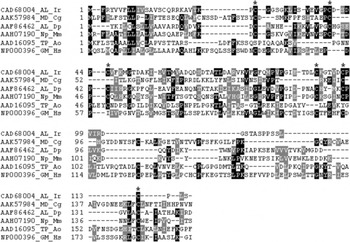
Der-p2 allergen-like protein is a member of the ML domain protein family and includes a Der-p2-like domain (Marchler-Bauer and Bryant, Reference Marchler-Bauer and Bryant2004). Alignment of I. ricinus protein with several members from different groups of the ML protein family showed a low similarity, defining only a few regions with higher homology. Conserved cysteine residues were revealed in aligned sequences at specific positions. The highest level of similarity of I. ricinus Der-p2 allergen-like protein was shown in group II of the ML domain protein family, while no similarity between it and the selected members from other groups (I, III, IV) was detected (Fig. 1).
Alignment of Ixodes ricinus Der-p2 allergen-like protein (CAD68004) and other members from all four groups of ML protein family. Group I: MD-2 protein of Cricetulus griseus (GenBank AAK57984), group II: Der p 2 allergen from Dermatophagoides pteronyssinus (AAF86462), Mus musculus Npc2 protein (AAH07190), group III: PG/PI-TP of Aspergillus oryzae (AAD16095), group IV: GM2 activator from Homo sapiens, (NP000396). Conserved cysteine residues are marked by asterisks. Black boxes with white letters show identical residues, and grey boxes with black letters show chemically similar residues.

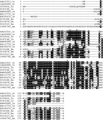
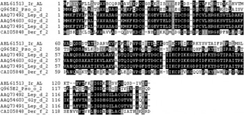
BLASTP, GenBank, and Gene Index Project analyses of the translated product of the Der-p2 allergen-like protein gene revealed similarity to several tick proteins from I. ricinus, I. scapularis and Rhipicephalus microplus and to different mite allergens (members from the group II of ML protein family from organisms of the same subclass Acari, class Arachnida). The amino acid sequence of the I. ricinus Der-p2 allergen-like protein, submitted to the GenBank (ABL61513), showed the highest sequence identity to the allergen-like protein from I. ricinus (CAD68004) – 92% and to the Der-p2 allergen-like protein from I. scapularis – 88% (TC55436). Lower identities (42–58%) to other related proteins from I. scapularis and R. microplus were also revealed. An average similarity of 35% to allergens and their precursors from Psoroptes ovis (group 2 allergen Pso o 2 precursor, Q965E2), Lepidoglyphus destructor (type 2 allergens Lep d 2, AAQ73492, AAQ73491, AAQ73488, AAQ73490) or Glycyphagus domesticus (Gly d 2.03, AAQ54603) was detected (Figs 2 and 3).
Proteins obtained by comparison of Der-p2 allergen-like protein with other tick proteins using blastp analysis against GenBank, VectorBase and Gene Index project databases (http://blast.ncbi.nlm.nih.gov/Blast.cgi; http://iscapularis.vectorbase.org/index.php; http://compbio.dfci.harvard.edu/tgi/tgipage.html). Der-p2 allergen-like protein (ABL61513) shows the highest similarity (96%) with allergen-like protein of Ixodes ricinus (CAD68004), lower similarity with proteins of I. scapularis – 60–95% (TC55436, TC47248, EW945095, EEC13776, EEC06676) and with proteins of Rhipicephalus microplus – 65–72% (TC18108, TC16878, TC22513, CV443743).

Comparison of Ixodes ricinus Der-p2 allergen-like protein with selected house dust mite proteins from group II of ML protein family. The similarity with allergens and their precursors of Psoroptes ovis (group 2 allergen Pso o 2 precursor, Q965E2), Lepidoglyphus destructor (type 2 allergens Lep d 2, AAQ73492, AAQ73491), Glycyphagus domesticus (Gly d 2.03, AAQ54603) and Dermatophagoides farinae (Der f 2, CAI05848) ranges from 30 to 35%.

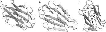
The tertiary structure of ML proteins was defined only for a few members of the protein family. The highest homology of I. ricinus Der-p2 allergen-like protein to known structures was defined for the major mite allergen from Dermatophagoides farinae Der f 2 (PDB 1 ahm) with 32·8% identity. Prediction of tick Der-p2 allergen-like protein structure revealed that it has an immunoglobulin fold (s-type). The tick allergen forms 2 plates of β-sheets, one is composed of 4 β-strands (residues 68–76, 79–83, 102–105, 121–129) and the other sheet consists of 3 β-strands (33–38, 53–61, 107–111). The mentioned structure is identical to that of Der f 2 (Bork et al. Reference Bork, Holm and Sander1994; Ichikawa et al. Reference Ichikawa, Hatanaka, Yuuki, Iwamoto, Kojima, Nishiyama, Ogura, Okumura and Inagaki1998) (Fig. 4).
Tertiary structure of Der f 2 allergen of Dermatophagoides farinae – 1 ahm (A) and predicted tertiary structure of Der-p2 allergen-like protein of Ixodes ricinus (B, C) with conserved cysteine residues marked.

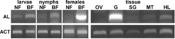
The results of RT-PCR on the detection of the stage expression pattern of the Der-p2 allergen-like protein gene show that its expression is strongly induced by a bloodmeal in all developmental stages (larva, nymph and adult) of the tick. This observation is confirmed by analysis of the time expression using total RNA from unfed and engorged females at different feeding stages. Der-p2 allergen-like protein is slightly expressed in unfed ticks. The expression of Der-p2 allergen-like protein starts on the first day of feeding and reaches its maximum on the third day. The level remains stable until full engorgement (data not shown). Analysis of the tissue-specific pattern of the gene expression revealed that gut and haemolymph are the only sites of its expression. An expression level of the actin gene (positive control) was constant throughout the whole experiment (Fig. 5).
Stage and tissue specific Der-p2-like gene expression analysis by RT-PCR. AL – Der-p2 allergen-like protein, ACT – actin; NF – unfed ticks, BF – engorged ticks; OV – ovaria, G – gut, SG – salivary glands, MT – Malphigian tubes, HL – haemolymph.

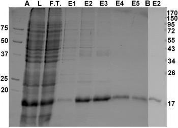
We produced a recombinant Der-p2 allergen-like protein in a prokaryotic expression system. The PCR product of 387 bp for Der-p2 allergen-like protein gene (without a signal peptide) was expressed as a 16 kDa 6xHis tag fusion protein (Fig. 6). MS analysis of the purified recombinant protein confirmed its identity to the predicted translated protein sequence of Der-p2 allergen-like protein gene from I. ricinus by 54·5% amino acid sequence coverage (data not shown).
Purification of recombinant Der-p2 allergen-like protein. A: L – bacterial lysate, F.T. – flow-through, E1-E5 – elution fractions, marker – Precision Plus Protein Standards (Bio-Rad, USA); B: fraction E2 in PBS used in further tests, marker – PageRuler Prestained Protein Ladder (Fermentas, Lithuania).

Analysis of the antimicrobial activity of I. ricinus Der-p2 allergen-like protein against E. coli or M. luteus using the double-layer technique did not reveal any effect on bacterial growth.
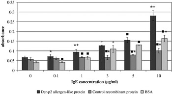
Because of the relatively high similarity of Der-p2 allergen-like protein to Der f 2 and other mite allergens that interact with IgE, we tested the IgE-binding reactivity of the recombinant tick Der-p2 allergen-like protein by ELISA. BSA and a recombinant protein produced by E. coli were used as controls. After incubation of Der-p2 allergen-like protein and IgE, the detected signal increases according to the concentration of antibody used (Fig. 7). Statistically significant differences (P<10−5) were observed between all tested groups – protein samples, IgE concentrations and combination of proteins and used IgE concentrations. The results obtained after incubation of the Der-p2 allergen-like protein with IgE are significantly different from those of both control proteins in the case of 1 and 10 μg/ml IgE concentrations; in the case of other concentrations of IgE the results significantly differ between Der-p2 allergen-like protein and BSA (0·1 μg/ml IgE) and between the Der-p2 allergen-like protein and the control recombinant protein (3 and 5 μg/ml IgE).
IgE binding profiles to recombinant Der-p2 allergen-like protein, control recombinant protein produced in E. coli and BSA. It was confirmed that there are no statistically significant differences between proteins not incubated with antibody. Statistically significant differences were found in other groups: * statistical significant difference from BSA,+statistical significant difference from control recombinant protein, ▪ statistical significant difference from Der-p2 allergen-like protein.

DISCUSSION
This is the first time that the gene encoding Der-p2 allergen-like protein from the hard tick Ixodes ricinus has been identified, isolated and characterized. Similar genes or their protein products have not yet been characterized in any other tick species. Protein homologues to I. ricinus Der-p2 allergen-like protein were found in I. scapularis EST database (Quackenbush et al. Reference Quackenbush, Cho, Lee, Liang, Holt, Karamycheva, Parvizi, Pertea, Sultana and White2001), but have not yet been isolated or characterized. A similarity search of sequential databases from other available tick species revealed the existence of the related proteins in I. scapularis and R. microplus. All detected proteins contain the ML domain. The presence of N-terminal signal peptide and its gene expression in the tick's gut during blood intake could be an indicator of protein occurrence in secretory cells and its possible secretion into the gut lumen, as was observed in I. ricinus allergen-like protein (Inohara and Nuñez, Reference Inohara and Nuñez2002; Mullen et al. Reference Mullen, Kennedy, Visintin, Mazzoni, Leifer, Davies and Segal2003; Jacot, personal communication). Visualization of the predicted tertiary structure of Der-p2 allergen-like protein on the basis of its similarity to Der f 2 allergen from D. farinae showed a structure with 2 anti-parallel beta-plated sheets typical for members of the ML protein family (Ichikawa et al. Reference Ichikawa, Hatanaka, Yuuki, Iwamoto, Kojima, Nishiyama, Ogura, Okumura and Inagaki1998; Derewenda et al. Reference Derewenda, Li, Derewenda, Dauter, Mueller, Rule and Benjamin2002; Marchler-Bauer et al. Reference Marchler-Bauer, Anderson, Derbyshire, DeWeese-Scott, Gonzales, Gwadz, Hao, He, Hurwitz, Jackson, Ke, Krylov, Lanczycki, Liebert, Liu, Lu, Lu, Marchler, Mullokandov, Song, Thanki, Yamashita, Yin, Zhang and Bryant2007). Three disulfide bridges stabilize the tertiary structure of ML proteins; Der-p2 allergen-like protein conserved cysteines are involved in disulfide bridge formation in the order of 1–6, 2–3, 4–5 as was observed in house dust mite allergens from group 2 (Mueller et al. Reference Mueller, Benjamin and Rule1998; Derewenda et al. Reference Derewenda, Li, Derewenda, Dauter, Mueller, Rule and Benjamin2002; Thomas et al. Reference Thomas, Smith, Hales, Mills and O'Brien2002). The presence of 3 disulfide bridges is considered to be important for recognition by IgE antibodies in the case of allergens (Mueller et al. Reference Mueller, Benjamin and Rule1998).
Tertiary structures of different ML protein family members were revealed only for allergens of house dust mites D. pteronyssus Der p 2 (Mueller et al. Reference Mueller, Benjamin and Rule1998; Derewenda et al. Reference Derewenda, Li, Derewenda, Dauter, Mueller, Rule and Benjamin2002), D. farinae Der f 2 (Ichikawa et al. Reference Ichikawa, Hatanaka, Yuuki, Iwamoto, Kojima, Nishiyama, Ogura, Okumura and Inagaki1998, Reference Ichikawa, Takai, Inoue, Yuuki, Okumura, Ogura, Inagaki and Hatanaka2005), bovine Npc2 protein (Friedland et al. Reference Friedland, Liou, Lobel and Stock2003), human MD-2 (Ohto et al. Reference Ohto, Fukase, Miyake and Satow2007), and human ganglioside M-2 (GM-2) activator protein (Wright et al. Reference Wright, Li and Rastinejad2000, Reference Wright, Mi, Lee and Rastinejad2005). The first 3 mentioned proteins belong to group II of the protein family. Similar to Der f 2, they are single-domain proteins of immunoglobulin fold. Proteins with such folding appeared to be involved in binding functions (Bork et al. Reference Bork, Holm and Sander1994) that indicate also the presence of a putative lipid-binding site of the Der-p2 allergen-like protein. Ligands of Npc2, GM-2 activator or MD-2 proteins have been found, recognizing their role as lipid or lipidic molecule-interacting proteins (Mahuran, Reference Mahuran1998; Shimazu et al. Reference Shimazu, Akashi, Ogata, Nagai, Fukudome, Miyake and Kimoto1999; Friedland et al. Reference Friedland, Liou, Lobel and Stock2003). This feature indicates the possible interaction with lipidic parts containing structures of different microorganisms as well as their possible roles in innate immunity.
The majority of allergens can cause allergic reactions with different levels of severity. They are able to interact with IgE antibodies, stimulate their production as well as induce T-cell responses. In mites, the major groups of allergens known to interact with IgE are those of groups 1 and 2, but this ability was also observed in some allergens from groups 4, 10, 11, 14, 15, 17, 18 and 19 (Thomas et al. Reference Thomas, Smith, Hales, Mills and O'Brien2002). IgE binding is known not only for mite allergens but also for arthropod allergens from other groups, such as insects or ticks (Rolla et al. Reference Rolla, Nebiolo, Marsico, Guida, Bigo, Riva and Zanotta2004; Shin et al. Reference Shin, Jeong, Hong and Yong2009). Allergen Arg 1, identified in the tick A. reflexus, was characterized as the histamine-binding protein from the lipocalin protein family (Hilger et al. Reference Hilger, Bessot, Hutt, Grigioni, de Blay, Pauli and Hentges2005). The Der-p2 allergen-like protein that we identified in I. ricinus belongs to the ML protein family that is similar to lipocalins by the presence of disulfide bridges, N-terminal signal peptide or by the ability to bind hydrophobic molecules. Several members of both protein families are known to be able to interact with compounds involved in the host immune reaction (Akerstrom et al. Reference Akerstrom, Flower and Salier2000; Inohara and Nuñez Reference Inohara and Nuñez2002; Beaufays et al. Reference Beaufays, Adam, Decrem, Prévôt, Santini, Brasseur, Brossard, Lins, Vanhamme and Godfroid2008). Unfortunately, more information is available about tick lipocalins than for the tick ML proteins. To date, lipocalins were identified and/or characterized in A. reflexus, A. monolakensis, Ornithodoros moubata, O. savignyi, Rhipicephalus appendiculatus, Dermacentor reticulatus, I. scapularis, and finally in I. ricinus (Keller et al. Reference Keller, Waxman, Arnold, Schultz, Condra and Connolly1993; Paesen et al. Reference Paesen, Adams, Harlos, Nuttall and Stuart1999; Sangamnatdej et al. Reference Sangamnatdej, Paesen, Slovak and Nuttall2002; Mans et al. Reference Mans, Louw and Neitz2003, Reference Mans, Andersen, Francischetti, Valenzuela, Schwan, Pham, Garfield, Hammer and Ribeiro2008; Hilger et al. Reference Hilger, Bessot, Hutt, Grigioni, de Blay, Pauli and Hentges2005; Beaufays et al. Reference Beaufays, Adam, Decrem, Prévôt, Santini, Brasseur, Brossard, Lins, Vanhamme and Godfroid2008). Members of the ML protein family were found in the ticks O. parkeri, O. coriaceus, D. variabilis, R. appendiculatus, R. microplus, I. scapularis and I. ricinus (Francischetti et al. Reference Francischetti, Sá-Nunes, Mans, Santos and Ribeiro2009). Proteins similar to Der-p2 allergen-like protein were found in I. scapularis and in R. microplus. Their clear function is not yet known but it is possible that they are involved in the interaction with microorganisms entering the tick body during feeding. They can directly bind their surface structures carrying lipidic parts. In this way, they can label the microorganism that can be further processed by immune mechanisms of the tick.
Testing IgE binding by Der-p2 allergen-like protein, followed by comparison of the tertiary structure of the tick allergen to the house dust mite allergen of D. farinae, Der f 2, and also of D. pteronyssinus, Der p2, that possesses 2 main localized IgE epitopes (Ichikawa et al. Reference Ichikawa, Hatanaka, Yuuki, Iwamoto, Kojima, Nishiyama, Ogura, Okumura and Inagaki1998; Mueller et al. Reference Mueller, Benjamin and Rule1998; Derewenda et al. Reference Derewenda, Li, Derewenda, Dauter, Mueller, Rule and Benjamin2002), was performed to reveal the possible existence of similar epitopes in the studied tick protein. Although not all residues showed similar biochemical character, we examined whether tick allergen binds IgE and revealed a detectable interaction. Expression of Der-p2 allergen-like protein gene in the gut and haemolymph defines the place of interaction. Der-p2 allergen-like protein could be involved in the interaction of different blood-transmitted compounds or microorganisms. Specific IgE was documented in patients after a bite by I. ricinus, I. holocystus, I. pacificus, R. appendiculatus or A. reflexus (Acero et al. Reference Acero, Blanco and Bartolomé2003; Rolla et al. Reference Rolla, Nebiolo, Marsico, Guida, Bigo, Riva and Zanotta2004). However, the studied tick Der-p2 allergen-like protein is not an allergen in the sense of triggering an allergic reaction. Showing high sequential and structural similarity to mite allergens (with 30–35% similarity) it might possess a different functional activity that has not yet been defined. Der-p2 allergen-like protein could be involved in the neutralization of IgE antibodies received with blood from repeatedly tick-bitten people. This could be another mechanism of clearing the host antibodies by the tick rather than involving IgG-binding proteins, according to the mechanism known from Rhipicephalus appendiculatus (Wang et al. Reference Wang, Paesen, Nuttall and Barbour1998; Wang and Nuttall, Reference Wang and Nuttall1999). The predicted 3-D structure of I. ricinus Der-p2 allergen-like protein showed the presence of a cavity between the plates formed by β-sheets for lipid molecule binding. This feature was confirmed for several members of the ML protein family. Although characterization of Der-p2 allergen-like protein is not yet complete, the protein most probably participates in the recognition and transport of lipids from blood, or in the recognition of surface patterns created by lipidic molecules of microorganisms.
ACKNOWLEDGEMENTS
We are grateful to Jan Erhart for his help during the work with ticks, to Dr Miroslav Šulc (Institute of Microbiology AS CR, Prague) for his help with protein MS analysis, to Dr Iva Schödelbauerová for her help with statistical analysis, and to Jason Dean for his help in linguistic editing of the manuscript.
FINANCIAL SUPPORT
This work was financially supported by the Grant Agency of the Czech Republic (grant 524/06/1479), Ministry of Education of the Czech Republic (MSM 6007665801, LC06009) and grant Z60220518 (research project of the Institute of Parasitology AS CR).