Colonization of the lungs and secondary bacterial infections are well-known complications of influenza infection and are important contributors to morbidity and mortality worldwide. The microbiological interactions of influenza and bacteria causing pneumonia were first described from samples of the 1918 influenza pandemic Reference Muir and Wilson1–Reference Sheng, Chertow and Ambroddio4 and have been described in both pandemic Reference Schwarzmann, Adler, Sullivan and Marine5–Reference Lee, Wu and Lee9 and seasonal influenza. Reference Opatowski, Baguelin and Eggo10,Reference Metersky, Masterson, Lode, File and Babinchak11 Bacterial pathogens associated with influenza include Staphylococcus aureus, Reference Murray, Robinson and White8,Reference Metersky, Masterson, Lode, File and Babinchak11–Reference Yang, Gao and Chen13 Streptococcus pneumoniae, Reference Palacios, Hornig and Cisterna14–Reference Shrestha, Foxman, Weinberger, Steiner, Viboud and Rohani16 Streptococcus pyogenes, Reference Scaber, Saeed, Ihekweazu, Efstratiou, McCarthy and O’Moore17,Reference Chertow and Memoli18 and Haemophilus influenzae. Reference Brundage19 Less frequently associated pathogens include Pseudomonas aeruginosa, Reference Su, Lee, Liu, Chuang, Chen and Lee20 Legionella pneumophila, Reference Joseph, Togawa and Shindo15,Reference Rizzo, Caporali and Rota21 Neisseria meningitides, Reference Jacobs, Viboud and Tchetgen22 and Mycoplasma pneumoniae. Reference Dhanoa, Fang, Hassan, Kaniappan and Rajasekaram23
Complex interactions between microbes facilitate bacterial colonization and increase the risk of bacterial pneumonia. The biology of influenza virus, 24 bacteria, and host interaction is linked to a loss of lung repair function Reference Cauley and Vella25 and damage to basal epithelial cells, leading to increased bacterial attachment and apoptosis. Reference van der Sluijs, van der Poll, Lutter, Juffermans and Schultz26 Other factors that play a role in the pathogenesis of coinfection include poor neutrophil recruitment and other immune system alterations that decrease bacterial clearance. Reference van der Sluijs, van der Poll, Lutter, Juffermans and Schultz26–Reference Rynda-Apple, Robinson and Alcorn28 Bacterial coinfection in hospitalized patients could be caused by community pathogens already present on admission or later by nosocomial pathogens acquired in the hospital.
Recently published guidelines from the Infectious Diseases Society of America/American Thoracic Society (IDSA/ATS) for the treatment of CAP recommend that inpatients diagnosed with influenza also receive empiric antibiotics because bacterial coinfection is common. Reference Metlay, Waterer and Long29–Reference Uyeki, Bernstein and Bradley31 They recommend that treatment be directed at typical CAP pathogens, although they recognize that patients with influenza may be at increased risk for S. aureus. To help guide the choice of empiric antibiotic therapy for patients with influenza, we evaluated the frequency of bacterial coinfection and the distribution of bacterial pathogens over time in a national sample of inpatients during nonpandemic years. To better understand the impact of bacterial coinfection on influenza outcomes, we also compared outcomes of influenza patients with and without coinfection.
Methods
Patient population
We included adults aged ≥18 years admitted with CAP, tested for influenza within 3 days of admission and discharged between July 1, 2010, and June 30, 2015, from 179 US hospitals participating in the Premier Perspective Database. Because the database contains no protected health information, the Institutional Review Board of the Cleveland Clinic determined that the study was exempt from review and approval. During this period, no pandemic influenza outbreaks were reported by the US Centers for Disease Control and Prevention (CDC). Reference Rolfes, Foppa and Garg32 Pneumonia was identified using an International Classification of Disease, Ninth Revision, Clinical Modification (ICD-9-CM) algorithm, together with a charge code for chest imaging and antimicrobial treatment on the first hospital day. Reference Klompas, Imrey and Yu33 Patients were excluded if they were transferred from another facility, had cystic fibrosis, had a length of stay ≤1 day, if they were chronically ventilator dependent, or if they had secondary diagnosis codes for nonpulmonary infections. We also excluded patients with positive urine cultures in the first 3 hospital days.
Patient data
For each patient, we collected demographics (age, sex, and race), insurance status, hospital admission in the previous 6 months, current admission source, length of stay, discharge disposition, and the following comorbidities: chronic pulmonary disease, congestive heart failure, diabetes mellitus, immunosuppression, obesity, cancer, pulmonary circulation disease, paralysis, peripheral vascular disease, liver disease, and end-stage renal disease on dialysis. Immunosuppression was defined based on receipt of immunosuppressant medications (eg, chemotherapy or steroids ≥20 mg of prednisone per day) in the first 2 hospital days or based on ICD-9 codes for neutropenia, hematological malignancy, organ transplant, or acquired immune-deficiency syndrome (AIDS). We also noted daily charges for medications, laboratory tests, and other treatments, such as mechanical ventilation. Lastly, we collected hospital-level variables, including hospital size, academic status, region, and urbanity.
Laboratory assessment
Microbiological tests were collected from Safety Surveillor, a microbiology data system shared by the Premier hospitals. We included all patients who underwent an influenza test in the first 3 hospital days, irrespective of its result and who had bacterial culture results from the first 14 hospital days. To differentiate between bacterial coinfection and superimposed hospital-acquired pneumonia, Reference van der Sluijs, van der Poll, Lutter, Juffermans and Schultz26 we divided the bacterial cultures into those obtained on hospital days 1–3 (community onset) and those obtained on days 4–14 (hospital-acquired). Reference Uyeki, Bernstein and Bradley31 We included blood cultures and respiratory cultures (ie, sputum, tracheal aspirate, broncho-alveolar lavage) but excluded nonpneumonia pathogens (eg, Enterococcus spp and Candida spp). Patients with positive pneumococcal urinary antigen were considered to have S. pneumoniae infection.
Outcomes
We examined the distribution of bacteria among patients with a bacterial pathogen isolated. We also evaluated in-hospital outcomes related to severity of illness, including late deterioration (transfer to ICU, invasive mechanical ventilation, treatment with vasopressors on day 2 or later), and in-hospital mortality. As a proxy for other complications, we also evaluated length of stay and cost.
Statistical analysis
We used frequencies, proportions, and Pearson χ Reference Chien, Klugman and Morens2 tests for categorical variables, and means, standard deviations and Student t tests for continuous variables to describe and compare (1) the prevalence of bacterial pathogens between patients with negative and positive influenza tests and (2) among those with positive influenza tests, the demographic characteristics, insurance status, comorbidities, initial severities (eg, ICU, intermittent mechanical ventilation [IMV], vasopressor, and any oral medications) and hospital characteristics between patients with and without bacterial coinfections. Among patients with a bacterial pathogen identified, we compared the proportions of each organism between patients with positive and negative influenza tests. We tested for differences in these proportions, controlling for multiple comparisons, using a single multiple degree-of-freedom Wald test for the vector of differences between the marginal proportions of patients culturing each organism. Reference Grizzle, Starmer and Koch34,35 These analyses were performed for patients with a bacterial organism isolated within 3 days of admission and again for patients with organisms isolated on hospital days 4–14. Finally, we used mixed logistic regression for dichotomous outcomes (in hospital mortality, transfer to ICU, IMV, or vasopressor on day 2 or later) and gamma generalized linear mixed models with log link for continuous outcomes (length of stay and costs) to compare patients with and without bacterial coinfections. All models included hospital effect as random intercepts and were adjusted for demographic characteristics, US census regions (Midwest, Northeast, South, and West), hospital characteristics (number of beds, teaching hospital, and urban hospital), type of insurance (ie, Medicare, Medicaid, Managed care, commercial, other), and comorbidities (based on the work of Elixhauser et al Reference Elixhauser, Steiner, Harris and Coffey36,Reference Gagne, Glynn, Avorn, Levin and Schneeweiss37 ). All analyses were conducted using SAS version 9.4 software (SAS Institute, Cary, NC).
Results
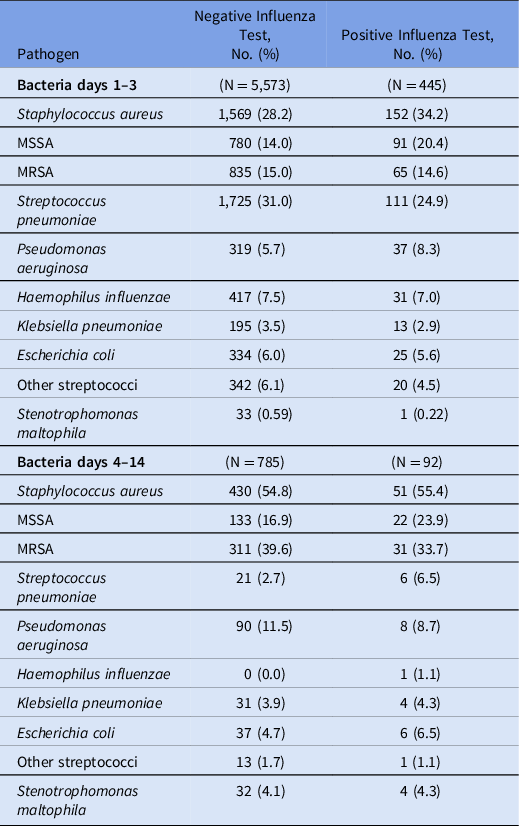
Among 38,665 patients hospitalized with CAP and tested for influenza in the first 3 hospital days, 4,313 (11.2%) tested positive for influenza. Of the patients with influenza, 3,964 (91.9%) also had a bacterial culture within 14 days, including 3,783 (87.7%) with blood cultures and 1,599 (37.1%) with respiratory cultures. Among patients without influenza, 94.3% had blood cultures and 47.9% had respiratory cultures. The rest of the analyses refer to patients with culture results or a positive urinary antigen. In the first 3 hospital days, patients testing positive for influenza were less likely than those testing negative to have an identified bacterial infection (10.3% vs 16.2%; P < .001), including among patients admitted to the ICU (7.2% vs 12.2%; P < .001). Table 1 shows the frequencies of pathogens by influenza status. In the first 3 days (community onset infections), patients with influenza were more likely than those without to have Staphylococcus aureus (34.2 vs 28.2%; P = .007), and they were commensurately less likely to have Streptococcus pneumoniae (24.9 vs 31.0%; P = .008). Of S. aureus isolates, the proportion that were methicillin resistant was lower among influenza-positive patients than among influenza-negative patients (42.8 vs 53.2%; P = .01). Frequency of S. aureus by culture source appears in the Supplementary Table (online). Pseudomonas aeruginosa and Haemophilus influenzae were found in 6%–8% of patients in both groups. After hospital day 3, representing hospital-acquired infection, most positive cultures in both groups yielded S. aureus (55% in both groups), and H. influenza was virtually absent. The distributions of recovered pathogens, including the fractions of MRSA among S. aureus cultures (72% vs 61%), did not differ significantly by influenza test result (P = .65).
Distribution of Bacterial Pathogens by Influenza Status and Timing of Cultures

Note. MSSA, methicillin-susceptible Staphylococcus aureus; MRSA, methicillin-resistant S. aureus.
Table 2 compares characteristics of influenza patients with community-onset bacterial coinfection to those without coinfection. Influenza patients with bacterial coinfection were younger (aged 66.7 vs 69.0 years; P = .006) and more likely to be male (55.3% vs 47.6%; P = .002), had higher combined comorbidity scores (3.2 vs 2.7; P < .001), and more often had had a prior admission within the previous 6 months (7.4% vs 4.3%; P = .03). Most comorbidities, including obesity, chronic pulmonary disease, heart failure and diabetes, were not associated with coinfection. However, patients with coinfection were more likely to have immunosuppression (18.9% vs 13.7%; P = .003) and liver disease (4.3% vs 2.2%; P = .006). They also had more severe illness on presentation, as demonstrated by higher rates of initial admission to the ICU (37.1% vs 22.4%; P < .001), use of mechanical ventilation (17.8% vs 6.5%; P < .001), and vasopressor medications (13.9% vs 3.8%; P < .001).
Characteristics of Influenza Patients With and Without Community-Onset Bacterial Coinfection a

Note. SNF, skilled nursing facility; ICU, intensive care unit; IMV, intermittent mechanical ventilation; SD, standard deviation.
a Differences are very highly statistically significant (P < .001) for all variables, by χ Reference Chien, Klugman and Morens2 test for dichotomous or nominal or t test for continuous or ordinally scored variables, except race (P = .11), smoking (P = .077), admission from a skilled nursing facility (P = .39), and hospital teaching status (P = .29) and location (urban vs rural, P = .87). Statistical significance does not imply clinical significance.
Compared to other influenza patients, those with community-onset bacterial coinfection had worse outcomes, including higher rates of clinical deterioration (late admission to ICU, and late IMV and vasopressor use), higher inpatient mortality, longer length of stay, and higher costs (Table 3). After adjustment for demographics, insurance status, comorbidities, and hospital characteristics bacterial coinfection was associated with increased odds of in-hospital mortality (aOR, 3.00; 95% CI, 2.17–4.16), transfer to ICU on day 2 or later (OR, 2.83; 95% CI, 1.98–4.04), vasopressors (OR, 3.63; 95% CI, 2.53–5.19), and IMV (OR, 3.23; 95% CI, 2.29–4.57). Coinfection was also associated with higher cost (risk-adjusted mean multiplier, 1.77; 95% CI, 1.59–1.96) and longer LOS (risk-adjusted mean multiplier, 1.48; 95% CI, 1.37–1.61).
Outcomes of Influenza Patients With and Without Community-Onset Bacterial Coinfection a

Note. IQR, interquartile range (Q1–Q3); ICU, intensive care unit; IMV, intermittent mechanical ventilation.
* Second day or after.
a Differences are very highly statistically significant (P < .001) for all variables, by χ Reference Chien, Klugman and Morens2 test for dichotomous variables and Wilcoxon rank sum test for length of stay and cost. Statistical significance does not imply clinical significance.
Discussion
In this retrospective cohort study featuring a large sample of US patients hospitalized with CAP and influenza, we found that 10.3% had community-onset bacterial coinfection based on blood or respiratory cultures. This estimate, based on the 91.9% of patients who had bacterial testing, likely represents a slight overestimate because untested patients would be expected to have a lower rate of positive tests. Patients with bacterial coinfection were more likely to be infected with MSSA and slightly less likely to have S. pneumoniae than patients without influenza. There were no differences in rates of other pathogens, most notably MRSA. These differences were present in the first 3 days of admission but not in later cultures, reflecting the impact of nosocomial pathogens. Lastly, bacterial coinfection was associated with substantially worse outcomes, including higher mortality, greater need for mechanical ventilation, and higher costs.
These findings have implications for the use of empiric antibiotics among patients with pneumonia who test positive for influenza. Older guidelines from the Infectious Disease Society of America (IDSA) Reference Uyeki, Bernstein and Bradley31 and the CDC recommended that in addition to receiving antiviral treatment, patients who present initially with severe disease (extensive pneumonia, respiratory failure, hypotension, and fever) or who deteriorate or do not improve after 3–5 days of antiviral treatment should receive empiric treatment for bacterial coinfection. Specifically, they recommend the use of broad-spectrum antibiotics that include coverage of MRSA. Reference Uyeki, Bernstein and Bradley31 In contrast, the more recent ATS/IDSA guidelines for CAP Reference Metlay, Waterer and Long29 recommend that all patients hospitalized with confirmed influenza should be treated with the usual empiric antibiotics for CAP, reserving anti-MRSA therapy for patients with other known risk factors. Our findings support the latter recommendation because bacterial coinfection was common, but MRSA was not. Moreover, patients with influenza were less likely than other patients to have any positive bacterial test, so the absolute risk of MRSA among influenza patients was lower than among noninfluenza patients. Of all influenza patients who underwent initial bacterial testing, <2% had cultures that grew MRSA.
Bacterial coinfection during pandemics is often characterized by superinfection with S. aureus. Reference Murray, Robinson and White8,Reference Metersky, Masterson, Lode, File and Babinchak11–Reference Yang, Gao and Chen13 This was particularly true of the 1918 pandemic and the 2009 H1N1 pandemic. One study of 207 adults admitted to 35 ICUs in the United States with bacterial coinfection during the 2009 pandemic, found that 45% was due to S. aureus, and 62% of the isolates were methicillin resistant. Reference Rice, Rubinson and Uyeki38 Both proportions were substantially higher than what we observed in nonpandemic years in a mixed population of both ICU and non-ICU patients. Patient susceptibility to bacterial coinfection or superinfection increases between 4 and 14 days after onset of influenza symptoms. Reference Shrestha, Foxman, Weinberger, Steiner, Viboud and Rohani16,Reference van der Sluijs, van der Poll, Lutter, Juffermans and Schultz26,Reference Grabowska, Högberg, Penttinen and Svensson39,Reference Morris, Cleary and Clarke40 We attempted to differentiate between community-onset coinfection present at the time of diagnosis and superinfection occurring later by analyzing cultures in the first 3 days separately from those obtained later. When we did this, we found no difference in the spectrum of bacterial pathogens detected in cultures taken on days 4–14 between patients with and without influenza, suggesting that the kinds of superinfection seen with some pandemic strains did not occur in our sample. Instead, influenza patients were susceptible to the same sorts of nosocomial infections as patients without influenza. As a result, influenza patients who demonstrate clinical deterioration after 3 days should be treated empirically for the usual hospital-acquired pathogens.
The most recent IDSA guidelines recommend that all patients hospitalized with influenza pneumonia also receive empiric antibacterial treatment. Reference Metlay, Waterer and Long29 Our finding that >10% of influenza patients had a bacterial coinfection supports these recommendations. This finding also raises the question of whether it is possible to identify a subset of influenza patients most likely to have bacterial coinfection to limit unnecessary antibacterial prescribing. Although patients with chronic heart and respiratory conditions, as well as obesity, may be more prone to coinfection, we did not observe these associations. Reference Chertow and Memoli18 Coinfected patients were more likely to have liver disease and immunosuppression, but both of these conditions were relatively rare, making it impractical to identify patients who need empiric therapy based on comorbidities alone. We did not assess whether certain clinical signs, radiologic features, or biomarkers (eg, procalcitonin) may help identify patients with coinfection.
Bacterial coinfection was associated with significantly worse outcomes, resulting in longer lengths of stay and higher costs. The fact that coinfection is associated with higher mortality rates has been previously reported. Reference Chertow and Memoli18,Reference Morris, Cleary and Clarke40,Reference Gill, Sheng and Ely41 Our analysis helps to differentiate the contribution of coinfection versus that of comorbidities, which are an important confounder of the relationship. Patients who were admitted with coinfection tended to have more comorbid conditions, but even after adjustment for these baseline characteristics, they had ≥3 times the odds of needing mechanical ventilation, vasopressors, and of death.
Our study has several limitations. Inasmuch as clinical trials and cohort studies of this issue are unlikely to occur, we used a large multi-institutional observational hospital discharge database that has frequently been used for research but lacks clinical data other than microbiological results. We included only patients who had both influenza testing and bacterial cultures; therefore, our results may be biased. However, of patients who underwent influenza testing, 92% also had at least 1 bacterial culture. Most of these were blood cultures, which are often negative, and the respiratory cultures were primarily sputum, which are often of poor quality. However, the spectrum of pathogens should not necessarily be affected by this, at least when comparing influenza patients to other patients with CAP. Additionally, we had no information on which antibiotics patients may have received prior to entering the hospital, and any antibiotics given prior to cultures being drawn could have affected the outcomes. Again, it is not clear that such antibiotics should affect influenza patients differently from noninfluenza patients. In our analysis of outcomes, we attempted to adjust for patient differences in comorbid conditions, but because the dataset lacked clinical measures such as vital signs, supplemental oxygen requirements, or laboratory values, there may have been some residual confounding. Lastly, we do not know when patients became infected with influenza, so our ability to separate out initial coinfection from delayed superinfection was limited. Nevertheless, we did find that the spectrum of pathogens differed after 3 days. Instead of seeing superinfection primarily with S. aureus, we saw a range of hospital-acquired pathogens that mirrored those of noninfluenza patients.
In conclusion, during nonpandemic years, influenza infection resulting in hospitalization is frequently accompanied by bacterial coinfection. Treatment with empiric antibiotics, at least for the first 2 days while awaiting maturation of clinical cultures and assessing patient response, is appropriate. Although coinfection is most common with S. aureus, most strains were susceptible to methicillin; therefore, coverage for MRSA is not warranted routinely. For patients who developed bacterial pneumonia later in their hospitalization, the bacterial spectrum resembled other nosocomial pneumonias. Patients with late deterioration should receive the same treatment as other patients with hospital-acquired infections.
Supplementary material
To view supplementary material for this article, please visit https://doi.org/10.1017/ice.2021.96
Acknowledgements
Financial support
The study was supported by the Agency for Health Research and Quality (AHRQ grant no. 5 R01 HS024277-05) to M.B.R.
Conflicts of interest
M.K. has received grant funding from the Centers for Disease Control and Prevention (CDC) and royalties from UpToDate for chapters on hospital-acquired pneumonia. A.D. has received research support from the AHRQ, research support from The Clorox Company; he serves as a consultant to Merck. M.Z. has received research support from Tetraphase Pharmaceuticals, Astellas, Lungpacer, Merck, Spero, The Medicines Company, and Melinta; she has also received consulting fees from Paratek, Arasanis, Shionogi, Pfizer, Nabriva, and Melinta. All other authors report no conflicts of interest relevant to this article.