Introduction
Endoscopic evaluation of the nasal sinuses was first reported in 1901, when a modified cystoscope was used for visualisation.Reference Draf1–Reference Kennedy, Zinreich, Rosenbaum and Johns3 Endoscopic sinus procedures have been highly successful in recent times, particularly for treating chronic rhinosinusitis and nasal obstruction.Reference Jones4, Reference Bhattacharyya5 While most sinonasal endoscopic procedures today are performed to treat local sinonasal cavity disease, endoscopes are now being deployed more routinely in more advanced surgical procedures, such as the treatment of midline anterior and lateral skull base lesions.Reference Stammberger6, Reference Stammberger7
Endoscope construction has developed throughout the twentieth century and into the twenty-first. Design types include rigid, semi-rigid and flexible endoscopes, each with its own advantages and limitations. Endoscopic procedures using traditional sinus endoscopes are currently limited by surgical field visualisation and procedural inconvenience. Only about one-third of the maxillary sinus can be manipulated because of the anatomical position. Although both antrostomy and angled scope attachments can improve visualisation, there may still be visual field deficits. In addition, current sinus endoscopes do not have an instrument channel, making biopsy collection and manipulation cumbersome.Reference Robey, O'Brien and Leopold8, Reference Gilain, Aidan, Coste and Peynegre9 The surgeon is often faced with the difficult task of juggling instrumentation to complete a basic biopsy procedure. Moreover, the currently available 4 mm and 2.7 mm rigid sinonasal endoscopes may cause patient discomfort.Reference Stankiewicz10 This limits their utility for basic procedures under local anaesthesia in the office setting.
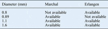
Recently, semi-rigid sialendoscopes (Marchal and Erlangen types) have been introduced for minimally invasive treatment of salivary gland diseases, and they have helped to revolutionise the treatment of these conditions.Reference Marchal and Dulguerov11 These endoscopes have smaller diameters, are semi-rigid and have a working channel which can be used for biopsy or drug delivery (Table I). The semi-rigid construction has inherent flexibility that increases the visual field to allow visualisation of a larger mucosal surface area. Moreover, biopsy sites can be targeted more precisely and swabs more reliably taken for bacterial culture. In addition, a working channel built into the sialendoscope allows the passing of instrumentation, which is convenient for interventional procedures. Other investigators have explored the use of sialendoscopes in the middle ear as a method of transtympanic drug delivery.Reference Peters, Lin, Arriaga, Nuss, Schaitkin and Walvekar12 Similarly, sialendoscopes may be inserted into the paranasal sinuses as a vehicle for drug delivery. This could allow the precise, directed delivery of pharmaceutical agents to treat inflammatory sinonasal disease.
Diameters of commonly used sialendoscopes*

*Only the 1.6 mm sialendoscope can pass grasping or biopsy forceps through the working channel
This study characterised the efficacy of current sialendoscopes in the sinonasal cavity in a cadaveric model.
Materials and methods
A cadaveric study was performed in a tertiary care academic setting. Sinus endoscopy was performed on 10 fresh-frozen cadaver heads by both senior authors, one with rhinology specialty training and the other with head and neck oncology training. The heads were minimally dissected and the maxillary ostia were enlarged. Endoscopes were then reintroduced and the study was performed.
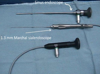
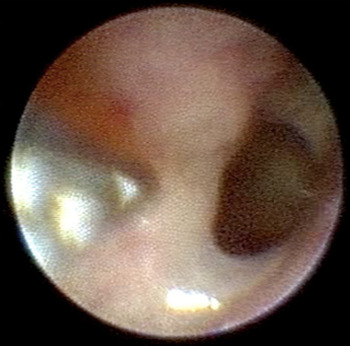
The Marchal 1.3 mm sialendoscope (0.4 mm diameter working channel) and the Erlangen sialendoscopes (1.1 mm and 1.6 mm external diameters, with 0.45 mm and 0.8 mm diameter working channels, respectively) were used for visualisation and performing interventions in the paranasal sinuses (Figure 1). Mucosal biopsies were obtained using flexible grasping forceps with double action jaws designed for use with each sialendoscope (Figure 2). Images and video documentation were obtained using a standard Karl Storz camera and video tower. Light was set to 100 per cent.
Photograph showing a 1.3 mm Marchal sialendoscope alongside a traditional sinonasal endoscope.

Photograph showing flexible grasping forceps being introduced into the sphenoid ostium, using the 1.6 mm Erlangen sialendoscope. Surgical instrumentation can be reliably incorporated through the working channel on the sialendoscope. The image shows the best resolution possible using this sialendoscope.

Outcomes included ostia identification, sinus cannulation, success of mucosal biopsy collection and image clarity. The outcomes were graded on a 1–4 scale: 1, poor visualisation; 2, just visible enough to comfortably manipulate; 3, easily visible enough to comfortably manipulate; and 4, excellent visualisation. All data were tabulated (Table II).
Outcome scores for different sialendoscopes by size*

*1, inadequate; 2, somewhat adequate; 3, very adequate, but less than can be achieved with current nasal endoscopes; 4, comparable view to the one afforded by a nasal endoscope
Results
The ability of Erlangen (1.1 mm and 1.6 mm) and Marchal (1.3 mm) sialendoscopes to identify the maxillary sinus ostium, identify the sphenoid sinus ostium, cannulate the maxillary sinus ostium, perform a mucosal biopsy and produce a clear image was evaluated (Table II). Both Marchal and Erlangen sialendoscopes demonstrated reliable visual field performance, with the Erlangen 1.6 mm sialendoscope providing the optimal maxillary ostium visualisation. All sialendoscopes were efficacious in cannulating the sphenoid sinuses; there was no appreciable difference among them. These sialendoscopes were found to be similarly effective in the sphenoid sinus both pre- and post-dissection. The 1.3 mm Marchal and 1.6 mm Erlangen sialendoscopes provided superior image clarity.
Discussion
Modern surgical paranasal sinus endoscopy has several limitations.Reference Draf1 While traditional sinus endoscopes produce high quality visual images, their rigid construction restricts the visual field to a small area of the mucosal surface. The rigid construction also causes patient discomfort. Furthermore, traditional nasal endoscopes are not equipped with instrument access ports. Therefore, the surgeon often has to switch equipment attachments several times during the procedure, while taking breaks to re-anesthetise patients when the discomfort becomes unbearable.
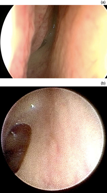
Improved sialendoscope design has aimed to overcome these limitations. While traditional sinonasal endoscopic instrumentation provides the optimal visual clarity, anatomical landmarks in the sinonasal cavity can be reliably visualised with the sialendoscope (Figure 3). In addition, the visual field is broadened as a result of increased flexibility, and there is no need to stop the procedure to change visualisation attachments. Furthermore, the sialendoscope has an access port for instrumentation to allow fast, precise biopsy collection and directed sampling for bacterial culture. Both of these factors decrease the procedure time.
Photographs showing the sphenoid ostium visualised with (a) a 1.3 mm Marchal sialendoscope and (b) a 4 mm traditional sinonasal endoscope.

Sialendoscope use in the sinonasal cavity might be ideally suited for practitioners whose practice includes both nasal endoscopy and sialoendoscopy because the smaller gauge sialendoscope functions in both anatomical orifices. In this study, dissections were performed by both senior authors with different specialist training. Performing these procedures is thus more than feasible for practitioners with solid endoscopic skills.
A principal weakness of the sialendoscopes tested in this study was the lack of adequate maxillary ostia visualisation without dissection. However, this was mitigated by antrostomy, after which visualisation was possible. Mucosal biopsy within the maxillary sinus was not possible prior to dissection; however, the procedure was possible within the sphenoid sinus. As expected, the least distorted images of the maxillary ostia were obtained using larger diameter sialendoscopes.
This cadaveric study evaluated the potential of interventional sialendoscopy for sphenoid and maxillary sinus examination. The range of function, image quality and interventional options afforded by the sialendoscope within the sphenoid sinus were found to be excellent. All agents could be delivered effectively through the working channel and intrasphenoidal biopsies could also be performed with biopsy and/or grasping forceps. Although this study did not directly examine patient tolerance, we hypothesise that the semi-rigid and smaller gauge construction will improve patient comfort. In this study, interventional sialendoscopy of the maxillary sinus was most effective after maxillary antrostomy.
In addition, the ease of evaluating deeper areas of the nasal cavity is improved with smaller calibre endoscopes. In this study, the sialendoscopes could be easily used to examine the post-nasal space in cadavers with nasal septum deviations, some of which were significant. By passing the sialendoscope along the floor of the nose, either along or under a prominent maxillary crest or under the inferior turbinate, the post-nasal space can be easily evaluated.
While the cost of sialendoscopes is currently higher compared with nasal endoscopes, it is likely that the cost will reduce as their use becomes more widespread.Reference Peters, Lin, Arriaga, Nuss, Schaitkin and Walvekar12 Sialendoscopes are currently used for many procedures and have possible applications in transtympanic drug distribution. In addition, the 1.6 mm sialendoscope could enable post-surgical debridement in sphenoid sinus.
• Traditional sinus endoscopy has limited functionality, outcomes and patient satisfaction
• Endoscopic sinus procedures are routinely used to treat rhinosinusitis and nasal obstruction
• Sialendoscopes have a potential use in trans-tympanic drug delivery
• Sialendoscopes are a minimally invasive method of post-operative drug delivery and/or biopsy for all sinuses
Modifications to current sialendoscopes that would optimise their use in the sinonasal cavity for minimally invasive endonasal procedures include (1) an additional working channel, (2) improvements in mechanical design to provide robust optics and (3) improvements in tip flexibility to optimise endoscopic manoeuvring.
Conclusion
The limitations of traditional sinus endoscopy have prompted investigations into ways to improve functionality, outcomes and patient satisfaction. Sialendoscopes can be used for visualising and performing interventions in the sinonasal cavity, but are mainly limited to the sphenoid sinus. They may also be considered a minimally invasive method of drug delivery and/or biopsy in the post-operative setting for all sinuses. Improved design could lead to the more widespread adoption of sialendoscopes for use in the head and neck. Future investigations should include randomised trials to compare the reliability of procedures performed and biopsies obtained with traditional sinus endoscopic instrumentation and sialendoscopic instrumentation.