Introduction
Endothelin 1 is a vasoconstrictive peptide consisting of 21 amino acids. It was originally isolated from the culture supernatant of porcine aortic endothelial cells by Yanagisawa in 1988.Reference Yanagisawa, Kurihara, Kimura, Tomobe, Kobayashi and Mitsui1 Endothelin 1 is widely distributed in cardiovascular and noncardiovascular tissues. It has many biological functions and plays a significant role in vasoconstriction and the regulation of fluid volume and ion balance. Endothelin-1-like activity has been demonstrated in the inner ear;Reference Jinnouchi, Tomiyama, Pawankar, Ikezono and Yagi2, Reference Jinnouchi, Tomiyama and Pawankar3 however, the role of endothelin 1 in maintaining cochlear homeostasis is not clear.
The lateral wall of the cochlea is important in the maintenance of cochlear homeostasis. The distribution and significance of endothelin 1 in the cochlear lateral wall requires further study.
In order to understand the microenvironment of the cochlea and how it is maintained, the present study investigated the distribution of endothelin 1 activity in the guinea pig cochlear lateral wall, using immunohistochemistry.
Materials and methods
Healthy guinea pigs weighing 350–400 g were used. All procedures concerning animals were approved by the animal care and use committee at the Huaxi Medical School of Sichuan University.
After anaesthesia with intraperitoneal pentobarbitone (30 mg/kg) injection, the animals were sacrificed using 200 ml of 0.9 per cent saline administered via cardiac puncture, followed by 4 per cent paraformaldehyde until the neck became stiff.
The temporal bones were removed and fixed overnight (the same fixative was used for all specimens). After decalcification with 10 per cent ethylene diamine triacetic acid for two weeks, the specimens were embedded in paraffin and serially sectioned into 5 µm thick slices. Tissue sections were then deparaffinised, rehydrated, treated with 3 per cent H2O2 for 10 minutes, immersed in 0.01 M citrate buffer (pH 6.0), microwaved for 5–10 minutes (90–98°C) to retrieve the antigen and then exposed to 5 per cent goat serum for 30 minutes (37°C) to block any nonspecific reaction. Rabbit anti-endothelin 1 polyclonal antibody (diluted 1∶1200; Peninsula Laboratories, San Carlos, CA, USA) was then added and incubated at 37°C for 120 minutes and at 4°C overnight. Tissue sections were then treated with biotin-labelled goat anti-rabbit immunoglobulin (Ig) G (Beijing Zhongshan, Golden Bridge Biotechnology Co. Ltd., Beijing, China) at 37°C for 60 minutes, followed by adding avidin–biotin–peroxidase complex (Beijing Zhongshan, Golden Bridge Biotechnology Co. Ltd., Beijing, China), incubating at 37°C for 60 minutes, reacting with DAB/H2O2 solution for 5–10 minutes and then counterstaining with haematoxylin. Specimens were then examined under an Olympus light microscope (Olympus Co. Ltd., Tokyo, Japan).
In the negative control experiment, sections were processed using the same protocol, except that the primary antibody was replaced by phosphate buffered saline. In the positive control experiment, sections of guinea pig trachea were incubated with the same primary antibody (since endothelin 1 immunoreactivities have been demonstrated in trachea).Reference Kizawa, Kotake, Kusama, Saito and Murakami4
Results
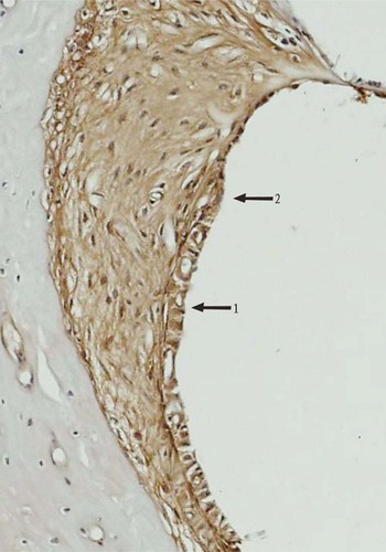
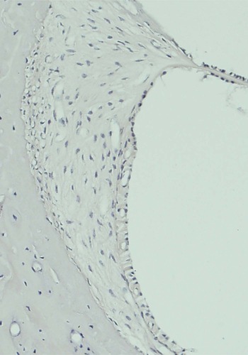
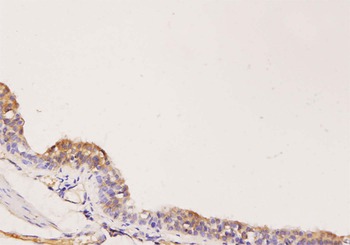
Endothelin-1-like activity was found to be extensively distributed in the cells of the cochlear lateral wall. Different degrees of positive reactivity were demonstrated in the marginal cells, spiral prominence epithelial cells, outer sulcus cells, stria vascularis capillaries, basal cells and spiral ligament fibrocytes (Figure 1). In the negative control, no immunostaining could be found in any area (Figure 2). In the positive control, immunoreactivity was observed in the guinea pig tracheal epithelia, in agreement with published data (Figure 3).
Endothelin-1-like activity was demonstrated in the stria vascularis (arrow 1), the spiral prominence epithelial cells and outer sulcus cells (arrow 2), and the spiral ligament fibrocytes (ABC; ×200).

No immunostaining was observed in the cochlear lateral wall cells from negative control specimens (ABC; ×200).

Immunoreactivity was observed in guinea pig tracheal epithelium (i.e. positive control specimen) (ABC; ×400).

Discussion
The lateral wall of the cochlea is composed of the stria vascularis, spiral prominence epithelial cells, outer sulcus cells, spiral ligament fibrocytes and capillaries. The cochlear lateral wall plays an important role in production and resorption of endolymph, ion transportation, and generation of the endocochlear potential. Therefore, the cochlear lateral wall is very important in maintaining the homeostasis of the cochlear microenvironment.
The present study showed that endothelin-1-like activity was extensively distributed in the tissues of the cochlear lateral wall, including the marginal cells, basal cells, spiral prominence epithelial cells, outer sulcus cells, spiral ligament fibrocytes and stria vascularis capillaries. These findings indicate that endothelin 1 may play an important role in the physiological and pathological function of the cochlear lateral wall.
Our findings differ from those reported by Jinnouchi et al., who found endothelin-1-like activity to be present only in the marginal cells and not in other cells in the stria vascularis.Reference Jinnouchi, Tomiyama, Pawankar, Ikezono and Yagi2 This may have been due to the different kind of primary antibodies used. The primary antibody used in our study was rabbit anti-endothelin 1 polyclonal antibody, while that used by Jinnouchi et al. was mouse anti-human endothelin 1 monoclonal antibody.
The endolymph in the cochlea duct has an ionic composition characterised by large amounts of K+ and small amounts of Na+. A highly positive endocochlear potential has also been found in cochlear endolymph. The unique ionic composition together with the high endocochlear potential are essential to normal hearing. The stability of the ionic composition and the endocochlear potential depend on the active transportation of ions by Na-K-adenosine triphosphatase (ATPase) and Na-K-2Cl, which present in many cells of the cochlear lateral wall.
Marginal cells are located on the surface of the stria vascularis, directly facing the endolymph fluid. The Na-K-ATPase and Na-K-2Cl transporter molecules present in the basolateral plasma membrane of the marginal cellsReference Goto, Oshima, Ikeda, Ueda and Takasaka5, Reference Nakazawa, Spicer and Schulte6 work together to transport K+ from the interstitial space and secrete it into the endolymph, across the apical membrane of the marginal cells. The apical membrane of the outer sulcus cells faces the endolymph, and the main functions of these cells include absorbing K+ and Na+ from endolymph and transporting K+ to the spiral ligament via the Na-K-ATPase located in the basolateral plasma membrane.Reference Marcus and Chiba7, Reference Schulte and Adams8 The presence of many gap junctions between the basal cells and the type I fibrocytes would be favourable for ion transportation from cell to cell. Some types of fibrocytes in the lateral wall display Na-K-ATPase and Na-K-2Cl transporter molecules,Reference Goto, Oshima, Ikeda, Ueda and Takasaka5, Reference Schulte and Adams8 providing evidence for a role in ion transportation.
It has been reported that endothelin 1 plays a role in the regulation of Na-K-ATPase and Na-K-2Cl transporter.Reference Prasanna, Dibas, Hulet and Yorio9, Reference Gupta, Ruderman, Cragoe and Sussman10 The present study demonstrated endothelin-1-like activity in marginal cells, spiral prominence epithelial cells, outer sulcus cells and spiral ligament fibrocytes. Therefore, we consider that endothelin 1 may regulate ion transportation and influence the generation of endocochlear potential, through its effect on the activity of Na-K-ATPase and Na-K-2Cl transporter.
The stability of endolymph fluid volume is influenced by many factors, including the endolymph flow and ionic composition. Any change in the composition and volume of endolymph will affect normal hearing. The cochlear lateral wall regulates the secretion of Na+ and H2O and so maintains fluid balance. The present study showed endothelin-1-like activity to be extensively distributed amongst the cells of the lateral wall. This suggests that endothelin 1 might affect the homeostasis of the cochlear endolymph volume.
• Endothelin 1 is a vasoactive peptide with many biological functions
• This study investigated the distribution of endothelin 1 in the guinea pig cochlea
• Endothelin 1 is widely distributed in the guinea pig cochlea, suggesting that it may have a role in maintaining cochlear haemostasis
Atrial natriuretic peptide is a diuretic, natriuretic hormone. This hormone has been reported to be extensively distributed in the stria vascularis.Reference Chen, Wang, Liu and Qiu11 The present study shows endothelin 1 to be localized in the same regions as atrial natriuretic peptide. This supplies evidence that endothelin 1 stimulates the release of atrial natriuretic peptide.Reference Skvorak, Nazian and Dietz12 We therefore suggest that the endothelin 1 of these cells may affect the homeostasis of endolymph through its action on atrial natriuretic peptide.
Conclusion
Endothelin 1 is extensively distributed in the cells of the cochlear lateral wall and may function as an important regulator for maintaining the homeostasis of the cochlear microenvironment. However, the mechanism of action of endothelin 1 requires further study.
Acknowledgements
We should like to thank Dr Hui-Min An for counsel and advice. This project was supported by the Natural Science Foundation of China (30271408).