Case report
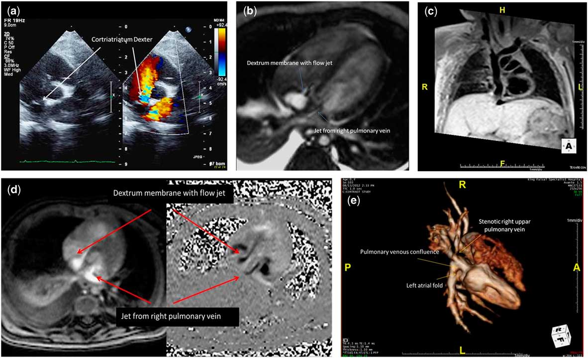
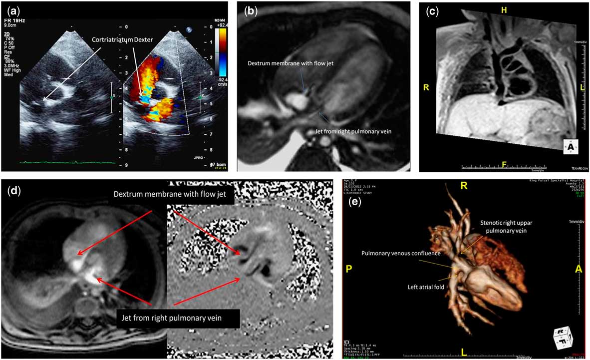
An 11-month-old girl, a product of a full-term gestation, presented to an outside emergency facility with abdominal distention and facial swelling. She was not in distress, had normal oxygen saturation and other vital signs, generalised oedema, ascites, and mild hepatomegaly. The cardiovascular system examination was within normal limits. Further investigation revealed lung congestion and bilateral pleural effusion, later confirmed as chylous.Reference Anderson, Baker, Redington, Rigby, Penny and Wernovsky 1 – Reference Niwayama 3 Echocardiogram showed obstructed persistent sinus venosus membrane dividing the right atrium into near-complete two compartments (Fig 1a and video 1). Pressure Doppler interrogation revealed a peak and mean gradient of 29 and 11 mmHg, respectively, across the membrane. A partial shelf was also seen separating the pulmonary vein confluence and the inflow of the mitral valve above the left atrial appendage with Doppler flow acceleration, peak and mean gradient of 12 and 5 mmHg, respectively. Cardiac magnetic resonance imaging (MRI) showed a tunnel-like structure formed by the free posterior wall of the right atrium on one side and the right venous valve remnant on the other, with very tight communication between the tunnel and the cavity, confirming the diagnosis of persistent sinus venosus membrane (Fig 1b–d and video 2, 3). It also demonstrated stenosis of the right upper pulmonary vein and confirming the echocardiogram finding of the divided left atrium as described above. In addition, ascites and pleural effusion were also confirmed. The technique and sequences were tailored to the suspected diagnosis.
Imaging modalities used to evaluate the anatomy. A 2D and colour Doppler image ( a ) showing the accelerated jet through the left atrial fold. White blood MR SSFP sequence ( b ) showing the jet through the left and right atrial membrane. MR black blood image ( c ) showing the extent of the right atrial membrane. MR flow phase and velocity encoded image ( d ) showing the two jets through the obstructive membranes. ( e ) Shows 3D reconstruction of cardiac magnetic resonance angiogram (MRA).
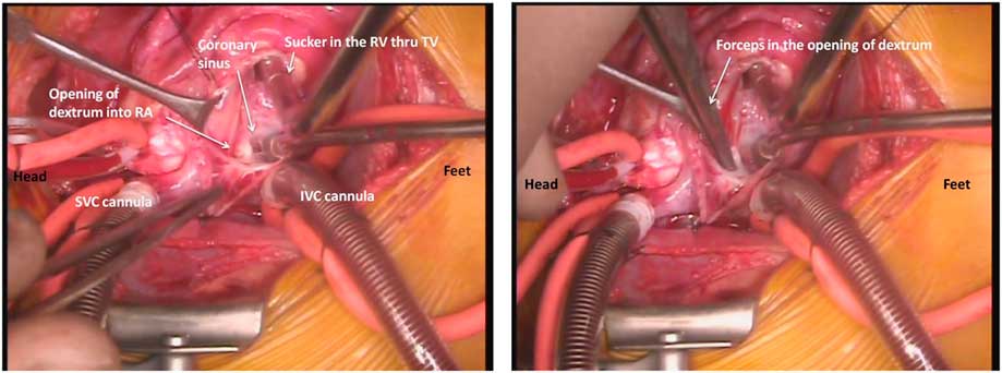
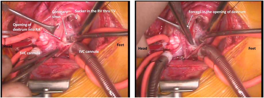
The patient underwent excision of the right atrial fibromuscular structure to relieve the caval obstruction. The left atrium was explored through a separate left atrial incision (Fig 2) where there was a fibromuscular structure related to right pulmonary veins, which was excised while no intervention was done to the right upper pulmonary vein stenosis.
Surgical images annotating the anatomy of the right atrium during surgical repair.
The patient had an uneventful postoperative recovery. A 4-month follow-up echocardiogram showed mild right upper pulmonary vein Doppler flow acceleration, with mean pressure gradient of 2–3 mmHg. There was trace of tricuspid regurgitation with peak systolic pressure gradient of 15 mmHg.
Discussion
The term cor triatriatum dextrum had first been described by Reference YaterYater,Reference Yater 4 whereas the first clinical description in the literature was given by Rossall and Caldwell,Reference Rossall and Caldwell 5 when they described near-complete obstruction of venous flow from the inferior caval vein to the right atrium in a 19-year-old patient. A review of 1940 heart specimens at cardio-pathological museum of the Children’s Hospital of Pittsburg identified 14 specimens with prominent remnants of the embryonic right valve.Reference Trento, Zuberbuhler and Anderson 6 Of those, two patients had divided right atrium, but only one was diagnosed preoperatively. Embryologically, the divided right atrium arises from the sinus venosus valve, which is composed of the right and left venous valves. The left venous valve joins the atrial septum secundum, whereas the right regresses to leave the Thebesian and Eustachian valves.Reference Gerlis and Anderson 7 The divided right atrium results from failure of regression of the right venous valve around the 12th embryonic week.Reference Trakhtenbroit, Majid and Rokey 8 However, there is variable degree of regression failure whereby we occasionally see, on echocardiogram, what we call prominent Chiari network without evidence of obstruction as noted by Trento et al.Reference Trento, Zuberbuhler and Anderson 6 The presence of intra-atrial obstruction can interfere with the normal development of the right side of the heart and thereby explains some of the associated lesions with hypoplasia of the right-heart structures.Reference Trento, Zuberbuhler and Anderson 6 , Reference Trakhtenbroit, Majid and Rokey 8
Clinical presentation depends on the degree of obstruction and associated lesions. It is more likely to be associated with hypoplasia or obstruction of the right side. The degree of intra-atrial obstruction could lead to early presentation with ascites, hepatic failure, and peripheral oedema. It can also lead to chylothorax such as in our case. The patient may present at a later age with cyanosis because of streaming of the blood across an atrial communication.Reference Raffa, Al-Ibrahim, Kayali and Sorefan 9 – Reference Jones and Niles 11 One case presented with recurrent syncope and sudden death because of a sail-like structure that obstruct the right ventricle inflow,Reference Jones and Niles 11 although others might be misdiagnosed because of misleading echocardiographic images confusing it with more common lesions such as Ebstein’s anomaly.Reference Salam, Gallacher and Uzun 12 The majority of cases with prominent Chiari network with no obstruction or minor obstruction will pass undetected.Reference Trakhtenbroit, Majid and Rokey 8 , Reference Yarrabolu, Simpson and Virani 13
Echocardiography is usually diagnostic and will suffice in most casesReference Alboliras, Edwards, Driscoll and Seward 14 to plan and proceed with the surgical management. However, associated lesions might require further imaging and haemodynamic information for better planning complex cases. In our case, we presented the role of cardiac MRI in providing both anatomic and haemodynamic information to better guide the surgical procedure of choice, especially that a combination of right- and left-sided venous obstruction is extremely unlikely and, to the best of our knowledge, has never been reported before in the literature. The role of cardiac MRI in defining pulmonary vein abnormalities is well described and is more accurate than echocardiography alone.Reference Masui, Seelos, Kersting-Sommerhoff and Higgins 15 In the divided right atrium, there is one previous publication describing the technique and imaging in a 70-year-old patient, who presented with features of congestive heart failure and peripheral oedema.Reference Joe, Poustchi-Amin and Woodard 16 The technique and sequences were modified in our case, as the technology has improved, and the size and weight of our patient are significantly smaller.
Treatment is mainly surgical. However, interventional palliation has also been reportedReference Savas, Samyn, Schreiber, Hauser and O'Neill 17 that can temporarily improve the patient general condition in preparation for surgery. Surgical correction is relatively straightforward and uncomplicated, barring the associated lesions that can coexist. Careful and adequate resection of the obstructing fibromuscular structure is crucial. The gradient may persist if inadequate resection is performed, and cavo-atrial wall perforation may occur with aggressive resection. Great deal of attention should be paid to the coronary sinus and atrio-ventricular node during resection. Repair of the associated cardiac anomalies is performed in a standard manner.
In conclusion, this report and review is intended to present three important points
-
1. To report previously unreported, left and right intra-atrial obstruction, and pulmonary vein stenosis.
-
2. The role of cardiac MRI in outlining the pathology and associated lesion.
-
3. Variable modes of presentation of such rare pathology through this case as well as literature review.
Acknowledgement
None.
Financial Support
This research received no specific grant from any funding agency, commercial, or not-for-profit sectors.
Conflicts of Interest
None.
Supplementary material
To view supplementary material for this article, please visit http://dx.doi.org/10.1017/S1047951114001073