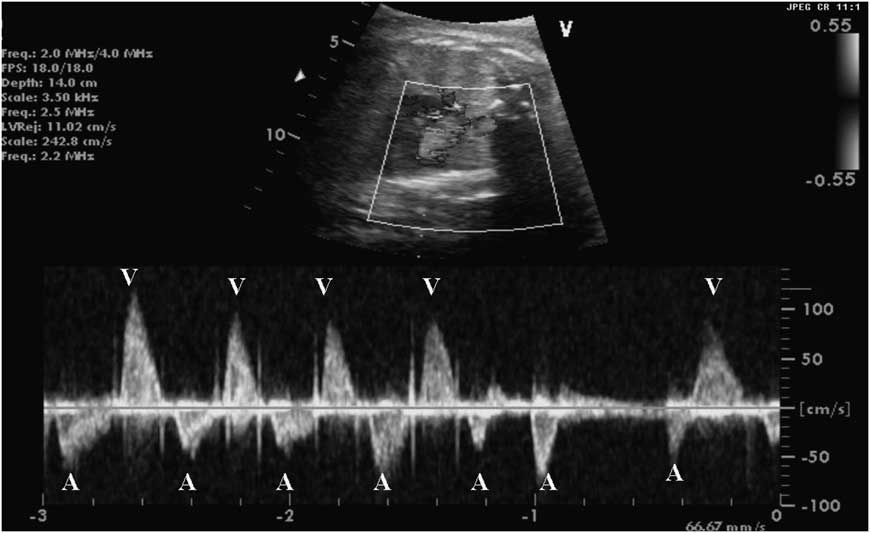
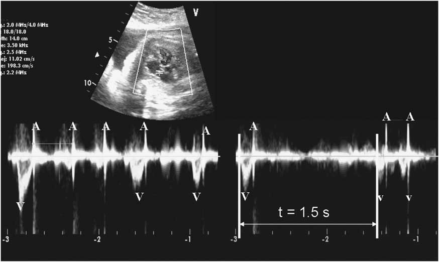
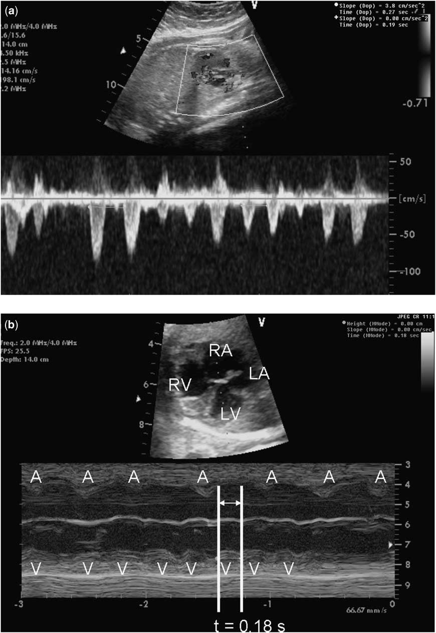
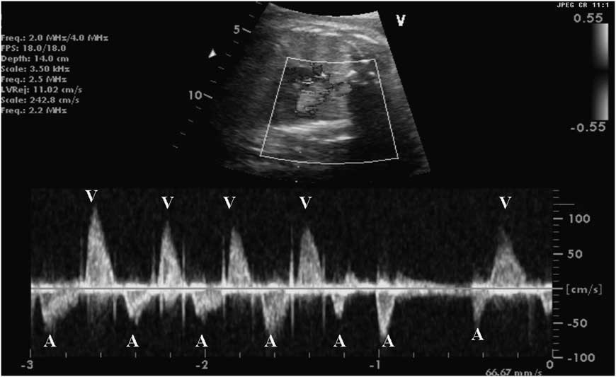
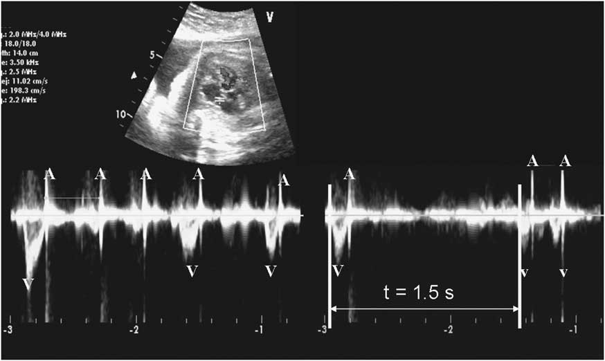
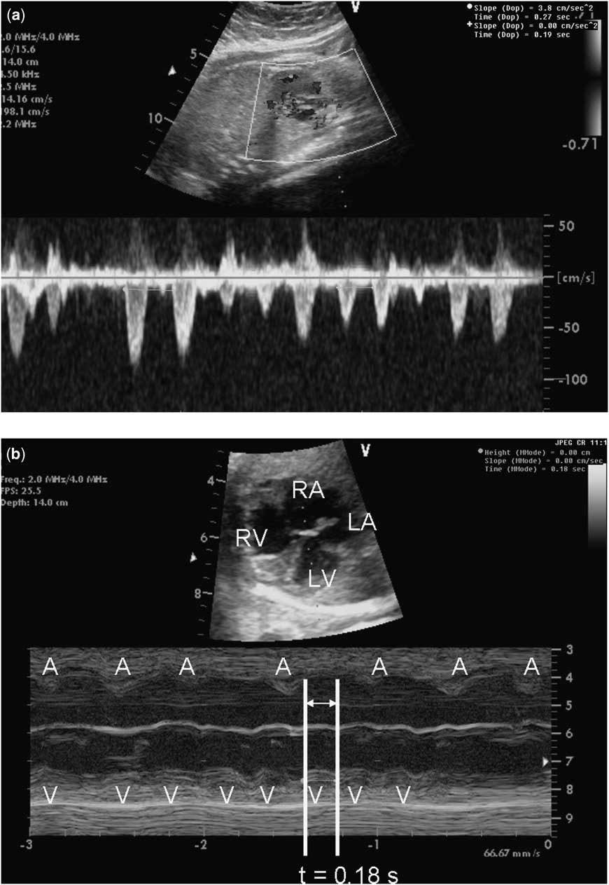
A 31-year-old woman with three children and no significant medical or family history was referred for foetal cardiac evaluation at 26 weeks’ gestation because of a 7-week history of an abnormal foetal heart rhythm. At 16 weeks’ gestation, the foetal heart rate and rhythm were normal at 150 beats per minute, but at 19 weeks the rhythm was detected to be irregular. The foetus was not compromised and no structural abnormalities were identified on foetal echocardiogram. Analysis of the rhythm using M-mode, pulsed Doppler interrogation of the mitral inflow–aortic outflow, and tissue Doppler of atrial and ventricular wall movement revealed an irregular rhythm with ventricular rate varying between 65 and 89 beats per minute. The atrial rate was faster, ranging between 120 and 130 beats per minute. There were periods of both atrioventricular synchrony and atrioventricular dissociation (Fig 1). During periods of atrioventricular synchrony, the mechanical atrioventricular interval ranged between normal at 120 milliseconds and prolonged at 150–160 milliseconds. The initial impression was that the foetus had sinus rhythm with variable periods of first- and second-degree block. Sjögren's syndrome antigen antibody titres were negative. Maternal and paternal electrocardiograms revealed sinus bradycardia, at rates of 40 beats per minute in the father and 50 beats per minute in the mother. There was no evidence for atrioventricular conduction abnormalities or QTc prolongation. At 30 weeks’ gestation, the rhythm appeared to be more chaotic with periods of both bradycardia and tachycardia. A new pericardial effusion was noted and the right ventricle appeared mildly dilated and hypertrophied, with tricuspid regurgitation. The bradycardia was secondary to atrioventricular block, with ventricular pauses of up to 1.5 seconds resulting in a low heart rate of 40 beats per minute (Fig 2). The tachycardia was noted to be nonsustained at a ventricular rate of 300 beats per minute with evidence of atrioventricular dissociation and a slower atrial rate, consistent with ventricular tachycardia (Fig 3). The combination of foetal ventricular tachycardia and sinus bradycardia with intermittent atrioventricular block raised concern for long QT syndrome.Reference Horigome, Nagashima and Sumitomo 1 The mother was admitted to the Labor and Delivery Unit for foetal monitoring while planning was initiated for specialised postnatal delivery room care and transport of the neonate. QTc-prolonging medications were avoided in the mother.
Aortic/mitral Doppler. There is AV synchrony at a rate of 140 initially. The 5th A may be either a retrograde A or a PAC. The 6th A is a non-conducted atrial beat and then a pause occurs. AV = atrioventricular; PAC = premature atrial contraction.
Aortic/mitral Doppler. There is no AV synchrony. There is a ventricular beat with a retrograde A or a blocked PAC and a subsequent 1.5-second pause. AV = atrioventricular; PAC = premature atrial contraction.
(a) Ventricular tachycardia seen on aortic Doppler at a rate of 315 beats per minute. (b) Ventricular tachycardia seen on M-mode of the RA and LV. There is ventriculoatrial dissociation with a ventricular rate of 330 beats per minute and an atrial rate of approximately 150 beats per minute. Note the RA and RV dilation on the four-chambered still image of the heart. RA = right atrium; LV = left ventricle; RV = right ventricle.
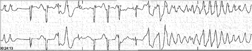
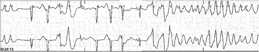
The baby was delivered by Caesarean section with a specialised delivery room team present for immediate care. The Complex Delivery Team included the foetal cardiologist, whose role at our institution is to organise the transitional care of foetuses with significant disease, the paediatric electrophysiologist, and a neonatologist who was scrubbed and waiting to place an umbilical venous catheter. Equipment for line placement, cardiac monitoring, defibrillation and resuscitation, and for administration of anti-arrhythmic medications was readied. On initial assessment of the neonate at less than 2 minutes of life, a rapid polymorphic ventricular tachycardia that appeared to be torsade de pointes (Fig 4) was noted. The baby was intubated, and a central umbilical venous line was placed. The ventricular tachycardia was suppressed with a bolus of lidocaine and she was prepared for transport.
Torsade de Pointes at birth.
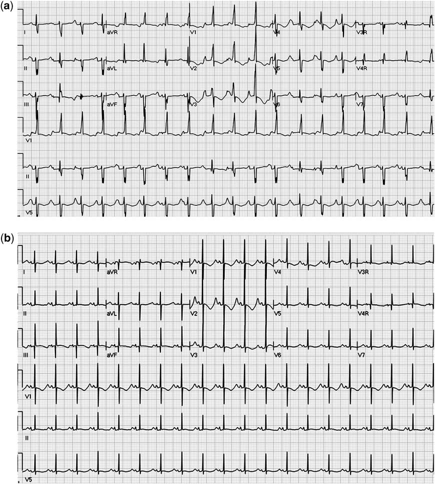
She arrived at the cardiac intensive care unit within 30 minutes of delivery. Cardiac evaluation revealed a structurally normal heart with normal biventricular function. Monitoring revealed repeated episodes of torsade de pointes and ventricular tachycardia (Fig 5a), which were treated with lidocaine, magnesium, and esmolol. Following anti-arrhythmic therapy, she remained in normal sinus rhythm with a markedly prolonged QTc of nearly 600 milliseconds on a 12-lead electrocardiogram. She had frequent ventricular ectopy, but no further ventricular tachycardia or torsade de pointes, and was transitioned to oral mexiletine and propranolol. After several days, she developed sinus bradycardia and intermittent second-degree atrioventricular block, likely secondary to the profound QT prolongation. Given the bradycardia, intermittent atrioventricular block, and potential risk for pause-dependent torsade de pointes, she underwent implantation of a single chamber epicardial pacemaker without complication. An implantable defibrillator was not chosen, given her weight of 2.2 kilograms, a good response to medications, and no need for cardioversion.
(a) 12-lead electrocardiogram at 30 minutes of life: this reveals accelerated idioventricular rhythm at a rate of 130 beats per minute with rare sinus capture beats. (b) 12-lead electrocardiogram at 3 months of life: the patient is being treated with mexiletine and propranolol but still demonstrates a markedly prolonged QTc of 510 milliseconds, as well as the ST segment typical of Long QT Type 3.
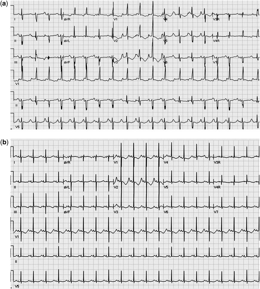
At the 3-month follow-up, she is doing very well on a combination of oral propranolol, mexiletine, and magnesium. Pacemaker interrogation has not revealed any further episodes of ventricular tachycardia. Subsequent electrocardiograms (Fig 5b) and Holter monitoring have shown persistence of QT prolongation with isolated ventricular ectopy and couplets but no torsade de pointes or ventricular tachycardia. Genetic testing was positive for Type 3 Long QT syndrome mutation (heterozygous for Arg1623Gln).
Discussion
Long QT syndrome is a rare but potentially lethal rhythm abnormality. Identification of prolongation of the QT interval is difficult in the foetus using current standard diagnostic techniques. Obtaining a foetal electrocardiogram through techniques such as magnetocardiography or signal averaging is possible, although it remains experimental and impractical for use in clinical practice.Reference Horigome, Iwashita, Yoshinaga and Shimizu 2
This case illustrates several key points regarding the diagnosis and management of long QT syndrome in the foetus and neonate. The findings of bradycardia and intermittent heart block in a foetus without a history of maternal anti-Ro/anti-La antibodies should raise suspicion for long QT syndrome.Reference Zhao, Strasburger, Cuneo and Wakai 3 , Reference Donofrio, Gullquist, O'Connell and Redwine 4 Sinus bradycardia and heart block may occur in any type of long QT syndrome mutation, depending predominantly on the severity of ventricular repolarisation delay. IKr, the delayed-rectifier potassium current, which is diminished in LQT2, is present in the conducting tissue, especially in infants. A decrease in IKr can cause conduction defects and bradycardia.Reference Webster and Berul 5 INa function is diminished in LQT3, causing prolonged sodium channel opening, which can cause even more profound bradycardia and heart block. In any of the LQT subtypes, functional 2:1 block can occur if ventricular repolarisation is very prolonged and results in a non-conducted P wave. The resultant combination of QT prolongation and bradycardia can then be the substrate for pause-dependent torsade de pointes. In addition to bradycardia and heart block, this patient had ventricular tachycardia in utero, which had an appearance of torsade de pointes after delivery. Ventricular tachycardia in utero is rare and should raise the suspicion for long QT syndrome, especially if no other findings on foetal echocardiogram suggest a primary cardiomyopathy or the presence of cardiac tumours. If ventricular tachycardia is suspected, a plan of care should be initiated to include maternal drug therapy with close foetal monitoring if the gestational age does not allow delivery. In our case, ventricular tachycardia and foetal compromise did not develop until 30 weeks of gestation, and the decision was made for early delivery as opposed to in utero treatment. The coordination of delivery room care should include a multidisciplinary collaboration with both neonatology and cardiologists with expertise in foetal medicine and in the treatment of complex arrhythmias. Clear delineation of the care plan and preparation of the delivery room to handle all anticipated outcomes assured the best chance of a successful transition and rapid treatment of the arrhythmia.
After initial stabilisation and medical management, our patient underwent pacemaker implantation to prevent significant pauses and bradycardia. This was later upgraded to an implantable defibrillator in the first year of life for protection against future life-threatening arrhythmias. In conclusion, this case highlights the unique aspects of intrauterine presentation of LQT3, with sinus bradycardia, atrioventricular conduction abnormalities due to profound repolarisation delay, and intermittent ventricular tachycardia. The recognition of this complex of rhythm abnormalities may aid in the preparation of successful transition to extrauterine life.