Absent pulmonary valve syndrome is a rare condition and is usually associated with tetralogy of Fallot. In some cases, absent pulmonary valve syndrome occurs, in the absence of tetralogy of Fallot, in patients with an intact ventricular septum. Absent pulmonary valve and intact ventricular septum can be complicated by tricuspid stenosis or atresia. We report a patient with absent pulmonary valve and intact ventricular septum involving functional near-tricuspid atresia caused by pulmonary regurgitation.
Case report
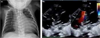
A newborn female was delivered by caesarean section at 35 weeks and 6 days of gestation because of fetal oedema. The infant’s birth weight was 2142 g, and her 1- and 5-minute Apgar scores were 4 and 8, respectively. Fetal echocardiography showed absent pulmonary valve and tricuspid valve dysplasia. The newborn required immediate intubation because of respiratory distress. Chest radiography showed cardiomegaly with a cardiothoracic ratio of 78% and no left lung aeration (Fig 1a). Echocardiography revealed main pulmonary artery dilatation, severe pulmonary regurgitation, little antegrade flow across the tricuspid valve, moderate tricuspid regurgitation, a patent ductus arteriosus, and an atrial septal defect. Blood flow across the tricuspid valve was minimal, giving the appearance of tricuspid atresia. The measurements (and Z score) of the pulmonary annulus, pulmonary artery branch, atrial septum defect, and pulmonary forward gradient were 10 mm (Z=+2.8), 7 mm (Z=+4.2), 7 mm, and 16 mmHg, respectively. In addition, the tricuspid valve diameter was 7 mm (Z=−6.2), with a maximum diastolic opening of <2 mm (Fig 1b). No major abnormalities were found in the right ventricular muscle or coronary arteries.
Preoperative views. (a) Chest radiograph shows cardiomegaly and absence of left lung aeration. (b) Echocardiography shows minimal tricuspid valve opening during diastole (arrow), severe tricuspid regurgitation during systole, main pulmonary artery dilatation, severe pulmonary regurgitation, and an atrial septum defect at 0 day of age. First palliation postoperative views. (c) Computed tomography shows left bronchial compression (arrow) at 4 days of age.

After birth, the patient underwent oxygen and nitric oxide treatment to maintain pulmonary flow; however, hypoxia persisted; subsequently, prostaglandin E1 was administered. Nonetheless, hypoxia remained; her oxygen saturation was 70%, and hypotension and anuria continued. Pulmonary antegrade flow was not increased, and blood flowed through a patent ductus arteriosus to the right ventricle, right atrium, left atrium, left ventricle, and finally to the aorta. This condition is known as the circular shunt.
At 1 day of age, main pulmonary artery ligation and bilateral pulmonary artery banding were performed; the patent ductus arteriosus was retained. Antegrade flow across the tricuspid valve was recognised postoperatively, with a 4-mm maximum diastolic opening. CT showed left bronchial compression due to the dilated pulmonary artery (Fig 1c).
At 11 days of the age, when the pulmonary vascular resistance had decreased, the patient underwent successful right ventricular outflow tract reconstruction by a hand-sewn bileaflet polytetrafluoroethylene valve and placement of a modified Blalock–Taussig shunt with a 3-mm graft. She was extubated at 17 days and gained weight. Chest radiograph at 42 days showed an increased air space in the left lung (Fig 2a). Moreover, at 42 days of age, echocardiography demonstrated that the tricuspid valve diastolic opening was 5 mm, with trivial regurgitation; pulmonary regurgitation was also trivial (Fig 2b). The patient was discharged at 67 days of age and is currently awaiting biventricular or one-and-a-half repair.
Second palliation postoperative views at 42 days of age. (a) Chest radiograph shows increased left lung aeration. (b) Echocardiography shows tricuspid valve diastolic opening of 5 mm.

Discussion
Absent pulmonary valve and intact ventricular septum is a rare malformation, with only 35 cases reported thus far, to our knowledge.Reference Litovsky, Choy and Park 1 , Reference Best, Vyas, Jaquiss and Sachdeva 2 The condition can be complicated by tricuspid atresia or stenosis and right ventricle dysplasia. The syndrome is associated with a poor prognosis. Litovsky et alReference Litovsky, Choy and Park 1 reported that 11 of 27 patients died within the 1st month of life and another nine patient died during childhood. Szwast et alReference Szwast, Tian and McCann 3 described a transplant-free survival rate of 20%, and the single-ventricle physiology strongly predicted heart transplantation. Estimating the degree of tricuspid stenosis is important for planning treatment and improving patient prognosis.
Nevertheless, estimating the degree of tricuspid stenosis is difficult unless pulmonary regurgitation is eliminated. In a case similar to the present one, Barbara et al reported that an aorto-right ventricular tunnel caused functional tricuspid atresia.Reference Burkhardt, Arnold, Schlensak, Bökenkamp and Stiller 4 In addition, the degree of tricuspid stenosis is closely related to single-ventricle or biventricular physiology. In the present case, because main pulmonary artery ligation was performed and pulmonary regurgitation was absent, the tricuspid valve diastolic opening could be estimated. Intraoperative estimation might also be effective if the main pulmonary artery is clamped or banded. Our patient did not demonstrate any major right ventricle abnormalities; however, in patients with such abnormalities, additional care is necessary. Previous investigators have described various degrees of right ventricular dysplasia and dysfunction.Reference Lato, Gembruch and Geipel 5 , Reference Kasahara, Aeba and Yozu 6 In such cases, main pulmonary artery ligation may cause additional right ventricular dysfunction. Whether main pulmonary artery ligation can be performed when the right ventricle does not function remains unknown.
Absent pulmonary valve and intact ventricular septum can also lead to bronchial compression caused by dilated pulmonary arteries. Some reports have described preoperative ventilation and severe respiratory distress as mortality risk factors.Reference Brown, Ruzmetov, Vijay, Rodefeld and Turrentine 7 , Reference Yong, Yim and Brizard 8 Pulmonary complications are also related to poor prognoses in patients with single-ventricle physiology. Thus, controlling pulmonary blood flow during the early neonatal period is advisable. In our case, bilateral pulmonary artery banding was performed to reduce pulmonary flow when the patient was 1 day old; because of pulmonary vascular resistance changes, especially during the early neonatal period, a modified Blalock–Taussig shunt was performed when the patient was 11 days old. In addition, we reconstructed the right ventricular outflow tract by a hand-sewn bileaflet polytetrafluoroethylene valve – the Nunn approach.Reference Nunn, Bennetts and Onikul 9 This strategy is reasonable for selecting a proper shunt and decreases the risk of high pulmonary blood flow.
In conclusion, functional near-tricuspid atresia may be caused by pulmonary regurgitation in patients with absent pulmonary valve and intact ventricular septum. The degree of tricuspid stenosis needs to be estimated to facilitate treatment and possibly improve patient prognosis.
Acknowledgements
None.
Financial Support
This research received no specific grant from any funding agency of from commercial or non-profit sectors.
Conflicts of Interest
None.