Introduction
Calcite, one of the three anhydrous crystalline polymorphs of calcium carbonate, is a particularly well-studied mineral system in the field of biomineralization and bioinspired mineralization due to its natural abundance as the building material for skeletal elements of many marine organismsReference Lowenstam and Weiner1 and its industrial importance as a filler in rubber, paints, and paper products, among others.Reference Brecevic and Kralj2 Geologically, calcite occurs as the principle component of limestone and chalk cliffs.Reference Scoffin3 Synthetic calcite crystals precipitated in the absence of additives adopt a regular rhombohedral morphology and fracture by cleavage along low-energy lattice planes. A wide range of living organisms, on the other hand, have found superb solutions to use such a notably brittle compound as a mechanical support.Reference Addadi and Weiner4 Prominent examples include red corals,Reference Vielzeuf, Floquet, Chatain, Bonnete, Ferry, Garrabou and Stolper5 sea urchins,Reference Seto, Ma, Davis, Meldrum, Gourrier, Kim, Schilde, Sztucki, Burghammer, Maltsev, Jäger and Cölfen6 mollusks,Reference Nudelman, Chen, Goldberg, Weiner and Addadi7,Reference Addadi, Joester, Nudelman and Weiner8 coccospheresReference van de Locht, Slater, Verch, Young, Haigh and Kröger9 (Figure 1a–d), and brittle stars.Reference Aizenberg, Tkachenko, Weiner, Addadi and Hendler10 A hallmark of many biominerals is their hierarchical architecture, where the structure is organized and optimized at several length scales ranging from the macroscopic to the nanometer and subnanometer level.Reference Fratzl and Weinkamer11 The remarkable control that organisms exert over the polymorph, morphology, size, and arrangement of mineral crystallites is well documented and commonly attributed to the deposition of the mineral phase in the presence of soluble as well as insoluble organic matrices.
Examples of calcitic biominerals acting as skeletal elements in marine organisms. (a) Red coral skeleton, (b) sea urchin spine (left) and test (right), (c) mollusk shell (Atrina rigida), and (d) coccosphere. (c) Reproduced with permission from Reference 8. © 2006 Wiley.

Another intriguing characteristic of biominerals is their enormous flexibility with respect to the incorporation of a wide range of (mostly organic) impurities as “guests”. In many cases, the coherence of the crystalline lattice of the inorganic host is preserved over large areas.Reference Albeck, Aizenberg and Addadi12–Reference Pokroy, Fitch, Marin, Kapon, Adir and Zolotoyabko14 Even more striking is the recent finding that characteristic defects in the mineral lattice introduced by the incorporation of impurities are frequently taken advantage of, because they enhance the mechanical properties of the hybrid biocomposites.Reference Kunitake, Mangano, Peloquin, Baker and Estroff15
Going beyond the mechanical performance, the material functions, which nature achieves by controlling mineral structures on a submicrometer length scale, hold more surprises. Recently, Li et al. investigated the structural origin of the blue coloration of prominent stripes on the shell of the blue-rayed limpet, Patella pellucida, and identified a calcite-based photonic architecture that combines a multilayer of periodically co-oriented mineral lamellae and an underlying layer containing a disordered array of colloidal light-absorbing particles.Reference Li, Kolle, Weaver, Ortiz, Aizenberg and Kolle16
The intricate structures observed in biogenic calcite have inspired a huge body of research aimed at creating synthetic calcite-based composite crystals with internal structures analogous to biominerals by adeptly combining organic and inorganic components and understanding their interplay to establish structure-property relationships. In this regard, the localization and distribution of intracrystalline organics within the inorganic host are of key importance for the mechanical behavior of the resulting composite material. In this article, we highlight recent developments in the characterization of the internal structure and related properties of biological and synthetic calcite single crystals with composite character.
Structural characteristics of biogenic calcite
The intricate hierarchical structures of biological calcite are discussed in the following text. Prismatic crystals from mollusk shells and the sea urchin spine were selected as model systems, both of which represent comparably large single crystals incorporating a substantial portion of foreign ions and organic materials. The complex ultrastructures of these organic–inorganic composites have been studied extensively using state-of-the-art analytical techniques.
Mollusk shells: Shatter-proof through hierarchical architecture
The shell of many marine bivalve mollusks such as Pinna nobilis (noble pen shell) or Atrina rigida (rigid pen shell, Figure 1c) consists of an inner mineral layer composed of aragonite (called nacre) and an outer layer formed from calcite (Figure 2a). The coexistence of these two polymorphs of calcium carbonate in well-defined domains within the same organism is intriguing, and it is widely believed that this remarkable control over the crystal structure of the mineral phase is achieved via specific macromolecules directing the deposition of the inorganic material.Reference Falini, Albeck, Weiner and Addadi17 The aragonitic nacre layer exhibits a structure based on well-aligned mineral tablets separated by an organic matrix. This is often referred to as a brick-and-mortar arrangement.Reference Nudelman, Gotliv, Addadi and Weiner13 The structure and properties of this iridescent and astonishingly tough material are the subject of extensive research efforts and have been reviewed in detail.Reference Sun and Bhushan18 In the present article, we focus on the calcitic layer, specifically discussing hierarchical levels from the macroscopic to the atomic scale (lattice structure), while placing special emphasis on the micro- and nanostructure. Since strong variations in structure and crystallinity have been observed for mollusk shell calcite from different species,Reference Olson, Metzler, Tamura, Kunz, Killian and Gilbert19 here we specifically consider minerals extracted from Atrina rigida and Pinna nobilis, two closely related mollusk species (taxonomically), as model systems for biogenic calcite single crystals.
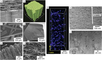
The hierarchical structure of prismatic crystals extracted from the shell of a mollusk. (a) A cross section of an Atrina rigida shell imaged by scanning electron microscopy. Prismatic calcite crystals (lower part) and nacre tablets (upper part) are seen. (b) Three-dimensional reconstruction of x-ray microtomography data obtained from the prismatic layer of Pinna nobilis. The elongated prismatic shape of the grains is discernible. (c–d) Electron micrographs of individual prisms extracted from Atrina rigida presenting views of the side facets (c) and the basal plane (d). (e) Selective removal of the mineral phase uncovers a network of organic fibers penetrating the prism. The periodicity indicates a cyclic growth pattern. (f) Mineral grains in the size range of approximately 50 nm are tightly associated with the organic network. (g) Electron tomographic reconstruction of a thin section of an Atrina rigida prism. Isolated disc-like patches of lower density (organic material) are shown in blue. The yellow arrow indicates the direction of the crystallographic c-axis. (h–i) Annular dark-field scanning transmission electron microscope images of thin slices of a prismatic crystal cut parallel (h) or perpendicular (i) to the c-axis. Dark areas correspond to low-density organic occlusions. (a, e, f) Reproduced with permission from Reference 7. © 2007 Royal Society of Chemistry. (b) Reproduced with permission from Reference 21. © 2014 Nature Materials. (g–i) Reproduced with permission from Reference 27. © 2011 Wiley.

Morphology and arrangement of the crystals in the calcitic layer
The elongated crystals constituting the calcite layer in the shell of both Pinna nobilis and Atrina rigida have a hexagonal prismatic shape typically measuring several hundreds of micrometers in length and 20–50 µm in diameter (Figure 2b–d).Reference Nudelman, Chen, Goldberg, Weiner and Addadi7 These prisms, whose crystallographic c-axes are oriented perpendicular to the shell surfaceReference Berman, Hanson, Leiserowitz, Koetzle, Weiner and Addadi20 and thus coincide with the long axis of the elongated crystals, are embedded in an organic interprismatic matrix mainly composed of glycine-rich proteins.Reference Nudelman, Chen, Goldberg, Weiner and Addadi7 Recently, Bayerlein et al. demonstrated, using advanced synchrotron microtomography (Figure 2b), that the evolution of the prismatic layer in the bivalve Pinna nobilis can be fully described by classical grain growth models, where the organism controls the thermodynamic boundary conditions in terms of the mobility and surface energy of the calcite/matrix interface.Reference Bayerlein, Zaslansky, Dauphin, Rack, Fratzl and Zlotnikov21
Each individual prism shows an x-ray diffraction pattern composed of narrow spotsReference Berman, Hanson, Leiserowitz, Koetzle, Weiner and Addadi20,Reference Gilow, Zolotoyabko, Paris, Fratzl and Aichmayer22,Reference Wolf, Lieberwirth, Natalio, Bardeau, Delorme, Emmerling, Barrea, Kappl and Marin23 with low mosaic spread.Reference Olson, Metzler, Tamura, Kunz, Killian and Gilbert19,Reference Berman, Hanson, Leiserowitz, Koetzle, Weiner and Addadi20 Astonishingly, while their scattering behavior can be associated with a nearly perfect single crystal of calcite, it has been found that the prismatic particles occlude around 0.3 wt% of intracrystalline organic material.Reference Nudelman, Chen, Goldberg, Weiner and Addadi7,Reference Metzger, Politi, Carbone, Bayerlein, Zlotnikov, Zolotoyabko and Fratzl24 Therefore, the incorporation of these impurities (soft occlusions) does not seem to have a detrimental effect on the continuity of the mineral lattice.
Microstructure of individual prisms
The intercalation of these organic molecules, however, significantly affects the internal structural architecture, and hence the mechanical properties of the prism.Reference Kunitake, Mangano, Peloquin, Baker and Estroff15 In order to understand the chemical and structural role of the “soft” occlusions, it is of great scientific interest to identify their location and distribution within the crystalline host material. On the length scale of micrometers, initial etching experiments, which are designed such that the mineral content is selectively removed, uncovered a fibrous network with alternating zones of higher and lower density of organic material in both Pinna nobilis Reference Dauphin, Cuif, Doucet, Salome, Susini and Williams25 and Atrina rigida.Reference Nudelman, Chen, Goldberg, Weiner and Addadi7 These characteristic bands, which are aligned perpendicular to the c-axis of the prism, have been generally associated with an oscillatory growth pattern of the calcite crystals in the shell. In Pinna nobilis, Dauphin et al. could correlate this periodic topographic pattern with a chemical zonation by using wavelength dispersive spectroscopy and x-ray absorption near-edge spectroscopy to map the elemental distribution of magnesium and sulphur, where the latter is associated with sulphated organic species preferentially accumulated at the growth lines.Reference Dauphin, Cuif, Doucet, Salome, Susini and Williams25 Besides a water-soluble fraction of intracrystalline organic material, which is discussed in detail later, Nudelman et al. identified the polysaccharide chitin as a major component of the water-insoluble fibrous network penetrating Atrina rigida prisms (Figure 2e).Reference Nudelman, Chen, Goldberg, Weiner and Addadi7 Atomic force microscopy of polished sectionsReference Dauphin, Cuif, Doucet, Salome, Susini and Willams26 and scanning electron microscopy (SEM) of partially etched prismsReference Nudelman, Chen, Goldberg, Weiner and Addadi7 visualized mineral particles in the size range of ∼50 nm, which appeared to be tightly associated with the organic material (Figure 2f), thus indicating the nanocomposite character of the prismatic crystals.
Nanostructure and distribution of intracrystalline organics within single crystals
Significant research efforts have recently been directed toward the analysis of the three-dimensional volume structure of individual prisms with nanometer resolution. Nondestructive techniques are preferentially applied in order to avoid artifacts arising from sample preparation. Li et al. used annular dark field scanning transmission electron microscopy (ADF-STEM) and ADF electron tomography to visualize organic–inorganic interfaces within prismatic single crystals extracted from Atrina rigida. Reference Li, Xin, Kunitake, Keene, Muller and Estroff27 Electron transparent slices cut along well-defined crystallographic directions of the prism were prepared by a focused ion beam, where the tomographic technique images the bulk structure of the composite material. The authors demonstrated that the occluded biomolecules were present in the form of isolated nanopatches (20–50 nm) distributed within a host of single crystalline calcite. These organic discs, which presumably correspond to aggregated biomacromolecules, were preferentially oriented perpendicular to the c-axis of the prisms, while their distribution in the basal plane was more uniform (Figure 2g–i).
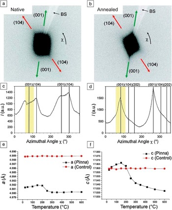
Such an alignment of platelet-shaped accumulations of intracrystalline organics with the (001)-type planes of the calcite lattice has also been observed by Gilow et al., who studied individual prisms from the species Pinna nobilis by combined small-angle x-ray scattering (SAXS) and wide-angle x-ray scattering (WAXS) under stepwise rotation of the crystal in a microfocused x-ray beam (Figure 3a–d).Reference Gilow, Zolotoyabko, Paris, Fratzl and Aichmayer22 This nondestructive technique provides quantitative three-dimensional structural information on both the nanometer-sized macromolecular occlusions (SAXS signal reflecting the diffuse scattering intensity around the origin of reciprocal space) and the calcite mineral lattice (diffraction spots in the WAXS regime) averaged over a comparably large sample volume in native (Figure 3a–c) and heat-treated (Figure 3b, d) prisms. In particular, orientation correlations between certain directions of the crystal lattice and the intensity distribution of the diffuse scattering signal arising from nanostructural heterogeneities could be identified. In these experiments, it was observed that pronounced intensity streaks in the SAXS signal, which correspond to directions of strong variations in electron density, are largely aligned with only two sets of lattice planes in the calcite unit cell. One set of streaks is aligned with the (001)-type planes, which are charged (as they are constituted from either calcium ions or carbonate ions in an alternating pattern), and the other set of streaks is aligned with low-energy neutral (104)-type planes. This observation suggests preferential adhesion of organic macromolecules specifically to these crystallite facets.Reference Gilow, Zolotoyabko, Paris, Fratzl and Aichmayer22
Nanostructure and lattice distortions in prismatic calcite from Pinna nobilis studied by x-ray scattering techniques. (a–d) Small- and wide-angle x-ray scattering (SAXS and WAXS) plots of individual calcite prisms. (a–b) Two-dimensional (2D)-detector images of simultaneously recorded SAXS and WAXS before (a) and after (b) annealing at 300°C. The SAXS signal (center of the 2D detector) is clearly anisotropic, and intensity streaks point toward the azimuthal position of specific Bragg spots (arrows), indicating non-homogenous incorporation of organic material aligned with the corresponding families of lattice planes. After annealing (decomposition of intracrystalline organics), the orientation of nanostructural features toward the [001]-direction of the mineral lattice becomes more pronounced. (c–d) Integral SAXS intensity plotted over the azimuthal angle χ before (c) and after (d) annealing. Drop lines indicate the angular position of (006) and (104) Bragg peaks. The yellow bar marks the region affected by the beamstop (BS). (e–f) Lattice parameters (a- and c-axes) calculated from high-resolution powder diffraction extracted from high-resolution powder diffraction data as a function of the annealing temperature. Significant anisotropic lattice distortions are seen with respect to a control sample. Mild annealing at temperatures above 200°C induces relaxation of the crystal lattice. (a–d) Reproduced with permission from Reference 22. © 2011 Wiley. (e–f) Reproduced with permission from Reference 14. © 2006 Elsevier.

Metzger et al. additionally performed a detailed investigation of the distribution of the diffuse scattering intensity near selected Bragg diffraction spots. While the reflections were isotropic in the pristine prisms, intense streaks along the [001]-direction appeared after mild annealing.Reference Metzger, Politi, Carbone, Bayerlein, Zlotnikov, Zolotoyabko and Fratzl24 In analogy to the SAXS-studies performed by Gilow et al., these experiments indicate that the charged (001)-lattice planes are particularly sensitive to structural modifications induced via the removal of the organic component by the heat treatment. Interestingly, the authors could correlate the observed rearrangement of the internal interfaces, which become notably smoother upon annealing, with a reduced hardness of the material in nanoindentation measurements. In this way, indirect evidence was provided that the presence of the intracrystalline biomolecules and the thus generated nanoscopic organic/mineral interfaces are of key importance for the mechanical behavior of the shell prisms.
Crystallographic structure in the mineral phase and chemical composition of intracrystalline organics
Based on high-resolution synchrotron powder x-ray diffraction data, Pokroy et al. have shown that the incorporation of intracrystalline organics affects the internal structure of mineral prisms considered herein down to the atomic scale, resulting in anisotropic distortions of the mineral lattice (as compared with geological calcite).Reference Pokroy, Fitch, Marin, Kapon, Adir and Zolotoyabko14 The observed strains were most pronounced along the c-axis of the calcite unit cell and relaxation occurred upon annealing, which led to the conclusion that thermally unstable organic molecules induce these significant lattice distortions (Figure 3e–f). Powder neutron diffraction further revealed a significant elongation of the carbon–oxygen bond length in Pinna nobilis as compared to its geological purely inorganic counterpart.Reference Zolotoyabko, Caspi, Fieramosca, Von Dreele, Marin, Mor, Addadi, Weiner and Politi28 The potential contribution of Mg2+ ions, which are often present in substantial amounts in biogenic calcite (2.65 wt% in Pinna nobilis),Reference Zolotoyabko, Caspi, Fieramosca, Von Dreele, Marin, Mor, Addadi, Weiner and Politi28 to the x-ray and neutron scattering signals was carefully corrected for by construction of calibration curves. In combination, these studies suggest that the intracrystalline biomacromolecules are tightly associated with the mineral phase, and their distribution with respect to the mineral lattice is nonuniform.
Numerous studies have addressed the chemical composition of intracrystalline organics. Besides the insoluble chitin matrix, as identified by Nudelman et al.,Reference Nudelman, Chen, Goldberg, Weiner and Addadi7 water-soluble proteins are involved in the biomineralization of mollusk shell calcite. The sequence of these macromolecules is now known to be particularly rich in amino acids bearing a negative charge such as aspartate and glutamate.Reference Evans29,Reference Marin, Amons, Guichard, Stigter, Hecker, Luquet, Layrolle, Alcaraz, Riondet and Westbroek30 Specifically, Marin et al. extracted two unusually acidic proteins—caspartin and calprismin—from calcite prisms of the species Pinna nobilis. In Atrina rigida, the Asprich family of acidic proteins was identified as a constituent of the soluble organic matrix present in the prismatic calcite crystals.Reference Gotliv, Kessler, Sumerel, Morse, Tuross, Addadi and Weiner31 It is widely believed that due to their chemical functionalization, acidic proteins are prone to electrostatic interaction with with charged planes of the calcite lattice, and are therefore often found aligned with the (001) faces of calcite crystallites. Wolf et al. has recently suggested the potential role of caspartin and calprismin in the formation of a transient amorphous mineral phase with liquid-like characteristics.Reference Wolf, Lieberwirth, Natalio, Bardeau, Delorme, Emmerling, Barrea, Kappl and Marin23
The sea urchin spine: A weapon made from chalk
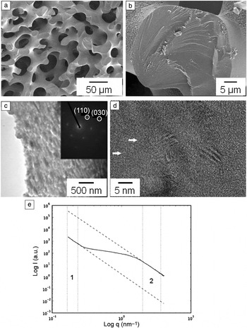
Another example of a biologically grown single crystal of calcite is the sea urchin spine (Figure 1b, left), whose presumed function is to provide protection from predators. The spines can grow to a length of centimetersReference Towe32 and appear spongy on a macroscopic scale.Reference Borzecka-Prokop, Weselucha-Birczynska and Koszowska33 Similar to the prismatic crystals extracted from mollusk shells, the spine of the sea urchin represents a single crystal with some degree of mosaicity when investigated by x-ray diffraction, where the crystallographic c-axis is aligned with the long axis of the spine.Reference Berman, Addadi and Weiner34,Reference Berman, Addadi, Kvick, Leiserowitz, Nelson and Weiner35 This is particularly astonishing when considering its porous microstructure and curved facets (Figure 4a–b), thus rendering the sea urchin spine one of the most intensely studied biomineral systems.Reference Seto, Ma, Davis, Meldrum, Gourrier, Kim, Schilde, Sztucki, Burghammer, Maltsev, Jäger and Cölfen6
Micrometer and submicrometer-scale structure of the sea urchin spine. (a) Scanning electron micrograph showing the spongy microstructure of a spine fractured perpendicular to the long axis. (b) Fracture surface imaged in higher detail revealing conchoidal fracture. (c) A substructure composed of elongated grains is seen in the transmission electron microscopy image obtained from a small section of the spine. Inset: selected area electron diffraction pattern comprising distinct diffraction spots. (d) High-resolution transmission electron microscopy shows defect structures (white arrows) around lattice fringes. (e) Small-angle x-ray scattering indicates a bimodal size distribution of the scattering objects, where larger objects (exceeding a size of 100 nm), which might represent the mineral grains observed in electron microscopy, dominate the scattering profile (Intensity (q) versus q, where q represents the modulus of the scattering vector) at low q (region 1). The intensity at higher q (region 2) indicates the presence of occluded organic material, with an estimated correlation length of 1.4 nm. (c–e) Reproduced with permission from Reference 6. © 2012 National Academy of Sciences.

Compared with mollusk calcite, the sea urchin spine typically contains a higher proportion of Mg2+ ions (3–15 mol%),Reference Magdans and Gies36 while a lower amount of organic material (0.02 wt%)Reference Aizenberg, Hanson, Koetzle, Weiner and Addadi37 is entrapped within the mineral host even though the exact composition depends on species and habitat.Reference Magdans and Gies36 The substitution of Ca2+ for Mg2+ (solid solution) appears to be a central strategy in strengthening the material, as magnesian calcite is known to show mechanical properties that are significantly different from pure calcite even when produced synthetically in the absence of other impurities.Reference Kunitake, Baker and Estroff38 In addition, the intracrystalline macromolecules have been discussed as a source for the characteristic conchoidal fracture surfaces of the spine (Figure 4b),Reference Borzecka-Prokop, Weselucha-Birczynska and Koszowska33,Reference Weiner and Addadi39 where the latter are reminiscent of fracture behavior usually associated with glass-like materials.Reference Addadi and Weiner4 Electron microscopy performed on such fracture surfaces as well as thin microtome-cut sections of the crystals further revealed a granular substructure composed of nanoparticle building blocks in the size range of 10–40 nm (Figure 4c).Reference Seto, Ma, Davis, Meldrum, Gourrier, Kim, Schilde, Sztucki, Burghammer, Maltsev, Jäger and Cölfen6
The ultrastructure of the sea urchin spine has recently been studied in depth by Seto et al. (Figure 4c–e), who provided evidence for coverage of the crystalline nanoparticle units with amorphous material and/or biomolecules.Reference Seto, Ma, Davis, Meldrum, Gourrier, Kim, Schilde, Sztucki, Burghammer, Maltsev, Jäger and Cölfen6 Based on this observation in combination with previous reports of amorphous calcium carbonate (ACC) being involved in the regeneration of broken spines,Reference Politi, Arad, Klein, Weiner and Addadi40 a growth model was suggested, which describes the formation of the coherently diffracting mineral structure as the concerted crystallization of a dense array of ACC particles. In this way, the crystalline phase retains a memory of the initially deposited amorphous material.Reference Seto, Ma, Davis, Meldrum, Gourrier, Kim, Schilde, Sztucki, Burghammer, Maltsev, Jäger and Cölfen6
Bioinspired concepts for controlling the internal structure of calcite single crystals
The sophisticated structures of biominerals in combination with their functionality have inspired a wealth of research activity aiming to translate their characteristics into synthetic materials with tailored properties. In turn, the study of simplified synthetic model systems with a reduced degree of complexity can provide new insights into the mechanisms of biomineralization.
Incorporation of small molecules, macromolecules, and particles into single crystals
There have been exciting recent reports concerning biomimetically synthesized calcite based hybrid single crystals, whose properties resemble aspects of the structural and mechanical properties seen in biogenic calcite. A wide range of “impurities” in different size regimes has been successfully introduced into calcite single crystal, including ions,Reference Davis, Dove, Wasylenki and De Yoreo41,Reference Mucci and Morse42 small molecules (∼1 nm),Reference Mucci and Morse42 water-soluble macromolecules (∼10 nm),Reference Wang, Cölfen and Antonietti44 , Reference Kim, Schenk, Ihli, Kulak, Hetherington, Tang, Schmahl, Griesshaber, Hyett and Meldrum45 micelle-forming block copolymers (∼20 nm),Reference Kim, Ganesan, Yang, Kulak, Borukhin, Pechook, Ribeiro, Kroger, Eichhorn, Armes, Pokroy and Meldrum46 gels (10–20 nm),Reference Asenath-Smith, Li, Keene, Seh and Estroff47,Reference Li, Xin, Muller and Estroff48 and even surface-functionalized organic/inorganic/metal nanoparticles (10 ∼200 nm).Reference Kulak, Yang, Kim, Armes and Meldrum49–Reference Kim, Ribeiro, Maillot, Ward, Eichhorn and Meldrum51
Water-soluble additives: Large and small
The most accessible and prevailing strategy for the generation of bioinspired composite structures is the precipitation of the mineral in the presence of water-soluble organic additives—a concept that has been explored extensively over the last decades.Reference Yu and Cölfen52,Reference Gower53 Here, inspiration is taken from the soluble organic matrix identified in biogenic calcite. Additives frequently applied include highly acidic polyelectrolytesReference Gower53 and block-copolymers,Reference Yu and Cölfen52 where the latter usually contain at least one block bearing carboxylate or sulfonate functional groups as a common active component.Reference Kim, Schenk, Ihli, Kulak, Hetherington, Tang, Schmahl, Griesshaber, Hyett and Meldrum45,Reference Meldrum and Cölfen54,Reference DeOliveira and Laursen55
However, additives are not limited to these components.Reference Schenk, Cantaert, Kim, Li, Read, Semsarilar, Armes and Meldrum56,Reference Cantaert, Kim, Ludwig, Nudelman, Sommerdijk and Meldrum57 A multitude of polyanionic and polycationic macromolecules with different chemistries and architectures have been identified to be excellent modulators of morphology and/or to act as growth-controlling agents of calcite crystals,Reference Yu and Cölfen52–Reference Chen, Qi, Tao, Zuckermann and De Yoreo58 and the versatile roles of different types of polyelectrolytes interfering with the calcium carbonate precipitation process at various stages have been classified.Reference Verch, Gebauer, Antonietti and Cölfen59,Reference Gebauer, Cölfen, Verch and Antonietti60 Studies addressing the nano/microstructures of synthetic calcite composites precipitated in the presence of commercial polyelectrolytes, polystyrene sulfonate (PSS),Reference Schenk, Zlotnikov, Pokroy, Gierlinger, Masic, Zaslansky, Fitch, Paris, Metzger, Cölfen, Fratzl and Aichmayer61 and poly(4-styrene sulfonate-co-maleic acid) (PSS-MA)Reference Kim, Schenk, Ihli, Kulak, Hetherington, Tang, Schmahl, Griesshaber, Hyett and Meldrum45 revealed that the distribution of polymers in mineral crystals (1–4 wt% occlusion level) is inhomogeneous, with preferential occlusion along the (001) and (104) lattice planes.
As opposed to macromolecules, which show cooperative effects of multiple functional groups, the occlusion of small molecules in calcite has been investigated only very recently. Borukhin et al. screened 20 common amino acids for their capacity to become incorporated into calcite crystals.Reference Borukhin, Bloch, Radlauer, Hill, Fitch and Pokroy43 They found substantial variation in the occlusion levels depending on the type of amino acid. A particularly strong interaction with the mineral phase was observed for aspartic acid, where this observation is in agreement with the composition of soluble biomacromolecules extracted from biogenic calcite (as discussed in the previous section). In general, charge, size, rigidity, and the relative pKa of the terminal groups in aqueous solution were suggested as governing factors for uptake into the crystalline host. Cysteine and glycine, however, as non-acidic amino acids, represent an unusual case in that they showed incorporation levels as high as those seen for aspartic acid.
Particles
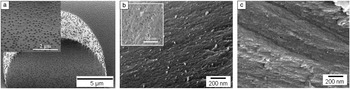
In many cases, the soluble additives entrapped in minerals form very small clusters of organic material providing low electron density contrast and hence are practically “invisible” in electron microscopic investigations using routine equipment. This substantially complicates their localization within the mineral host. A new methodology, which uses additives in particulate form, was developed in order to generate better defined organic–inorganic interfaces and to obtain more accurate information on the quantity and distribution of the occluded phase (Figure 5a–b). A variety of particles ranging from latex spheresReference Kim, Ribeiro, Maillot, Ward, Eichhorn and Meldrum51 over micelle-forming block copolymers,Reference Kim, Ganesan, Yang, Kulak, Borukhin, Pechook, Ribeiro, Kroger, Eichhorn, Armes, Pokroy and Meldrum46 to inorganic/metal nanoparticlesReference Kulak, Yang, Kim, Armes and Meldrum49,Reference Kulak, Semsarilar, Kim, Ihli, Fielding, Cespedes, Armes and Meldrum50 were successfully incorporated. The surface of the particles is functionalized with long, flexible charged polymer chains, which provide a strong binding capacity to crystal faces, thus playing a crucial role for particle incorporation. Micelle-forming copolymers in particular shared many common features with proteins, in terms of their composition, size, and shape (the folding structure of common proteins involved in calcite biomineralization is closer to a globular form than to a fully extended chainReference Li, Xin, Kunitake, Keene, Muller and Estroff27). This system has demonstrated a surprisingly high upper limit of particle occlusion (13 wt%) in the mineral single crystal (Figure 5b), exceeding the levels seen in other synthetic and biogenic systems. For example, latex particles with a diameter of ∼250 nm could be uniformly incorporated into calcite at an occlusion level of 9.3 wt% (Figure 5a), and the typical range of occlusion seen for soluble organic additives is 1∼4 wt%, with the exception of a 42-amino acid polypeptide, for which incorporation of 9.2 wt% in calcite was estimated.Reference Metzler, Tribello, Parrinello and Gilbert62
Scanning electron microscope images showing internal surfaces of synthetic calcite single crystals grown in the presence of polymer-based particles and gels. (a) Polystyrene latex particles (focused ion beam cross-sectioned), (b) block copolymer micelles (fractured), (c) in xyloglucan gel matrix functionalized with Fe3O4 nanoparticles (fractured). (a) Reproduced with permission from Reference 51. © 2010 Wiley. (b) Reproduced with permission from Reference 46. © 2011 Nature Materials. (c) Reproduced with permission from Reference 68. © 2014 Royal Society of Chemistry.

Gels
Another important synthetic strategy inspired by biomineralization is growing crystals in a gel medium.Reference Asenath-Smith, Li, Keene, Seh and Estroff47 As one of the oldest crystal growth methods, these processes have been well-studied using various gels, such as gelatin,Reference Metzler, Tribello, Parrinello and Gilbert62–Reference Nindiyasari, Fernandez-Diaz, Griesshaber, Astilleros, Sanchez-Pastor and Schmahl64 silica,Reference Fernandez-Diaz, Astilleros and Pina66 polyacrylamide,Reference Grassmann and Löbmann67 and agarose.Reference Li, Xin, Muller and Estroff48 Conceptually, gels were revitalized as a suitable model system to study crystal growth constrained by a viscous biological medium and to learn about interactions between the insoluble matrix and the crystal (detailed investigations on calcite crystals grown in gels are further discussed in the Hendley et al. article in this issue).
Combinations of synthetic approaches: Better together?
Combinations of different strategies have also been explored in order to achieve control over the distribution of the guest phase and for introducing diverse structures and functionalities into the single crystal. As an example of bringing particle and gel incorporation together, crystals were grown within a xyloglucan gel pre-functionalized with metal-oxide nanoparticles.Reference Kim, Schenk, Walsh, Kulak, Cespedes and Meldrum68 As an advantage of this approach, the nanoparticles could be nicely dispersed in the mineral phase due to their attachment to the gel network, while no special surface treatment of the particles was required. However, a relatively low level of occlusion was achieved in comparison to a system of free particles with adequate surface functionalization.Reference Kulak, Semsarilar, Kim, Ihli, Fielding, Cespedes, Armes and Meldrum50 This is possibly due to restricted transport of particles onto crystal growth fronts in a viscous mediumReference Kim, Schenk, Walsh, Kulak, Cespedes and Meldrum68 (Figure 5c). By growing calcite in agarose gel attached to self-assembled monolayers as substrates, Li et al. achieved both occlusion of an organic network and controlled orientation of macroscopic calcite crystals.Reference Li and Estroff69
Soft organic occlusions affect internal structures and mechanical properties of calcite crystals
The structural and compositional characteristics of composite materials are often directly linked to their mechanical properties, such as hardness and modulus as well as toughness. In contrast to the external morphologies and macroscopic structures of crystals grown in the presence of organic additives, their internal microstructures have been studied far less intensively. Conventional electron microscopy analyses have shown that many calcite crystals grown in an organic matrix share common features previously seen in biogenic calcite, for example, rough fracture surfaces (Figure 5b–c), and highly strained internal structuresReference Kim, Schenk, Ihli, Kulak, Hetherington, Tang, Schmahl, Griesshaber, Hyett and Meldrum45,Reference Kim, Ganesan, Yang, Kulak, Borukhin, Pechook, Ribeiro, Kroger, Eichhorn, Armes, Pokroy and Meldrum46,Reference Okumura, Suzuki, Nagasawa and Kogure70 (details of the electron microscope characterization of biogenic and bioinspired calcite are reviewed by Hendley et al. in this issue).
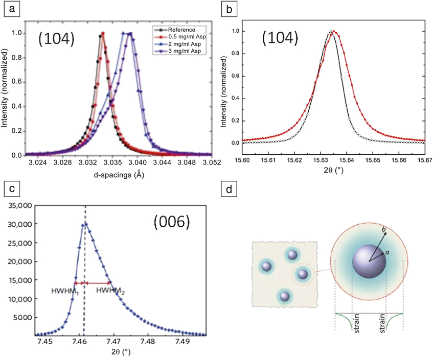
A series of early studies using single crystal diffraction techniques on both synthetic and biogenic calcite revealed that a significant reduction in the coherence length is caused by the anisotropic occlusion of intracrystalline macromolecules within the crystals.Reference Albeck, Aizenberg and Addadi12,Reference Berman, Addadi and Weiner34,Reference Berman, Addadi, Kvick, Leiserowitz, Nelson and Weiner35,Reference Aizenberg, Hanson, Koetzle, Weiner and Addadi37 More recent structural analyses based on synchrotron powder x-ray diffraction (XRD) data have provided new insights into the diverse effects that various synthetic additives exert on the microstructure of the composite as well as the crystalline lattice in the mineral phase.Reference Borukhin, Bloch, Radlauer, Hill, Fitch and Pokroy43,Reference Kim, Schenk, Ihli, Kulak, Hetherington, Tang, Schmahl, Griesshaber, Hyett and Meldrum45,Reference Kim, Ganesan, Yang, Kulak, Borukhin, Pechook, Ribeiro, Kroger, Eichhorn, Armes, Pokroy and Meldrum46,Reference Schenk, Zlotnikov, Pokroy, Gierlinger, Masic, Zaslansky, Fitch, Paris, Metzger, Cölfen, Fratzl and Aichmayer61 For this purpose, the profile shape (peak width contains information on the microstrain as well as the size of coherently scattering domains) and the maximum position (related to macrostrain) of the diffraction peaks have been studied in depth. Considering the effects of small molecules on the crystal structure, Borukhin et al. conducted high-resolution XRD analysis of calcite/amino acid systems discussed earlier and observed that significant lattice distortions (macrostrains) were introduced in the mineral phase by the occlusion of acidic amino acids, where such behavior had not been seen in other synthetic calcite/organics composite systems (Figure 6a).
Powder x-ray diffraction profile of synthetic calcite crystals with organic occlusions. (a) Profiles of the (104) reflection obtained from calcite crystals precipitated with aspartic acid. The effects of different amounts of aspartic acid on the peak positions are demonstrated. In red, blue, and violet—calcite powders grown from solutions with the addition of 0.5, 2, and 3 mg/ml aspartic acid, respectively, compared to the (104) reflection of pure calcite (black). (b) Profiles of the (104) reflection obtained from composite crystals precipitated with poly(4-styrenesulfonic acid-co-maleic acid) sodium salt)(red) showing broadening in the peak compared to that of pure calcite (black). (c) Profile of the (006) reflection obtained from calcite grown with block copolymer micelles. The peak is anisotropically distorted (described by two different HWHMs). (d) Schematic representation of the model used to evaluate the effect of particle incorporation on the crystal lattice, where a particle of radius a causes a strain gradient in the lattice to a distance b from its center (strain-affected zone). The green curve depicted below the particle and its strain-affected zone describes the distribution of compressive strain (in green). (a) Reproduced with permission from Reference 43. © 2012 Wiley. (b) Reproduced with permission from Reference 45. © 2014 Nature Publishing Company. (c–d) Reproduced with permission from Reference 46. © 2011 Nature Materials.

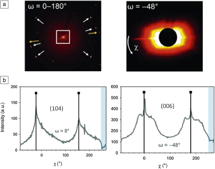
For calcite crystals grown in the presence of the acidic polyelectrolyte PSS-MA, a broadening of the diffraction peaks was observed that was attributed to microstrain caused by occlusions of the additive within the crystal matrix. Negligible shifts in the peak positions indicated the absence of uniform macrostrains (Figure 6b).Reference Kim, Schenk, Ihli, Kulak, Hetherington, Tang, Schmahl, Griesshaber, Hyett and Meldrum45 SAXS analysis of calcite precipitated in the presence of another acidic polyelectrolyte, the PSS homopolymer, revealed internal structural motifs on the nanometer level and provided an indication for the existence of fractal-like rough interfaces between the organic and inorganic components.Reference Schenk, Zlotnikov, Pokroy, Gierlinger, Masic, Zaslansky, Fitch, Paris, Metzger, Cölfen, Fratzl and Aichmayer61 The diffuse scattering signal, which presumably arises from intracrystalline PSS macromolecules, appeared to align with the (001) and (104) planes of the calcite lattice, suggesting preferential adhesion of organic material to these two families of lattice planes (Figure 7a–b).Reference Schenk, Zlotnikov, Pokroy, Gierlinger, Masic, Zaslansky, Fitch, Paris, Metzger, Cölfen, Fratzl and Aichmayer61
Microbeam small- (SAXS) and wide-angle x-ray scattering (WAXS) of single calcite-PSS crystals. (a, left) Two-dimensional SAXS/WAXS scattering pattern recorded during a 180° rotation of an individual calcite-PSS crystal. The anisotropic SAXS signal in the center of the image (white frame) indicates the presence of a pronounced nanostructure in the hybrid crystal. Diffraction spots are observed in the wide-angle region (white arrows: (104) plane, yellow arrows: (006) reflections). (a, right) The SAXS pattern was recorded at a fixed rotation angle (ω). The intensity streak points to (006) lattice planes of calcite (scale bar = 1 nm−1). The azimuthal angle χ is illustrated by the white arrow. (b) Profiles of the intensity of the scattering signal versus the azimuthal angle χ are plotted for two distinct sample rotation angles ω, where maxima appear in the anisotropy of the SAXS signal (blue bar = angular range affected by the beam stop). Reproduced with permission from Reference 61. © 2012 Wiley.

Considering the mechanical properties of bioinspired composite crystals, nanoindentation experiments on calcite/PSS particles showed varying degrees of hardness correlating with the polymer content. An inhomogeneous distribution of the polymer impurity (enriched by a factor of 2.5 in the inner part) resulted in a lower hardness polymer-enriched particle core (H = 2.0 ± 0.25 GPa) in comparison to the mineral-enriched exterior (H = 2.7 ± 0.4 GPa ).Reference Schenk, Zlotnikov, Pokroy, Gierlinger, Masic, Zaslansky, Fitch, Paris, Metzger, Cölfen, Fratzl and Aichmayer61 A similar observation has also been made for calcite occluding 9.3 wt% of polystyrene particles (H = 1.71 ± 0.44 GPa versus pure calcite H = 2.49 GPa).Reference Kim, Ribeiro, Maillot, Ward, Eichhorn and Meldrum51 The mechanical properties of these types of composite crystals therefore simply reflect the compliancy of the soft materials and the rigidity of the mineral phase.
On the other hand, calcite incorporating block copolymer micelles, which act as pseudo proteins, shows unique structural features at both the microscale and the atomic level. In contrast to pure calcite crystals and other hybrid crystals that give rise to isotropically shaped diffraction peaks, the peaks obtained from this micelle composite crystal were highly asymmetric, demonstrating the existence of a compressive strain gradient in the mineral phase (Figure 6c–d).Reference Kim, Ganesan, Yang, Kulak, Borukhin, Pechook, Ribeiro, Kroger, Eichhorn, Armes, Pokroy and Meldrum46 In fact, calcite single crystals incorporating a significant amount of “soft” micelle-forming block copolymers block copolymers show a similar trend to biogenic calcite, in that they get harder (H = 3.0 ± 0.1 GPa) than pure calcite crystals (H = 2.6 ± 0.1 GPa). Thus, this material has been referred to as an artificial biomineral. This unexpected behavior is thought to be due to the extensive heterogeneous strain-field around the occluded micelles preventing the dislocation movement in the crystals.Reference Kim, Ganesan, Yang, Kulak, Borukhin, Pechook, Ribeiro, Kroger, Eichhorn, Armes, Pokroy and Meldrum46
It is not yet fully understood how organic impurities, which are relatively large with respect to the dimensions of the crystalline lattice of the inorganic host, can enter into the tight crystal matrix without any significant disruption. Moreover, the molecular basis for the unusual effect of hardening inorganic materials by “soft” occlusions remains to be further studied.
Conclusions and outlook
Intriguing similarities between biogenic and synthetic single crystals of calcite have been identified by thorough structural characterizations—particularly with respect to the nanoscale organization of these materials, where it appears common that intracrystalline organic material is preferentially occluded along specific lattice directions of the mineral host. Even anisotropic lattice distortions, indicating intimate interaction between organic and inorganic components, were observed in both naturalReference Pokroy, Fitch, Marin, Kapon, Adir and Zolotoyabko14 and syntheticReference Mucci and Morse42 systems. However, reproducing such hierarchical architectures, with complex structural patterns ranging from the nanoscopic up to the macroscopic level in vitro remains a challenge.
In view of synthetic approaches, the ability to correlate structural variations in composite material, with the type and amount of the occluded additives and unraveling the mechanisms by which their incorporation proceeds, will enable us to build a more complete picture of how and why specific organic additives affect the host crystal structure and how these effects translate into macroscopic properties. Much remains to be learned about the exact interplay between the organic and inorganic components as well as the role of the internal interfaces in biogenic nanocomposites. The structural principles of matrix-mediated crystal growth will certainly facilitate the development of design strategies, which yield synthetic materials with mechanical properties superior to those seen in naturally occurring biominerals.
Acknowledgments
This work was supported by the Engineering and Physical Sciences Research Council via grants EP/J018589/1and EP/H005374/1 (YYK), and Programme Grant ASS EP/I001514/1 which funds the Materials Interface with Biology Consortium. A.S.S. further acknowledges financial support from the Deutsche Forschungsgemein-Schaft. The authors would like to thank Fiona Meldrum and Sabine Ludwigs for valuable advice and inspiring discussions. The authors are grateful to Alex N. Kulak for recording SEM images of Atrina rigida prisms and sea urchin skeletal elements. Andreas Verch is acknowledged for kindly providing an electron micrograph of a coccosphere.