Introduction
Pleistocene remains of Megalonychidae from the intertropical region of Brazil are rare, and their taxonomy has undergone considerable revision. Pioneering efforts were undertaken by Lund (Reference Lund1839, Reference Lund1840), the Danish paleontologist and “father of Brazilian paleontology” (Cartelle, Reference Cartelle2002), in the area of Lagoa Santa (Minas Gerais) regarded the sloth remains that he collected as belonging to Platyonyx oweni Lund, Reference Lund1839 and Platyonyx bucklandi Lund, Reference Lund1839. Based on numerous subsequently recovered remains, Lund (Reference Lund1846) corrected his identifications to Scelidotherium oweni (Lund) Lund, Reference Lund1846 and Scelidotherium bucklandi (Lund) Lund, Reference Lund1846.
Winge (Reference Winge1915) revised Lund’s Lagoa Santa material, which had by then been sent to Denmark, and recognized two scelidotheriine species, Catonyx giganteus Winge, Reference Winge1915 and Scelidotherium magnum Burmeister, Reference Burmeister1886. The valid names for these species were recognized by Cartelle et al. (Reference Cartelle, De Iuliis and Lopes2009) as Catonyx cuvieri (Lund) McDonald, Reference McDonald1987 and Valgipes bucklandi (Lund) Cartelle, De Iuliis, and Lopes, Reference Cartelle, De Iuliis and Lopes2009, respectively.
Hoffstetter’s (Reference Hoffstetter1954) work on the Lagoa Santa xenarthrans reconsidered the material that Lund and Winge had studied and arrived at opinions contrasting with those of the earlier researchers. Hoffstetter (Reference Hoffstetter1954) disagreed with Winge’s (Reference Winge1915) conclusions on the postcranial remains that the latter had recognized as the scelidotheriine C. giganteus; instead, the former considered these remains as belonging to a megalonychid and created a new combination for them, Ocnopus gracilis (Lund) Hoffstetter, Reference Hoffstetter1954 (see Cartelle et al., Reference Cartelle, De Iuliis and Lopes2009 for further details). This author also considered an incomplete calcaneum, which Gervais (Reference Gervais1874) had identified as Valgipes deformis Gervais, Reference Gervais1874, as megalonychid. Cartelle et al. (Reference Cartelle, De Iuliis and Lopes2009), based on more complete material including nearly complete skeletons of the two species, were able to determine that both “O. gracilis” and “V. deformis” are scelidotheriines and, indeed, conspecific. These authors demonstrated that the valid name for this species is V. bucklandi.
Based on very incomplete material from the Brazilian state of Ceará, Paula Couto (Reference Paula Couto1980) erected Xenocnus cearensis Paula Couto, Reference Paula Couto1980 and created for its reception the megalonychid subfamily Xenocninae. This species is invalid, as its holotype is a carpal, the unciform of Eremotherium laurillardi (Lund, Reference Lund1842), and not, as Paula Couto had surmised, the astragalus of a megalonychid. Clearly, the subfamily is also invalid. Paula Couto’s (Reference Paula Couto1980) assignment of a mandibular fragment and incomplete molariform teeth to the species cannot be supported based on these remains, given that their state of preservation and incompleteness preclude confident identification (Cartelle et al., Reference Cartelle, De Iuliis and Lopes2009). Consequently, remains from the Brazilian state of Piauí assigned to this species by Guérin et al. (Reference Guérin, Faure, Simões, Hugueney and Mourer-Chauviré2002) and Guérin and Faure (Reference Guérin and Faure2006) are incorrect.
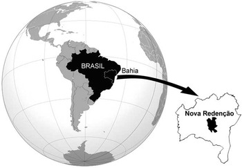
Cartelle et al. (Reference Cartelle, De Iuliis and Pujos2008) and De Iuliis et al. (Reference De Iuliis, Pujos and Cartelle2009), respectively, erected two Pleistocene megalonychid species from the intertropical region of Brazil: Ahytherium aureum Cartelle, De Iuliis, and Pujos, Reference Cartelle, De Iuliis and Pujos2008 and Australonyx aquae De Iuliis, Pujos, and Cartelle, Reference De Iuliis, Pujos and Cartelle2009, the remains of which were recovered from the submerged cave Poço Azul (municipality of Nova Redenção, Bahia, Brazil, Fig. 1). The skeleton (except for the manus) and dentition of the first species are almost completely known (Cartelle et al., Reference Cartelle, De Iuliis and Pujos2008). The second species is not as completely known as the first, but the skull and several other skeletal elements have been described and figured (De Iuliis et al., Reference De Iuliis, Pujos and Cartelle2009). The two species are the only megalonychids so far known from the intertropical region of Brazil. The current report complements the initial description by De Iuliis et al. (Reference De Iuliis, Pujos and Cartelle2009) in describing new material from Poço Azul of Au. aquae (Fig. 2.1–2.3). In addition, reference is made to material recovered from the Amazonian state of Rondônia (Fig. 3) that, we propose, belongs to this species.
Map showing location of the Poço Azul cave, Nova Redenção County, Chapada Diamantina, Brazil.

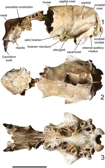
Skull of Australonyx aquae (holotype MCL 23315/01). (1) left lateral (anterior toward left, dorsal toward top); (2) dorsal (anterior toward left); and (3) ventral (anterior toward left). Scale bar represents 100 mm.

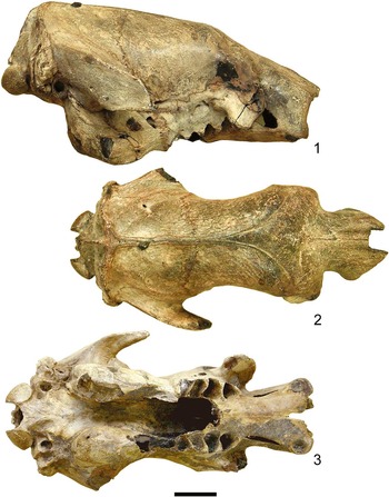
Skull of Australonyx aquae from the Brazilian state of Rondônia (1) in right lateral (anterior toward top right, dorsal toward top); (2) dorsal (anterior toward right), and (3) ventral (anterior toward right) views. Scale bar represents 50 mm.

Material and methods
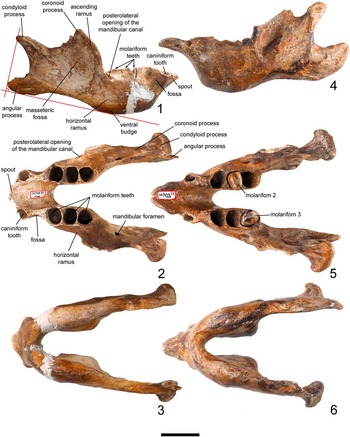
From among the material awaiting preparation in the paleontological collections of MCN PUC, we noted a nearly complete jaw that was recovered from the same locality that yielded the material of Au. aquae described by De Iuliis et al. (Reference De Iuliis, Pujos and Cartelle2009; Fig. 2). Although this element was overlooked during the latter publication, it is clear, as explained below, that it belongs to the same individual, MCL 23315, designated as holotype of this species. This dentary is cataloged as MCL 23315/4 (Fig. 4.1–4.3). The mandible is well preserved, missing only small portions of the symphysis and the angular and coronoid processes. This mandible is compared mainly with six specimens of the sympatric megalonychid Ah. aureum, each in different states of preservation, with the most comprehensive comparisons made to MCL 22834/04 (Fig. 4.4–4.6), the holotype of this species. Also, it is compared with the mandible of several other fossil megalonychids, including Acratocnus Anthony, Reference Anthony1916, Megalocnus Leidy, Reference Leidy1868, Megalonyx Harlan, Reference Harlan1825, Neocnus Arredondo, 1961, and Parocnus Miller, Reference Miller1929.
Mandible of Australonyx aquae (1–3, specimen MCL 23315/04) in (1) right lateral (anterior toward right, occlusal edge toward top); (2) occlusal (anterior toward left); and (3) ventral (anterior toward left) views; and of Ahytherium aureum (4–6, specimen MCL 21834/04) in (4) left lateral (anterior toward left, occlusal edge toward top); (5) occlusal (anterior toward left); and (6) ventral (anterior toward left) views. Scale bar represents 50 mm.

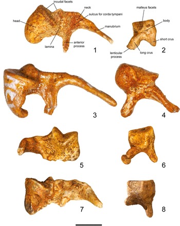
De Iuliis et al. (Reference De Iuliis, Pujos and Cartelle2009) provided a cursory description of the malleus and incus of Au. aquae. A more comprehensive description is provided here (Fig. 5.1–5.3), with comparisons to the middle ear ossicles of the megatheriid E. laurillardi (Fig. 5.4–5.5; MCL 32819), the megalonychid Neocnus dousman MacPhee, White, and Woods, Reference MacPhee, White and Woods2000 (described and figured by Gaudin, Reference Gaudin2011), the nothrotheriid Nothrotherium maquinense (Lund) Lydekker, Reference Lydekker1889 (Fig. 5.5–5.6; MCL 23315/02), and the mylodontid Valgipes bucklandi (Fig. 5.7–5.8; MCL 4293/01). As noted, reference is made to a megalonychid skull recovered in Rondônia (Fig. 3). Preliminary observations and comparisons, based on images of this skull made available to us by M. Cozzuol (Fig. 3), suggest that it belongs to Au. aquae.
Middle ear ossicles of Brazilian ground sloths, malleus in lateral view (1, 3, 5, and 7) and incus in lateral view (2, 4, 6, and 8). (1, 2) Australonyx aquae (Megalonychidae) left malleus (1, MCL 23315/02) and left incus (2, MCL 23315); (3, 4) Eremotherium laurillardi (Megatheriidae) left malleus (3, MCL 32819) and right incus (4, MCL 32831, reversed from the original); (5, 6) Nothrotherium maquinense (Nothrotheriidae) right malleus (5, MCL 21731, reversed from the original) and left incus (6, MCL 21731); (7, 8) Valgipes bucklandi (Mylodontidae scelidotheriine) left malleus (7, MCL 4293) and left incus (8, MCL 4293). Scale bar represents 50 mm.

Systematic paleontology
The following abbreviations are used in the text for institutional and anatomical acronyms:
C: upper caniniform; c: lower caniniform; M: upper molariform; m: lower molariform; MCL: paleontological collection of MCN PUC; MCN PUC: Museu de Ciências Naturais, Pontifícia Universidade Católica de Minas Gerais, Belo Horizonte, Brazil; Mt: metatarsal; ZMC: Zoologische Museum, Copenhagen, Denmark.
Mammalia Linnaeus, Reference Linnaeus1758
Xenarthra Cope, Reference Cope1889
Pilosa Flower, Reference Flower1883
Tardigrada Latham and Davies in Forster, Reference Latham and Davies1795
Megalonychidae Gervais, Reference Gervais1855
Genus Australonyx De Iuliis, Pujos, and Cartelle, Reference De Iuliis, Pujos and Cartelle2009
Type species
Australonyx aquae De Iuliis, Pujos, and Cartelle, Reference De Iuliis, Pujos and Cartelle2009 by monotypy.
Australonyx aquae De Iuliis, Pujos, and Cartelle, Reference De Iuliis, Pujos and Cartelle2009
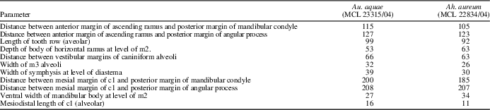
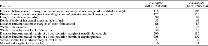
Mandibular measurements of Australonyx aquae (MCL 23315/04) and Ahytherium aureum (MCL 22834/04)
All measurements are in millimeters.
Holotype
MCL 23315, representing a well-preserved individual including parts of the skull, much of the axial skeleton, forelimb and hind limb elements, and many elements of the pes.
Material
Mandible MCL 23315/4.
Occurrence
Nova Redenção, Bahia, Brazil; late Pleistocene.
Description
In the dentary of Au. aquae (MCL 23315/04; Fig. 4.1–4.4, Table 1) the posterior margins of the mandibular condyle and the angular process project posteriorly to approximately the same level in lateral view. The angular process is not particular high dorsoventrally and the convexity of its lateral surface and the concavity of its medial surface are not pronounced. A shallow masseteric fossa is present and the ventral bulge of the horizontal ramus occurs below m2-m3. The posterolateral opening of the mandibular canal lies lateral to the anterior margin of the base of the ascending ramus. The basal length of the coronoid process is nearly equal to the distance between the posterior margin of the ascending ramus and the posterior margin of the mandibular condyle. Reflecting the large size of the caniniform, the lateral projection of the caniniform alveolar wall is pronounced, so that a deep fossa lies between this projection and the more posterior projection of the alveolar wall of m1 (that is, effectively at the level of the diastema). In dorsal view the lingual margins of the caniniform and molariforms are nearly aligned and the dorsal margin of the dentary between c1 and m1 (i.e., at the diastema) is thin mediolaterally. In ventral view the ventral margins of the dentary below the molariforms are narrow and the symphysis is wide. The medial surface of the dentary is nearly vertical, with ample space between the tooth rows.
Only the C1s of Au. aquae are known (De Iuliis et al., Reference De Iuliis, Pujos and Cartelle2009), as the upper molariforms and the lower dentition were not recovered. The c1 alveoli suggest that c1 was a large tooth, similar in size to C1, longitudinally rectilinear (i.e., not curved), oval and narrowed distally in section (Fig. 4.2). The form of C1 (Fig. 2.1, 2.3) indicates that wear on c1 was pronounced, producing a wedge-shaped occlusal portion, the apex of which is mesial in C1 and distal in c1. The lingual margins of c1 and the molariforms are aligned, as already noted, and the left and right tooth rows are parallel to each other (Fig. 4.2).
The skull recovered from the Brazilian state of Rondônia (see Discussion; Fig. 3) is nearly complete, although the nasals are apparently broken anteriorly. The morphology, proportions and ontogenetic stage are clear from the images and the accompanying scale bar. Based on sutural closure and the development of the sagittal crest, the Rondônia individual was probably slightly older than the holotype of Au. aquae (MCL 23315, Fig. 2), as is also suggested by the former’s slightly larger size. The Rondônia skull preserves the palate and inflated pterygoid sinuses (Fig. 3.1, 3.3). Of these regions the holotype of Au. aquae preserves only the roof of the m3 and m4 alveoli and the base of the pterygoid sinuses (Fig. 2.1, 2.3). The positions of these regions, however, coincide nearly exactly between the two specimens. This applies to other features, such as the anatomically complex posterolateral and posteroventral regions (=basicranium), which encompass multiple cranial foramina and the otic region. One difference between the specimens is that the preorbital constriction is slightly more pronounced in the Rondônia skull (Figs 2.3, 3.3).
In the malleus of Au. aquae the head (MCL 23315/02; Fig. 5.1, Table 1) is smooth, dorsally convex and anteriorly narrowed. The concave articular facet for the incus lies posteriorly and consists of dihedral planes meeting at nearly 90o. The area of the dorsal part is approximately thrice that of the ventral part. A shallow depression is present medially on the mallear neck to accommodate passage of the chorda tympani. The manubrium is narrowed ventrally and curves slightly anteriorly (Fig. 5.1). The medial surface proximal to the neck bears a muscular process that served for the attachment of the tendon of the tensor tympani muscle. The anterior process is about a third as long as the manubrium.
The articular surface of the incus (MCL 23315/02; Fig. 5.2, Table 1) comprises a flattened part and a concave part, with the latter about one-third the size of the former. The long crus is slightly curved medially and bears a lenticular process for articulation with the stapes distally. The short crus, projecting posteriorly, is thinner and tapers toward its tip.
Comparisons
The inflated pterygoid sinuses (Fig. 3.1, 3.3) at the base of the pterygoid that are present in Au. aquae resemble those in Choloepus, Megalocnus, and N. maquinense (see Naples, Reference Naples1982; Cartelle and Fonseca, Reference Cartelle and Fonseca1983; Patterson et al., Reference Patterson, Turnbull, Segall and Gaudin1992). The dentary of Au. aquae is immediately distinguishable from that of the megalonychids Parocnus and Megalocnus, in which the angular process is large and relatively dorsal in position. As well, the most mesial tooth in Megalconus is incisiform rather than caniniform. The dentary of Megalonyx is also easily distinguished from that of Au. aquae. In the former the angular process is considerably larger, the most mesial tooth is mesiodistally elongated and worn nearly flat, and the symphysis is not drawn anteriorly into a symphyseal spout.
The ventral bulge of the horizontal ramus is more strongly developed in Au. aquae than in Ah. aureum and occurs below m1-m2, rather than m2-m3 (Fig. 3.1). Among other megalonychids, the conformation of the ventral bulge resembles that in Acratocnus, Megalonyx, and Neocnus, athough in the last two genera the angular process is farther ventral. In Au. aquae and Ah. aureum the bulge is more pronounced, resembling more that of Megalonyx.
In contrast to Au. aquae (MCL 23315/04; Fig. 4.1–4.3, Table 1), the angular process of Ah. aureum clearly extends more posteriorly than does the mandibular condyle. This is also true of Megalonyx and Parocnus, whereas Acratocnus, Megalocnus, and Neocnus resemble more the condition in Au. aquae. The angular process of Ah. aureum is also more robust and higher than in Au. aquae, and its lateral convexity and medial concavity are more pronounced (Fig. 4.4–4.6). Au. aquae differs in form of the condyle from these other megalonychids, including Ah. aureum, in being notably tapered posteriorly in lateral view.
The posterolateral opening of the mandibular canal lies just medial to the ascending ramus of the dentary in Ah. aureum, rather than lateral as in Au. aquae, (Fig. 4.1, 4.2). In the other megalonychids the opening lies lateral to the base of the ascending ramus but is often farther ventral, so it appears removed from the ascending ramus and opens on the lateral surface of the dentary (see De Iuliis et al., Reference De Iuliis, Pujos, Toledo, Bargo and Vizcaíno2014). In Au. aquae the basal length of the coronoid process nearly equals the distance between the posterior margin of the ascending ramus and the posterior margin of the mandibular condyle, but in Ah. aureum the basal length of the coronoid process clearly exceeds the distance between the posterior margins of the ramus and mandibular condyle (Fig. 4.4, 4.5). The first condition applies also to Megalocnus, Megalonyx, Neocnus, and Parocnus, whereas the second condition occurs in Acratocnus.
Among Acratocnus, Megalocnus, Neocnus, and Parocnus the form of c1 of Acratocnus most resembles that of Au. aquae and Ah. aureum, although it is stouter and more triangular in section. In Megalocnus and Parocnus the most mesial tooth is crescentic in section and not sharply worn. In dorsal view the area of the caniniform alveolus in Au. aquae is approximately twice that of Ah. aureum (Fig. 4.1, 4.4), but about half that in Acratocnus (see Anthony, Reference Anthony1926: plate 38). The latter resembles more Au. aquae in a narrow dorsal surface of the dentary between c1 and m1 (i.e., at the diastema), but more Ah. aureum in that the lingual margin of the c1 alveolus is more laterally positioned, rather than nearly aligned with the lingual margins of the molariform alveoli as in Au. aquae. The wear pattern on the occlusal surface of C1 and c1 in Au. aquae is similar to that in Ah. aureum and Acratocnus, producing a strongly wedge-shaped occlusal portion. The shape of the molariform alveoli of these three genera are closely similar.
The fossa on the lateral surface of the dentary between c1 and m1 (i.e., at the diastema) is shallower in Ah. aureum (Fig. 4.5), partly reflecting a smaller caniniform (see below), than in Au. aquae. Anteroventrally the margin of the mandible of Ah. aureum describes a regularly curved arc that ends as a spout-like projection (Fig. 4.4), whereas in Au. aquae the anteroventral ventral margin is more vertically oriented for about half its length (Fig. 4.1). This suggests, even though the most anterior portion of the symphysis is not preserved, that the symphysis would have been less narrow and anteriorly-projecting that in Ah. aureum.
The ventral margins of the dentary below the molariforms are considerably narrower in Au. aquae than in Ah. aureum. In ventral view, the medial surface of the dentary is more obliquely oriented dorsomedially in Ah. aureum (Fig. 4.3). In consequence, the space between the tooth rows and the width of the symphysis are greater in Au. aquae.
The malleus of Au. aquae (MCL 23315/02; Fig. 5.1) is easily distinguished from that of the megalonychid Neocnus dousman (see Gaudin, Reference Gaudin2011: Fig. 6). For example, in Au. aquae the anterior projection of the mallear head is more pronounced, the arc between the anterior process and manubrium is more closed, and the muscular process, located at the base of the manubrium, projects more strongly.
The smooth, dorsally convex, and anteriorly narrowed mallear head (MCL 23315/02; Fig. 5.1) of Au. aquae lacks the pronounced hook-like process that extends anteroventrally from the mallear head in E. laurillardi (MCL 32.819; Fig. 5.3). In N. maquinense (MCL 21731; Fig. 5.5) the head, in addition to being short, is convex dorsally and anteriorly.
The form of the incudal facet, formed by two planes that meet at nearly 90o described above for Au. aquae, is similar in E. laurillardi and N. maquinense. In these last two species the area of the dorsal portion of this facet is slightly greater than that of the ventral portion, but in Au. aquae the area of the dorsal portion is approximately thrice that of the ventral. The malleus of V. bucklandi (MCL 4293; Fig. 5.7) is more robust than that of Au. aquae. Its anterior process is incomplete but is sufficiently well preserved to indicate that it was more robust than that of Au. aquae, in which the anterior process is thin and spike-like. The mallear head in E. laurillardi is more robust and ends in a small, dorsally-projecting process, which is incomplete, missing about 2 mm (Fig. 5.3).
The mallear neck bears a shallow depression medially to accommodate passage of the chorda tympani in Au. aquae, N. maquinense, and V. bucklandi, whereas in E. laurillardi a sulcus rather than a depression is present. The manubrium of the malleus of Au. aquae is narrowed ventrally and curves slightly anteriorly (Fig. 5.1). The muscular process for the attachment of the tendon of the tensor tympani muscle, on the medial surface proximal to the neck, is closely similar in E. laurillardi (Fig. 5.3), although it is proportionately more robust in Au. aquae and N. maquinense (Fig. 5.5).
The mallear articular surface of the incus of Au. aquae comprises a flattened part and a concave part. In E. laurillardi (MCL 32.831; Fig. 5.4) the flattened part is C –shaped, whereas the concave surface is about one-third the size of the former, similar to the proportion present in Au. aquae. As in Au. aquae, the distal end of the long crus bears the lenticular process (Fig. 5.4). The incus of N. maquinense (Fig. 5.6) closely resembles that of Au. aquae. V. bucklandi (MCL 4293/01; Fig. 5.8) preserves both the left and right incus. The left incus is missing the long crus and the right is missing the short crus. The latter is straighter, whereas the former is more robust and longer, and bears the lenticular process distally.
Discussion
The new mandibular remains described here (Fig. 4.1–4.3) belong to the same individual that was designated the holotype of Au. aquae by De Iuliis et al. (Reference De Iuliis, Pujos and Cartelle2009; Fig. 2) and therefore is cataloged with the same number, MCL 23315. The mandible articulates perfectly with the skull, though both are incomplete, and the two elements correspond in form and in the position of the caniniform and molariform alveoli.
Although the dentary of Au. aquae (Fig. 4.1–4.3) bears a strong general resemblance to that of Ah. aureum (Fig. 4.4–4.6), the elements differ in several features that allow them to be distinguished. As noted above, the anterior portion of the mandibular symphysis of Au. aquae (Fig. 4.1, 4.2) is broken, but it is clear that compared to the dorsal surface of the symphysis, the anterior portion was narrower than in the mandible of Ah. aureum; the area of the caniniform alveolus in Ah. aureum (Fig. 4.5) is about half that in Au. aquae (Fig. 4.2); the width between the lateral walls of the caniniform alveoli in Au. aquae exceeds the length of the molariform series and nearly equals the distance between the ventral margin of the angular process and the dorsal margin of the mandibular condyle (Fig. 4.1), whereas in Ah. aureum, the width between the caniniform alveoli nearly equals the length of the molariform series (Fig. 4.5). These, as well as other marked differences between the dentaries MCL 23315/04 and MCL 22834/04 described above, reflect the generic and specific distinctions between Au. aquae and Ah. aureum and reinforce the differences between these contemporaneous and sympatric species from the intertropical region of Brazil that were noted by De Iuliis et al. (Reference De Iuliis, Pujos and Cartelle2009).
The skull recovered over a decade ago from fluvial deposits in Rondônia (Fig. 3) was compared with the skull material of Au. aquae from Poço Azul (Fig. 2). These skulls differ only in that the preorbital constriction is slightly more pronounced in the Rondônia skull (Fig. 3.2, 3.3), a feature that may be the result of intraspecific variation, age differences, and/or post-mortem preservation. Such differences have been well documented in other sloths (e.g., Cartelle, Reference Cartelle1992; De Iuliis, Reference De Iuliis1996; Esteban, Reference Esteban1996; McDonald, Reference McDonald2006). There is also a difference in the dorsal profile of the skulls, in that MCL 23315/01 is deeper dorsoventrally at it very anterior tip (Fig. 2.1), but this is almost certainly due to the missing anterior portion of the nasals in the Rondônia skull. However, in other respects, such as size, proportions, sutural patterns, and other detailed aspects of morphology, the Rondônia skull resembles that described by De Iuliis et al. (Reference De Iuliis, Pujos and Cartelle2009) so strongly that it is difficult to arrive at any conclusion other than that the skulls are conspecific. This skull is not available for formal systematic work because it is not housed in an institutional repository, but is, rather, in the possession of an individual who is not associated with a research institution. It cannot, therefore, be assigned formally to Au. aquae. Nonetheless, we think that it is prudent to note the strong similarities between this specimen and that from Poço Azul and its almost certain specific identity, thereby signaling its existence in the published literature, with the hope that it soon will be deposited into an institution so that it may be more fully described. In addition, the existence of this skull expands considerably the known geographic range for Au. aquae, which was formerly restricted to the state of Bahia.
Conclusions
The mandibular remains of Au. aquae described here belong to the same individual (MCL 23315) designated by De Iuliis et al. (Reference De Iuliis, Pujos and Cartelle2009) as holotype of this species, and must therefore be considered part of the holotype.
Specific assignment of the mandibular remains is based on size, precise articulation with the skull remains, correspondence between upper and lower caniniform and molariform alveoli, and the recovery of the mandible from the same locality as the skull.
Conspecificity between the material described by De Iuliis et al. (Reference De Iuliis, Pujos and Cartelle2009) and a skull from Rondônia is proposed. Although the latter cannot be formally assigned to Au. aquae because it is not curated in an institutional repository, its existence expands the known range of this species.
Of the eleven known pilosan species from the Pleistocene of Brazil, only two, Nothrotherium maquinense and Au. aquae, possess inflated pterygoids.
There is overall similarity between the malleus of the megalonychids Au. aquae and Neocnus dousman, although several minor differences were noted above. Comparisons of the malleus and incus among Au. aquae, Neocnus dousman, the megatheriid E. laurillardi, and the nothrotheriid N. maquinense indicate morphological differences that distinguish them and reflect the commonly-recognized separation of Megalonychidae, Megatheriidae, and Nothrotheriidae. In the past, species of the latter have been assigned to one or the other of the first two clades.
Acknowledgments
We are grateful to M. Cozzuol (Universidade Federal de Minais Gerais, Brazil) for providing the image of the skull from Rondônia, which permitted us to establish its specific identity, and to B. Garzon (Pontifícia Universidade Católica de Minas Gerais, Brazil) for his help with the images of the MCL material presented here. The research for this project was partially supported by PICT 2010-1805 (Agencia, Argentina) to FP. We are grateful to the efforts of the two reviewers, whose comments significantly improved the original manuscript.