Introduction
Cardiovascular magnetic resonance imaging is an integral part of the cardiovascular diagnostic process in paediatric patients with acquired and CHD. With the rapid emergence of new sequences and diagnostic techniques, cardiovascular magnetic resonance is increasingly used to significantly improve clinical patient management. Reference Dorfman, Geva and Samyn1,Reference Fogel, Anwar and Broberg2 However, in younger paediatric patients and those unable to cooperate for both clinical and non-clinical reasons, cardiovascular magnetic resonance can be challenging as these patients often struggle to lie still during image acquisition and are unable to follow breathing commands. Motion and irregular breathing adversely impact cardiovascular magnetic resonance image quality and can make accurate quantitative cardiovascular measurements unattainable. Cardiovascular magnetic resonance image acquisition in these patients is therefore typically performed with the use of general anaesthesia or deep sedation. In a single-centre study, both general anaesthesia and deep sedation have been found to be safe for cardiovascular magnetic resonance imaging, Reference Fogel, Weinberg and Parave3 but the general anaesthesia group consisted of more critically ill patients which makes the comparability of the general anaesthesia and deep sedation groups more difficult. Reference Fogel, Weinberg and Parave3 Other studies showed that the use of general anaesthesia in hospitalised patients is a risk factor for adverse events, Reference Dorfman, Odegard, Powell, Laussen and Geva4 and that in paediatric patients with CHD who receive general anaesthesia for cardiovascular magnetic resonance, the complication rate is higher than in the general paediatric population due to the underlying cardiac condition. Reference Stockton, Hughes, Broadhead, Taylor and McEwan5 For sedation, propofol has been found to be an effective and efficient drug; Reference Srinivasan, Turmelle, Depalma, Mao and Carlson6,Reference Mallory, Baxter and Kost7 however, side effects might be more common compared to phenobarbital. Reference Mallory, Baxter and Kost7
For this study, the following hypothesis was formulated: with a standardised approach, deep sedation and general anaesthesia are both safe and efficient for cardiovascular magnetic resonance imaging in paediatric patients with acquired and CHD. To test this hypothesis, hospital records in two different hospitals with large paediatric cardiovascular magnetic resonance programmes were retrospectively analysed.
Methods
Patients
All consecutive children and adolescents of 0–18 years of age who had deep sedation for a cardiovascular magnetic resonance examination at the University Hospital Schleswig-Holstein (Kiel, Germany) between 2010 and 2020 and all paediatric patients who underwent a cardiovascular magnetic resonance examination under general anaesthesia at the Royal Brompton Hospital (London, United Kingdom) between 2013 and 2022 were retrospectively reviewed and included.
Data were collected using cardiovascular magnetic resonance reports, anaesthetic charts, and medical records. Demographic data, cardiac diagnoses, the severity of congenital cardiac defects (defined as simple, moderate, and severe), Reference Warnes, Liberthson and Danielson8 comorbidities, and history of any cardiology or cardiac surgical procedures were all recorded. Adverse events were documented. Hypotension was defined as a decrease in more than 20% of the baseline blood pressure. Hypothermia was present if the body temperature decreased below 36°C.
The study was approved by the ethics committee of the Medical Faculty of the Christian Albrechts University, Kiel (approval date: 29/09/2021, reference number: D 597/21). The research has been conducted in accordance with the Declaration of Helsinki. Parents or guardians signed a research consent in both hospitals.
Sedation protocol
For cardiovascular magnetic resonance under deep sedation, fasting guidelines similar to those described by the American Society of Anesthesiologists were used. Reference Bertram, Sauer and Haas9,10 Midazolam was given as premedication followed by initiation of sedation with bolus doses of propofol. Sedation was maintained with continuous infusion of propofol using a magnetic resonance-compatible infusion pump (MRidium®, IRadimed Corporation, Florida, United States of America). Phenobarbital, ketamine, or chloral hydrate was given in a minority of cases. Electrocardiogram monitoring, as well as monitoring of oxygen saturation and blood pressure, was performed with a magnetic resonance-compatible monitoring system (Expression MR, Philips Healthcare) throughout the examination. Supplemental nasal oxygen was given at a rate of 2–3 l/min. The response to sedation was measured using the Richmond Agitation-Sedation Scale (RASS). Reference Sessler, Gosnell and Grap11 During the examinations, a paediatric cardiologist with experience in cardiovascular magnetic resonance, a paediatrician or paediatric cardiologist with experience in paediatric intensive care medicine, and a technician were present.
General anaesthetic protocol
Detailed internal guidelines for patients undergoing cardiovascular magnetic resonance under general anaesthesia were followed before, during, and after the cardiovascular magnetic resonance scan under general anaesthesia. Patients were admitted on the day of the cardiovascular magnetic resonance scan unless they had additional risks that required admission the night before, for example, patients with shunt-dependant pulmonary blood flow. These patients also received intravenous fluids overnight on a case-by-case basis. Another group of patients undergoing general anaesthesia cardiovascular magnetic resonance were inpatients on the ward or ICU requiring cardiovascular magnetic resonance under general anaesthesia during admission.
Premedication with midazolam was considered in all patients other than small infants. Induction of anaesthesia was performed in the cardiovascular magnetic resonance anaesthetic room using either an inhalation technique with sevofluorane or intravenous technique. Following use of a non-depolarising neuromuscular-blocking agent, predominantly rocuronium, all patients were intubated in the anaesthetic room to facilitate multiple breath-holding sequences required during cardiovascular magnetic resonance scans. Anaesthesia was maintained with sevoflurane or isofluorane, and paralysis was maintained using a non-depolarising neuromuscular-blocking agent intermittently as required during the procedure. In non-ventilated ICU patients and those not requiring additional procedures under general anaesthesia, muscle relaxation was reversed using neostigmine/glycopyrrolate or sugammadex once the scan was complete. A magnetic resonance conditional Ohmeda ventilator on the Aestiva MRI anaesthetic machine which operates in static fields of less than 30 mT (300 Gauss) was used in all patients. The circuit has an extra-long inspiratory limb that loops back in the control room through the waveguide, specifically designed to facilitate breath-holds during the scan. Magnetic resonance conditional monitoring using the Philips Expression MR400 monitor is used from the start of the anaesthetic in the anaesthetic room. The Association of Anaesthetists of Great Britain and Ireland standard monitoring was adhered to with electrocardiogram, non-invasive blood pressure, oxygen saturation, and gas analysis, including capnometry, oxygen analysis, and agent concentration. Continuous temperature measurement was performed using the FlexTEMP ll sensor. All patients received clear fluid (5–15 mls/kg) during the anaesthetic.
Cardiovascular magnetic resonance scan
Cardiovascular magnetic resonance scanner
Deep sedation scans were performed using a 3T scanner (Ingenia, Philips Healthcare), and general anaesthesia scans were performed using a 3T scanner (Magnetom Vida, Siemens Healthineers) or 1.5T scanner (Magnetom Sola, Siemens Healthineers). Standard sequences were used with the protocols tailored to the clinical question. Gadolinium contrast agent for contrast angiography and/or late gadolinium enhancement was used in a subset of patients.
Statistical analyses
The software R version 4.0.3 12 was used for the statistical analyses. All tests were performed two-sided, and a significance level of 0.05 was chosen. Categorical data are shown as absolute frequencies and percentages, whereas continuous data were non-normally distributed and are presented as median with interquartile range. For the univariable analysis, Fisher’s exact test was applied for categorical data and the Wilcoxon rank-sum test for continuous data. A logistic regression analysis was performed to adjust for additional influence variables. The outcome was an adverse event (yes/no). Influence variables were group (deep sedation vs general anaesthesia), weight, body mass index, oxygen saturation, and the Warnes score. This analysis was applied with all variables included in the model, and additionally a backwards selection with a p-value threshold of 0.05 was performed.
Moreover, a propensity score analysis with matching was conducted. For this, the R package MatchIt was utilised. Reference Ho, Imai, King and Stuart13 The propensity score was calculated using a logistic regression model and covariables (weight, body mass index, oxygen saturation, and Warnes score).
Results
Demographic data and diagnoses
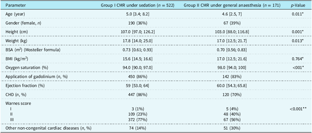
Five hundred twenty-two patients were in the deep sedation group and 171 in the general anaesthesia group. Detailed demographic data are shown in Table 1 and age distribution separately in each group in Figure 1. Most of the patients had CHD (86% in deep sedation group and 70% in general anaesthesia group). The overview of the cardiac diseases in each group broken down to congenital and non-congenital diseases is depicted in Figure 2. Seventy per cent of the congenital conditions in the deep sedation group and 56% in the general anaesthesia group were of great complexity by Warnes classification.
Patient characteristics

Table 1 Long description
The table compares patient characteristics between two groups: Group I CMR under sedation with 522 patients and Group II CMR under general anesthesia with 171 patients. It includes parameters such as age, gender, height, weight, body surface area, body mass index, oxygen saturation, application of gadolinium, ejection fraction, congenital heart disease, Warnes score, and other non-congenital cardiac diseases. Notable trends include significant differences in age, height, weight, oxygen saturation, and Warnes score between the two groups.
p-Values correspond to univariable analysis.
*Wilcoxon rank-sum test.
**Fisher’s exact test.
Age distribution for patients undergoing sedation ( a ) and general anaesthesia ( b ) GA = general anaesthesia.

Figure 1 Long description
The bar graph compares the age distribution of patients undergoing sedation and general anaesthesia. The graph consists of two separate bar charts labeled (a) and (b). Chart (a) represents the sedation group with a total of 522 patients. It shows three vertical bars: the first bar represents infants with 19 patients, the second bar represents patients aged 1 to 6 years with 293 patients, and the third bar represents patients older than 6 years with 210 patients. Chart (b) represents the GA group with a total of 171 patients. It also shows three vertical bars: the first bar represents infants with 20 patients, the second bar represents patients aged 1 to 6 years with 107 patients, and the third bar represents patients older than 6 years with 44 patients. The x-axis labels the age groups, and the y-axis labels the number of patients. The bars are colored blue, and each bar has a black label indicating the number of patients in that age group. All values are approximated.
Distribution of CHD in the sedation ( a ) and general anaesthesia groups ( b ) and non-CHD in the sedation ( c ) and general anaesthesia groups ( d ) at the Royal Brompton Hospital and University Hospital Schleswig-Holstein. ARVC = arrhythmogenic right ventricular dysplasia; GA = general anaesthesia; ccTGA = congenitally corrected transposition of great arteries; COA = coarctation of aorta; DCM = dilated cardiomyopathy; DORV = double-outlet right ventricle; HCM = hypertrophic cardiomyopathy; HLHS = hypoplastic left heart syndrome; LVNC = left ventricular non-compaction; PA = pulmonary atresia; TOF = tetralogy of fallot; TGA = transposition of great arteries; VSD = ventricular septal defect; PIMS = paediatric inflammatory multisystem syndrome.

Figure 2 Long description
The image contains four bar graphs. The first graph shows the distribution of congenital heart diseases in the sedation group with 485 cases. The conditions include hypoplastic left heart syndrome, tetralogy of fallot, transposition of great arteries, and others. The second graph displays the distribution of congenital heart diseases in the general anaesthesia group with 120 cases, listing conditions like tetralogy of fallot with ventricular septal defect, double-outlet right ventricle, and aortic valve disease. The third graph illustrates non-congenital heart diseases in the sedation group with 36 cases, including coronary heart disease, cardiomyopathy, and hypertrophic cardiomyopathy. The fourth graph presents non-congenital heart diseases in the general anaesthesia group with 51 cases, covering dilated cardiomyopathy, coronary heart disease, and paediatric inflammatory multisystem syndrome. Each graph uses bars to represent the number of cases for each condition.
In the deep sedation group, the most common cardiac defect was hypoplastic left heart syndrome (n = 203, 39%), followed by tetralogy of Fallot (n = 61, 12%) and transposition of the great arteries (n = 36, 7%). Most patients had a single ventricle and had previously undergone completion of the Fontan circulation (n = 246, 47%). Forty-one patients had associated syndromes such as VACTERL association (n = 7), trisomy 21 (n = 8), DiGeorge syndrome (n = 15), Noonan syndrome (n = 1), Turner syndrome (n = 2), Williams syndrome (n = 2), Goldenhar syndrome (n = 3), Klinefelter syndrome (n = 1), Rubinstein–Taybi syndrome (n = 1), and Kabuki syndrome (n = 1).
In the general anaesthesia group, at the time of cardiovascular magnetic resonance, 22 (18%) of the patients with CHD had an unrepaired condition, 41 (34%) were on single-ventricle pathway, and 57 (48%) had a history of complete repair. Sixty-one (36%) patients in the general anaesthesia group had associated comorbidities and/or syndromes, neurodevelopmental delay of unknown genetic cause (n = 18), autism spectrum disorder (n = 7), trisomy 21 (n = 6), DiGeorge syndrome (n = 6), Noonan syndrome (n = 6), Williams syndrome (n = 4), CHARGE association (n = 4), and 10 other genetic disorders.
Cardiovascular magnetic resonance scan
In the general anaesthesia group, 141 (82.5%) patients received gadolinium contrast agent with contrast angiography (104 patients) and/or late gadolinium enhancement (69 patients). Stress perfusion was performed in seven cardiovascular magnetic resonance studies (five positive). There were three positive studies in one patient with a stent in the left coronary artery who presented in cardiac arrest due to severe stenosis of the left coronary artery, one positive study in a patient after arterial switch repair with single coronary artery, and another positive study in a patient with anomalous origin of left coronary artery from right coronary sinus and intramural course.
Additional procedures
Fifty-five patients (32%) in the general anaesthesia group have undergone additional procedures following the cardiovascular magnetic resonance scan under the same general anaesthesia. Brain MRI was performed together with the cardiovascular magnetic resonance in 13 patients. Thirty-four patients underwent cardiac catheterisation, three insertion of implantable loop recorder, two transoesophageal echocardiogram, and two patients underwent non-cardiac intervention (dental extraction in one and operation of hydrocele in another one). One 8-year-old patient with autism underwent CT before right ventricular to pulmonary artery conduit balloon valvuloplasty to delineate coronary artery anatomy.
Deep sedation
Most patients (n = 517, 99%) received midazolam as premedication that was followed by propofol boluses and propofol infusion. Phenobarbital was used instead of midazolam in two patients and chloral hydrate in one patient. Ketamine instead of propofol was used in two patients. A RASS of 2 could be maintained during cardiovascular magnetic resonance. The average duration of the cardiovascular magnetic resonance scan was 95 ± 32 minutes.
Anaesthetic procedure
In the general anaesthesia group, anaesthetic data was available for analysis from 158 patients. General anaesthesia was induced using inhalation of sevofluorane in 89 patients and with intravenous drugs in 55 patients (54 propofol and 1 ketamine). The 14 remaining patients did not require induction as they were referred from ICU.
General anaesthesia was then maintained using intermittent positive pressure ventilation with sevofluorane in 95 patients and isofluorane in 63 patients.
Complications
Deep sedation group
Complications were documented in eight patients (1.5%). These complications comprised mild anaphylactic reactions in three patients, a severe coughing fit in one patient, increasing cyanosis in three single-ventricle patients, and suspected aspiration in one patient. None of the patients required admission to the ICU or had a significantly prolonged hospital stay.
The cardiovascular magnetic resonance scan was terminated early because of early awakening and agitation in 2.5% and coughing fits in 1.9%.
General anaesthesia group
Based on risk assessment before cardiovascular magnetic resonance, 13 patients (8%) were considered as high risk for the general anaesthesia: two patients had severe pulmonary hypertension, four severe obstruction to the pulmonary flow, three severe left ventricular outflow tract obstruction, and four significant left ventricular dysfunction.
Hypotension requiring some intervention was present in three patients (four scans) in the general anaesthesia group. Two patients out of these were considered as high risk for general anaesthesia.
A 15-month-old patient with unrepaired double-outlet right ventricle and multilevel pulmonary stenosis experienced a severe cyanotic spell on induction to anaesthesia. Therefore, the scan was abandoned. Following this, a semi-urgent bidirectional Glenn was performed. The patient underwent a successful cardiovascular magnetic resonance scan under general anaesthesia before full repair.
One patient (0.6%) had inadvertent endobronchial intubation. Minor hypothermia during the scan was present in 56 (33%) patients.
Management after the cardiovascular magnetic resonance scans
In the group of patients with CHD, 57 required surgical procedures and 55 continued with conservative management following the cardiovascular magnetic resonance under general anaesthesia. In the group of patients with non-CHD, cardiovascular magnetic resonance helped with the diagnostic process in 36 patients and excluded cardiomyopathy/myocarditis in 10. Altogether 13 patients required further imaging.
Comparison of adverse events between deep sedation and general anaesthesia groups
There were overall 14 adverse events (2%); 8 (1.5%) in the deep sedation group and 6 (3.5%) in the general anaesthesia group (excluding minor hypothermia in the general anaesthesia group). This difference was not statistically significant (p = 0.122). All other variables except body mass index showed a significant difference between the deep sedation and general anaesthesia group (Table 1). A logistic regression with outcome adverse event and influence variables group (deep sedation vs general anaesthesia), weight, body mass index, oxygen saturation, and Warnes score showed no significant results (p > 0.05 for all influence variables). When a backward selection was performed, no variable remained as significant in the model. A propensity score analysis was performed which generated matched data balanced with respect to weight, body mass index, oxygen saturation, and Warnes score in the two groups, deep sedation and general anaesthesia. Comparison of the adverse events showed again no significant difference between the two groups (p = 0.497), odds ratio (95% confidence interval: 1.88 (0.303,11.7)).
Discussion
We present the feasibility and safety of cardiovascular magnetic resonance performed under deep sedation or general anaesthesia in two paediatric cardiology tertiary centres.
The overall reported risk of severe injury or death for a healthy child having an MRI scan under general anaesthesia is extremely low, less than 1:100 000. 14 However, in children with underlying cardiac disease, this risk is significantly higher. The general anaesthetic can therefore pose more risk to patients with congenital and acquired heart disease than the cardiovascular magnetic resonance itself. Patient comorbidities, the severity of cardiac lesion, and functional status at the time of the general anaesthesia for any non-cardiac procedure are the main factors determining risk, rather than the type of procedure itself. Reference Faraoni, Zou, DiNardo and Nasr15 Using the CHD classification based on residual lesion and functional status (paediatric database of the American College of Surgeons National Surgical Quality Improvement Program, ACS NSQIP Pediatric), it has been shown that patients with major CHD (after repair with residual findings) or severe CHD (e.g. patients with uncorrected cyanotic CHD, with pulmonary hypertension or ventricular dysfunction requiring medication) have significantly higher incidence of overall mortality when compared to matched controls (3.9% vs 1.7%, p < 0.001 and 8.2% vs 1.2%, p = 0.001, respectively). Importantly, no difference has been noted in overall mortality between patients with minor CHD (e.g. fully repaired CHD (without residual haemodynamic findings and normal biventricular circulation) or with small atrial/ventricular septal defects without haemodynamic effect) and matched controls. Reference Faraoni, Zurakowski and Vo16
Other studies have shown that there are several particularly high-risk cardiac conditions which are associated with higher rate of complications related to anaesthesia when patients undergo non-cardiac procedures. Reference Taylor and Habre17 These include left ventricular obstructive lesions, single-ventricle lesions, pulmonary hypertension, and ventricular dysfunction often complicating all former conditions. Aortic stenosis accounts for 16% of cardiac arrests in patients with CHD but with 62% mortality rate from cardiac arrest. Williams syndrome, when associated with aortic stenosis, represents a particularly high-risk subgroup well known to be at extremely high risk of morbidity and mortality related to general anaesthesia. Patients with single-ventricle lesions are consistently identified as being at high risk. Single-ventricle physiology accounts for 19% of cardiac arrests in patients with CHD and is associated with a 35% mortality rate in infants and children prior to completion of total cavopulmonary connection. Systemic or suprasystemic pulmonary hypertension is a high-risk factor for adverse events under general anaesthesia. Major events (defined as perioperative mortality or life-threatening incidents requiring immediate treatment) were found to be associated with severity of pulmonary hypertension (OR 2.04; p = 0.006) but not with aetiology. Incidence of intra- or post-operative cardiac arrest has been reported in 2.7–5% of cases. Reference Bernier, Jacob, Collaco, McGrath-Morrow, Romer and Unegbu18
Ventricular dysfunction itself often complicates cardiac diagnoses and thus increases the risk of complications. In general, an ejection fraction of <25% is considered high risk. Although, there are no data to support specific levels of ejection fraction being associated with a specific level of risk, it appears that it is more important to treat patients individually and understand the context and their current status.
Our own data, collected between 2013 and 2022, show a low incidence of adverse events and lower than (4.4%) described in the literature. Reference Dorfman, Odegard, Powell, Laussen and Geva4 There are several potential reasons for this. This is a consultant-led and delivered service with a small group of trained staff regularly delivering care to this cohort. In the past, all patients referred for the cardiovascular magnetic resonance have been discussed in the departmental paediatric cardiology and cardiac surgery meeting which included attendance of cardiovascular magnetic resonance consultant and anaesthetist performing the procedure. After establishing safe processes and education of the referring paediatric cardiologists, discussion at these meetings is now generally limited to only high-risk patients. Patients known to be at extremely high risk are not scanned in the acute phase, and their cardiovascular magnetic resonance is either postponed or alternative safer imaging modalities are used (although the patients‘ complexity has increased over the years).
Internal hospital guidelines for patients undergoing cardiovascular magnetic resonance under general anaesthesia have been established compliant with the most recent published advice from the Medicines and Healthcare products Regulatory Agency, 19 the Association of Anaesthetists of Great Britain and Ireland 20,21 , and guidelines for the provision of anaesthetic services from the Royal College of Anaesthetists. 22 In addition, new guidance has been published by the Association of Paediatric Anaesthetists of Great Britain and Ireland, Royal College of Paediatrics and Child Health, and Royal College of Anaesthetics, detailing the process for informed consent in patients requiring diagnostic imaging procedures to be undertaken under general anaesthesia or sedation. This involves consideration of the risk for cardiovascular magnetic resonance prior to the scan and informed discussion with the parents and ensures cardiovascular magnetic resonance is only undertaken under general anaesthesia where there is likely to be clear benefit despite risks. 14
It is worth noting that a holistic approach is taken when patients undergo cardiovascular magnetic resonance scan under general anaesthesia, and other procedures and imaging methods might be undertaken at the same time. This way the whole diagnostic process can be completed under one general anaesthesia. Alternatively, cardiac catheterisation is performed after cardiovascular magnetic resonance either for diagnostic purposes or combined with an intervention.
Conclusion
We have shown that both deep sedation and general anaesthesia can be utilised for cardiovascular magnetic resonance scans in paediatric patients with a low rate of complication. This, however, requires a highly skilled team that adheres strongly to the safety policies set up by each hospital.
Acknowledgements
None.
Financial support
None.