Introduction
White sponge nevus (WSN) is a rare hereditary leucokeratosis that was first described by Hyde in 1909 (Ref. Reference Hyde1); the term white sponge nevus was first coined by Cannon in 1935 (Ref. Reference Cannon2). The onset of WSN is early in life, and both sexes are affected equally. Lesions of WSN are easily recognised and clinically important; the lesions appear as bilateral white spongy plaques, typically found on the buccal mucosa, and the patients report no painful symptomatology. The lesion can be found in other common sites, including the tongue, floor of the mouth and alveolar mucosa (Ref. Reference Frithiof and Bánóczy3). The disease are characterised by white, thickened, folded and spongy lesions of the oral mucosa, although the oesophageal, laryngeal, nasal and anogenital mucosa can also be affected (Ref. Reference Jorgenson and Levin4). The WSN plaques are considered benign because the lesions are asymptomatic and painless in many cases, although they may undergo alternate periods of remission and exacerbation due to infections. This disorder often manifests in early childhood and exhibits no gender preference. WSN occur on the surface of the skin, including the oral mucosa and anal mucosa, among other areas. The pathogenesis of WSN is linked to a disorder of the epithelium and epithelial cells. Electron microscopic imaging of the oral mucosa of patients diagnosed with WSN reveals cellular keratinisation in the mucosa. Furthermore, McGininis observed keratinising epithelium in those tissues by focusing on the ultrastructural features of the tissues (Ref. Reference McGinnis and Turner5). Richard et al. and Rugg et al. first reported that WSN was related to abnormalities in genes encoding keratin (KRT) (Refs Reference Richard6, Reference Rugg7).
KRT is an intermediate filament (IF) cytoskeletal protein. The human genome contains 54 genes encoding functional KRTs, 37 of which encode epithelial KRTs and 17 of which encode hair KRTs. KRTs can be divided into acidic and basic forms. Both types are coexpressed during the differentiation of epithelial tissues and are arranged in heterotypic pairs to form chains of laterally aligned coiled-coil structures (Ref. Reference Moll8). KRTs are types I and II IF proteins that form cytoskeletal networks within all epithelial cells. KRTs are expressed in pairs in a tissue and differentiation-specific fashion.
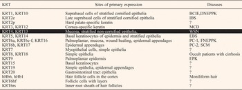
KRTs, which play an important role in the cellular cytoskeleton, are the largest subgroup of IF proteins and are typically found in skin and other epithelial tissues. To date, KRTs have been shown to play regulatory roles in electrolyte transport, post-translational modifications and to confer protection against degradation (Ref. Reference Majumdar9). KRTs can provide epithelia with mechanical support. Many KRT-related diseases have been reported (Table 1). KRT4 and KRT13 are the major differentiation-specific KRTs of the buccal mucosa and nasal, oesophageal and anogenital epithelia. The tissue distribution and nature of the lesions in patients affected by WSN suggest that mutations in KRT4 and/or KRT13 might be responsible for this disorder. Moreover, abnormal KRT mutations, such as KRT4 and KRT13, are closely linked to WSN (Ref. Reference Smith10). Both the oral and anogenital mucosae express type II KRT4 and its type I partner, KRT13. Furthermore, mutations of KRT4 and KRT13 were recently demonstrated to be the underlying cause of WSN.
KRT-related disease

WSN in the oral mucosa is an autosomal dominant genetic disease. In recent years, there have been many reports of new cases and mutations. Here, we summarise recent research concerning WSN in the oral mucosa, including clinical pathology, genetics, animal models, treatment, prognosis and future directions of WSN research.
Clinical report
To date, several hundreds of cases of WSN have been reported worldwide. WSN present as corrugated or velvety plaques on the oral, oesophageal or genital mucosa.
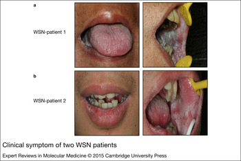
We examined two WSN patients who presented with white lesions on the lips, tongue and buccal mucosa (Fig. 1). Maahs found to exhibit bilateral asymptomatic lesions. The tissue changes associated with WSN can be present at birth or can manifest during childhood or adolescence, and this disorder occurs in females more frequently than in males (Ref. Reference Maahs11). Jornet presented a Spanish family consisting of a father and three boys affected with WSN. The proband, a 6-year-old boy, exhibited white lesions on the buccal mucosa. His 11-year-old brother exhibited lesions in the vestibule floor and gums, and the 4-month-old brother exhibited lesion on the lip frenulum and oral buccal mucosa (Ref. Reference Jornet12). The most frequent sites of WSN are the buccal and labial mucosa, with a rare appearance on the palate. No extra-oral lesions were found. Lucchese described three cases diagnosed as WSN following the clinical and histological criteria that exhibited unusual appearances. In one case, the lesion was also present in the vaginal area (Ref. Reference Lucchese and Favia13). Cheong reported the case of a 50-year-old Chinese woman with WSN signs on the buccal mucosa and genital mucosa (Ref. Reference Cheong14). Another case described a 28-year-old female with white plaques on the vulvar mucosa (Ref. Reference Cutlan15). A few other cases have been reported independently without familial backgrounds. For example, in one case, the parent reported that the family history was notable for a 7-year-old cousin with similar lesions, but nobody else in the family exhibited such findings (Ref. Reference Polcari and Chamlin16). A 33-year-old male with a 13-year history of white, soft and folded plaques on the buccal mucosae and surfaces of the tongue was reported by Dadlani et al. (Ref. Reference Dadlani17). Aghbali et al. report a case of WSN in a 21-year-old healthy Iranian male with no familial history who exhibited white bilateral lesion in the oral mucosa (Ref. Reference Aghbali18). To date, only a few reports of WSN have been related to other inherited diseases. Dalben et al. first report the case of a 10-year-old boy diagnosed WSN and Ectrodactyly–Ectodermal Dysplasia–Clefting (EEC) syndrome. Subsequent studies focused on the pathogenesis of both WSN and EEC syndromes (Ref. Reference Dalben19). Legler et al. report a rare case of a 16-year-old American boy with a woolly hair nevus on his left parietal scalp, an associated epidermal nevus along Blaschko's lines, and a white plaque on the central third of his ventral tongue who was clinically diagnosed as a WSN patient (Ref. Reference Legler20). Further WSN research has revealed an increasing number of familial cases. These cases confirm that WSN is an autosomal dominant hereditary condition. DeTomas et al. reported 16 WSN cases from six families (Ref. Reference DeTomas21). Martelli et al. report a three-generation family with WSN; clinical examination of family members revealed that of 23 descendants, 34.78% exhibited WSN features (Ref. Reference Martelli22). Another report presented a familial hereditary case of WSN in which two sisters (13 and 15 years old) exhibited white patches in the oral mucosa, tongue, lip and gum. Their mother exhibited similar lesions in her mouth when she was an adolescent (Ref. Reference Martins23). Samatha et al. reported a familial WSN case in which oral lesions were found in three generations of a family (Ref. Reference Samatha24). Kimura et al. described a Japanese family; the proband was an 11-year-old boy, and three other members of the family were affected by WSN (Ref. Reference Kimura25). Both genetic and environmental factors contribute to the development of WSN; Marrelli et al. described a father (38 years old) and son affected by WSN whose oral diseases were related to a Staphylococcus aureus infection (Ref. Reference Marrelli26). Lin et al. described two siblings affected by WSN who exhibited different clinical manifestations based on their smoking status. Theoretically, smoking may initiate exacerbation of WSN symptoms, but the authors reported that the degree of oral epithelial keratinisation among smokers was less than among non-smokers (Ref. Reference Lin27). All of these clinical reports describe the clinical manifestations of the disease and provide a solid foundation for aetiology research.
Clinical symptom of two WSN patients. (a) WSN patient-1 exhibited white lesions on the tongue and buccal mucosa; (b) WSN patient-2 exhibited white lesions on the lips and buccal mucosa.

Mutation report
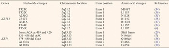
All of the reported mutations to date in exon 1A of the KRT4 and KRT13 gene are listed in Table 1. This disease may be hereditary. However, the pathogenesis of WSN remains unclear. Although the disease name refers to moles, the disorder is not associated with features of moles. Furthermore, the symptoms are caused by normal keratinisation (Ref. Reference Rugg7). Researchers have found that the abnormal expressions of several KRTs, mainly KRT4 or KRT13, play an important role in the generation of WSN (Ref. Reference Su28). In Terrinoni's study, the authors investigated a new KRT4 gene mutation (3 bp (ACA) insertion) and detected an amino acid insertion in exon 1A (Ref. Reference Terrinoni29). Another mutation identified was a heterozygous missense mutation 1345G > A in the KRT4 gene, which caused an amino acid substitution Glu449Lys in exon 2B (Ref. Reference Chao30). Zhang et al. described WSN in a Chinese family and identified two new KRT4 mutations. One mutation was 1829G > A, which is predicted to cause the amino acid substitution Glu520Lys. The second mutation was 2324A > G in the non-coding region (Ref. Reference Zhang31). Several other mutations of the KRTs have been close linked to WSN. The KRT4 gene mutation 1558G > A in the 2B region is predicted to change amino acid Glu520 to a Lys (Ref. Reference Shimizu32). Kimura et al. detected a mutation (3 bp AAC deletion) in the KRT4 gene (Asn160Del) (Ref. Reference Kimura25).
Richard et al. first identified the missense mutation 356T > C in the 1A region of the helical rod domain of KRT13 that led to Leu115Pro, which is linked to WSN (Ref. Reference Richard33). Rugg et al. identified two novel KRT13 mutations: a familial 344T > C mutation causing a Leu115Pro substitution and a familial 323T > C mutation causing a Met118Thr substitution (Ref. Reference Rugg34). The heterozygous missense mutation 335A > G was detected in exon 1 of the KRT13 gene and was predicted to cause the amino acid change Asn112Ser in the 1A domain of the KRT13 polypeptide (Ref. Reference Terrinoni35). Shibuya et al. identified a mutation at 332T > C, resulting in a Leu111Pro substitution (Ref. Reference Shibuya36). Nishizawa et al. analysed the KRT13 mutation 341G > A, which was predicted to cause the amino acid substitution Arg114His (Ref. Reference Nishizawa37). Liu et al. described a mutational analysis in familial and sporadic patients with WSN and identified the novel mutation 340C > T, causing the amino acid substitution Arg114Cys (Ref. Reference Liu38). In our study, we identified two heritable mutations in the KRT13 gene that were associated with the development of WSN in two Chinese families: 332T > C (Leu111Pro) and 340C > T (Arg114Cys) (Ref. Reference Cai39).
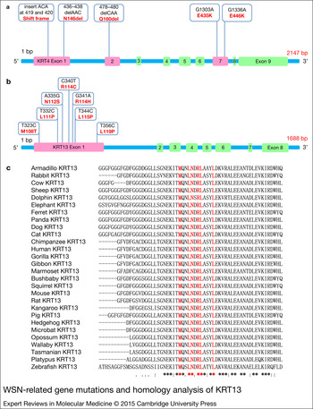
We have described five mutations in the KRT4 gene (Fig. 2a) and seven heritable mutations in the KRT13 gene (Fig. 2b) that are associated with WSN (Table 2). We analysed the homology of the KRT4 protein sequence from 43 species and the KRT13 protein sequence from 29 species, and we performed a multiple alignment of the KRT13 protein sequence from 29 species using the ClustalW program (http://www.genome.jp/tools/clustalw/). Some of the amino acids from exon1 of the KRT13 sequences of 29 species were relatively conserved (Fig. 2c). However, there were no highly similar fragments of the KRT4 protein sequences among the 43 species.
WSN-related gene mutations and homology analysis of KRT13. (a) There were five mutations in the KRT4 gene on exons 1, 2 and 7; (b) There were seven heritable mutations in the KRT13 gene on exon 1; (c) The homology analysis of the KRT13 protein sequence from 29 species. Some amino acids from exon 1 of the KRT13 sequences of the 29 species were relatively conserved.

WSN-related gene mutations

WSN animal model
The WSN animal model focuses on KRT4. Ness used KRT4-deficient mice to confirm that KRT4 is an important protein for internal epithelial integrity. In humans, KRT4 mutations lead to WSN, and KRT4-deficient mice may serve as ideal models for WSN to examine the changes in KRT4 when cells proliferate and differentiate (Ref. Reference Ness40). McGowan et al. identified a missense mutation in mutant animals (bright coat colour 1 (Bcc1)) that predicts an Asn154Ser amino acid substitution in the 1A domain of KRT4. Bcc1 recapitulates the histopathology and human genetics of WSN (Ref. Reference McGowan41). We should improve the development of animal models associated with WSN (KRT4 and KRT13). It is necessary to establish an ideal animal model for further research on WSN, which will provide a platform for drug screening and clinical treatment.
Treatment and prognosis
Although WSN patients experience no significant physical pain, they often complain of an altered texture to their mucosa or dissatisfaction with the appearance of the lesions. Many WSN patients undergo therapeutic treatments with nystatin, antihistamines, vitamins and mouth rinses. Azithromycin, tetracycline and penicillin have exhibited some clinical effects (Ref. Reference Songu42). However, there is no standard treatment protocol for WSN to date. A case of WSN exhibiting significant improvement following penicillin administration has been reported (Ref. Reference Alinovi43). McDonagh et al. first reported that tetracycline medicines were effective in four WSN patients (Ref. Reference McDonagh44). Another report stated that oral tetracycline rinse improved the symptoms of WSN (Ref. Reference Lim and Ng45). Subsequently, WSN was successfully treated with a tetracycline mouth rinse (Ref. Reference Becker46). Long-term low-dose systemic antibiotic therapy maintained the remission of WSN (Ref. Reference Lamey47). Victoria A acid can also inhibit keratosis formation (Ref. Reference Irvine and McLean48). Quintella et al. described a WSN patient who received orthodontic treatment in parallel with a sibling (Ref. Reference Quintella49). Otobe successfully treated a WSN patient with concurrent systemic lupus erythematosus using a topical tetracycline (Ref. Reference Otobe50). Four cases also improved with tetracycline mouth rinse (Ref. Reference Otobe51). To date, systemic antibiotics or local applications of retinoic acid have provided limited benefits, but both are poorly effective. Dufrasne et al. described a case of effective surgical resection, and the patient was free of recurrence 2 years later (Ref. Reference Dufrasne52). These methods together with the iPS technology may point to a treatment for this rare disease. WSN may be treated with chlorhexidine (Ref. Reference Satriano53). Meanwhile, WSN patients should perform a careful oral hygiene to reduce infection in the oral cavity. The proper diagnosis and treatment of this rare disease will require the combination of clinical history, clinical examination and pathologic findings.
Discussion
To date, several hundreds of cases of WSN have been reported worldwide, including in China (Refs Reference Chao30, 31, Reference Cai39), Japan (Refs Reference Kimura25, Reference Shimizu32), Italy (Refs Reference Lucchese and Favia13, Reference Marrelli26, Reference Terrinoni29), Spain (Ref. Reference Jornet12), Iran (Ref. Reference Aghbali18), Caucasia (Refs Reference Cutlan15, Reference McDonagh44), Brazil (Ref. Reference Martins23), Turkey (Ref. Reference Songu42) and Scotland (Ref. Reference Terrinoni54). The KRT4 and KRT13 gene mutations have been demonstrated to be involved in the pathogenesis of WSN. Because WSN in the oral mucosa is an autosomal dominant genetic disease, the percentage of the affected population in a family is generally greater than 30%. The hazards of this disease are much larger and more widespread than we previously appreciated.
In Table 2, we summarise the reported mutations of KRT4 and KRT13 in WSN patients. Because most KRT-related mutations occur in pairs (Refs Reference Rugg and Leigh55, Reference Cordon and McLean56), we expect that WSN-related genes will be limited to KRT4 and KRT13. However, new mutations in KRT4 and KRT13 are likely to be found in the near future. Our recent work suggests that the sequences of the KRT13 protein are highly conserved in the α-helix 1A domain from G102 to I136 in 23 diverse species, which represents the region in which all mutations of KRT13 have been found. Thus, this area represents a ‘mutation hotspot’ in the KRT13 gene. However, KRT4 does not exhibit a ‘mutation hotspot’. New mutations in this region will undoubtedly be identified, and the majority of genetic diagnosis will focus on this hotspot.
In a recently reported mouse model, the murine KRT4 gene was ablated by gene targeting. These animals exhibited a marked increase in mucosal cell division, as shown by staining for proliferating cell nuclear antigen (Ref. Reference Ness40). Precisely how the defective cytoskeleton in mucosal epithelial cells leads to the massive hyper-proliferative changes observed in WSN and the KRT4 knockout mice is unclear. Previous work revealed that when one member of a pair of KRTs is absent, its natural partner tends to be degraded. Due to differences between humans and mice, the regulation of the members of the KRT4 and KRT13 pair may be different. Studies of WSN disease and drugs are largely dependent on animal models. Genetically modified animal models provide important insights into the pathogenesis of human diseases. These models are also essential in developing new drugs. Classical mouse models have promoted significant developments in biomedicine. However, mice and humans are different in many aspects, including physiological traits and gene expression. Thus, mouse models cannot sufficiently mimic human diseases in some cases, and large animal models remain urgently needed (Refs Reference Verma57, Reference Walters58, Reference Li and Li59). Pigs have been recently used as models for human diseases because they are more similar to humans than mice in terms of anatomy, neurobiology, cardiac vasculature, gastrointestinal tract and genome (Ref. Reference Bendixen60). Pigs also present fewer ethical and economic concerns compared with primates. To date, several genetically modified pig models for human diseases have been produced, including models of Huntington's disease, Alzheimer's disease, spinal muscle atrophy, cardiovascular disease, diabetes mellitus (MODY3), diabetes mellitus type 2, retinitis pigmentosa, Stargardt-like macular dystrophy type 3 and breast cancer (Ref. Reference Fan and Lai61). With respect to the WSN disease animal model, further considerations are necessary because the molecular mechanism of this process in vivo remain incompletely understood. In our future work, we will use the CRISPR/Cas9 system to build a KRT13 T332C WSN transgenic pig model and explore the pathogenesis of WSN. We will apply phenotype reversal comprehensive treatment and drug screening to interpret and validate the molecular pathogenesis of WSN in vivo. This approach will provide an important reference for further exploration of the molecular mechanism of WSN as well as its clinical treatment and drug screening.
Lentiviruses using HIV-1 as a resource exhibit superiority over other transgenic technologies with respect to infection efficiency, stable expression, weak antigenicity, high degree of manoeuvrability, capability of containing large sizes of gene fragment, and infection of cells either in dividing and non-dividing phases (Refs Reference Wiznerowicz and Trono62, Reference Allie63). In gene therapy, numerous studies have confirmed that lentiviral vectors can be used for robust and highly efficient gene transfer into organs and tissues that are out of reach for many other gene delivery systems. However, there has been no related report about the therapy or study of WSN.
To develop better therapeutic trials for WSN, it is important to understand the consequences of the genetic mutations and molecular changes associated with WSN. Human-induced pluripotent stem cells (iPSCs) represent an excellent tool for this work with many advantages for research (Ref. Reference Takahashi and Yamanaka64). With the development of iPSC, many completed clinical trials have demonstrated the efficacy of mesenchymal stem cell (MSC) infusion for diseases, including acute myocardial ischaemia, stroke, liver cirrhosis, amyotrophic lateral sclerosis and graft-versus-host disease (Ref. Reference Wang65). However, there has been no related report about the application of WSN. We obtained WSN iPSC by transforming human WSN fibroblast cells and wild-type fibroblasts into iPSC, and the iPSC could be transformed into human epidermal cells for drug screening or mechanism research. Another cell research platform for WSN is MSCs. Recently, we have succeeded in isolating MSCs from patient gum tissue with the KRT13 point mutation (Ref. Reference Cai39). Many other biotechnologies can be used based on this cell platform, including mass spectrometry (Co-IP-MS), RNA-seq and weighted gene co-expression network analysis (Ref. Reference Xue66). The results would provide a valuable resource to dissect the gene regulatory mechanisms underlying the progressive development of WSN. This approach could identify genes downstream of KRT4 and KRT13, clarifying the pathogenesis of WSN. Meanwhile, our work suggests that the abnormal degradation of the KRT13 protein in WSN is likely associated with an abnormal ubiquitination process. Together, these efforts may contribute to molecular therapy for WSN.
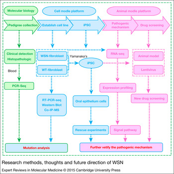
To the best of our knowledge, there remains no appropriate animal model or effective therapeutic drugs for WSN. The mechanism underlying WSN remains unclear. Almost all studies have focused on case reports or mutation reports. Prenatal diagnosis seems to be the only method to reverse the phenotype in affected families. We have described the research methods, thoughts and future research directions concerning WSN (Fig. 3). With further research into WSN, there is optimism that the problem will be solved in the next decade.
Research methods, thoughts and future direction of WSN.

Acknowledgements
This work was supported, in whole or in part, by the National Basic Research Programme (973 Programme, No. 2013CB967501), the Natural Science Foundation of Shanghai (No. 12ZR1434200), the Fundamental Research Funds for the Central Universities (No. 20120072110016), the Key Programme of Shanghai (No. 074119614) and the Shanghai Science and Technology Commission Programme (Nos124119A7400 and114119A3400).
Competing interests
The authors declare that they have no competing interests.