Introduction
Dilated cardiomyopathy is a common cause of heart failure and sudden cardiac death in children. Reference Lipshultz, Law and Asante-Korang1 Long-term follow-up and prognosis prediction are of crucial importance in clinical practice. It has been reported in the literature that the indicators for predicting the prognosis of children with dilated cardiomyopathy in clinical settings include a history of unexplained syncope, a family history of ventricular arrhythmia or sudden cardiac death, and the severity of heart failure, etc. Reference Reichart, Magnussen, Zeller and Blankenberg2–Reference Akhtar and Elliott4 Biomarkers such as B-type natriuretic peptide, N-terminal pro-brain natriuretic peptide, high-sensitivity C-reactive protein, soluble suppression of tumorigenicity 2 protein, transforming growth factor β, interleukin-1, interleukin-11, and the level of serum β2-microglobulin are also closely related to the prognosis of patients with dilated cardiomyopathy.
Electrocardiogram, with its characteristics of being economical, non-invasive, and simple, has become an important indicator for evaluating the prognosis of dilated cardiomyopathy. The occurrence of malignant ventricular arrhythmia, sudden cardiac death, and the progression of heart failure are the main events that affect the prognosis of patients with dilated cardiomyopathy. Reference Türe, Balık, Akın, Bilici and Nergiz5,Reference Vassallo, Cassidy, Kindwall, Marchlinski and Josephson6 Among the electrocardiographic markers, an increased negative value of the P-wave terminal force in lead V1 (PtfV1) is associated with a reduced left ventricular ejection fraction and sudden cardiac death Reference Tereshchenko, Henrikson and Sotoodehnia7 ; a decreased QRS amplitude is related to the adverse outcomes in patients with dilated cardiomyopathy Reference Merlo, Zaffalon and Stolfo8 ; the QRS duration is associated with fatal arrhythmias caused by the asynchrony of cardiac electrical activities Reference Amiya, Tanabe, Ikari, Nakajima and Hara9,Reference Konishi, Ohtani and Mizuno10 ; the corrected QT interval (QTc) and the corrected QT interval dispersion (QTcd) are often used to predict the risks of malignant arrhythmia and sudden cardiac death in patients with cardiomyopathy. Reference Nielsen, Graff and Rasmussen11,Reference Stewart12 Sabaz et al. Reference Sabaz, Akın, Bilici, Demir, Türe and Balık13 found that sinus tachycardia, frequent ischaemic changes in the ST segment on the electrocardiogram, and a prolonged QTc are risk factors for predicting the death of children with dilated cardiomyopathy.
The T wave represents the ventricular repolarisation process. The amplitude and abnormal morphology of the T wave are usually related to myocardial ischaemia. The changes in the QTc, QTcd, and the interval from the peak to the end of the T-wave Tp-Te/QT ratio are closely related to the asynchrony of ventricular myocyte repolarisation and the instability of cardiac electrical activities, which are of great significance for predicting adverse prognoses such as sudden cardiac death and malignant arrhythmia in children with dilated cardiomyopathy. Most previous studies have focused on the relationship between electrocardiographic markers such as the QT interval and QRS wave and the prognosis of patients with dilated cardiomyopathy. There are relatively few studies on the correlation between ventricular repolarisation-related electrocardiographic indicators such as Tp-Te/QT ratio, the amplitude of the T wave and the prognosis of patients with dilated cardiomyopathy, and the research conclusions have not yet reached a consensus. Electrocardiographic studies related to the prognosis of dilated cardiomyopathy in children are even rarer. This article will explore the value of electrocardiographic ventricular repolarisation parameters in estimating the prognosis of children with dilated cardiomyopathy.
Subjects and methods
Subjects
A retrospective analysis was conducted on the clinical data of 89 children who were hospitalised and diagnosed with dilated cardiomyopathy in the Department of Pediatric Cardiovascular Diseases of The Second Xiangya Hospital, Central South University from July 2008 to September 2023. There were 46 males and 43 females, with an age range of 0.17–17 [5.24 (4.32, 6.15)] years. After a follow-up of 5.16 (2.29, 8.34) years, 76 cases survived, and 13 cases died. The control group consisted of 80 children of the same age and sex who underwent physical examinations in The Second Xiangya Hospital, Central South University during the same period. The research was conducted according to the guidelines of the Declaration of Helsinki, and approved by the Ethics Committee of The Second Xiangya Hospital, Central South University [Ethical Audit No. Study 249(2022)].
Diagnostic criteria
The diagnoses of children with dilated cardiomyopathy all met the diagnostic criteria of the World Health Organization/International Society for Heart and Lung Transplantation. Reference Maron, Towbin and Thiene14 Cardiac diastolic dysfunction and systolic dysfunction caused by coronary artery disease, hypertension, valvular disease, or CHD were excluded. Reference Jefferies and Towbin15 Both the research group and the control group underwent echocardiography and electrocardiography examinations. The medical history of the children in the research group was inquired in detail. Some children in the research group underwent cardiac magnetic resonance imaging, urine and blood metabolic screening, and clinical whole-exome sequencing for monogenic genetic diseases. Among them, 20 children underwent clinical whole-exome sequencing for monogenic genetic diseases, and 14 cases had confirmed evidence of pathogenic genes related to dilated cardiomyopathy.
Electrocardiogram recording and measurement
Electrocardiogram recording
The supine synchronous 12-lead electrocardiogram was recorded using the MAC800 electrocardiograph from GE Medical System (China) Co., Ltd The gain was set as 1 mV = 10 mm, the paper speed was 25 mm/s, and no filtering device was used.
Electrocardiogram measurement
The electrocardiogram with clear waveforms was selected for measurement, with the starting part of the QRS wave as the horizontal line. The methods for determining the end point of the T wave were as follows: ①The T wave returns to the isoelectric line; ②The notch between the T wave and the U wave; ③The intersection point where the biphasic T wave finally returns to the isoelectric line. For each ECG index, three consecutive cardiac cycles with clear waveforms were selected for measurement, and the average value was taken. Reference Tan, Yi and Chen16 The measurement indexes included maximum QT interval (QTmax) and minimum QT interval (QTmin) of 12 leads, and corrected maximum QT interval (QTcmax) and corrected minimum QT interval (QTcmin) were obtained by correcting heart rate according to Bazett’s formula, and QT dispersion (QTd) and corrected QT dispersion (QTcd) were calculated as QTmax-QTmin and QTcmax-QTcmin respectively. Tp-Te refers to the interval from the peak of the T wave to the end of the T wave on the electrocardiogram, and the Tp-Te/QT ratio was calculated. T-wave amplitude: For the positive T wave, the amplitude was measured vertically from the upper edge of the isoelectric line to the peak of the waveform; for the negative T wave, the amplitude was measured vertically from the lower edge of the isoelectric line to the bottom of the waveform; for the biphasic T wave, it was the algebraic sum of the positive and negative amplitudes.
Follow-up
Children with dilated cardiomyopathy were all given comprehensive treatments including diuretics (hydrochlorothiazide, spironolactone, furosemide), angiotensin-converting enzyme inhibitors (enalapril), beta-blockers (metoprolol), cardiotonic agents (digoxin), anticoagulant treatments (aspirin, warfarin), and drugs for improving myocardial metabolism (levocarnitine). Reference Tan, Yi and Chen16,Reference Wang, Xu and Zou17 All subjects were followed for 5.16 (2.29, 8.34) years by outpatient/inpatient or telephone to determine clinical outcomes.
Statistical analysis
Data were analysed statistically using SPSS 26.0 software. For measurement data that conformed to a normal distribution, they were expressed as mean ± standard deviation(
 $\overline{x}$
± s), and the independent sample t-test was used for comparison between groups. For measurement data that did not conform to a normal distribution, they were expressed as interquartile range [M (P25, P75)], and the Mann–Whitney U rank sum test was used for comparison between groups. Counting data were expressed as number of cases (%), and the χ
2
test or Fisher’s exact test was used for comparison between groups. The receiver operating characteristic curve (ROC curve) was drawn to evaluate the prognostic estimation value of ventricular repolarisation for children with dilated cardiomyopathy. A p value less than 0.05 was considered statistically significant.
Results
Comparison between the research group and the control group
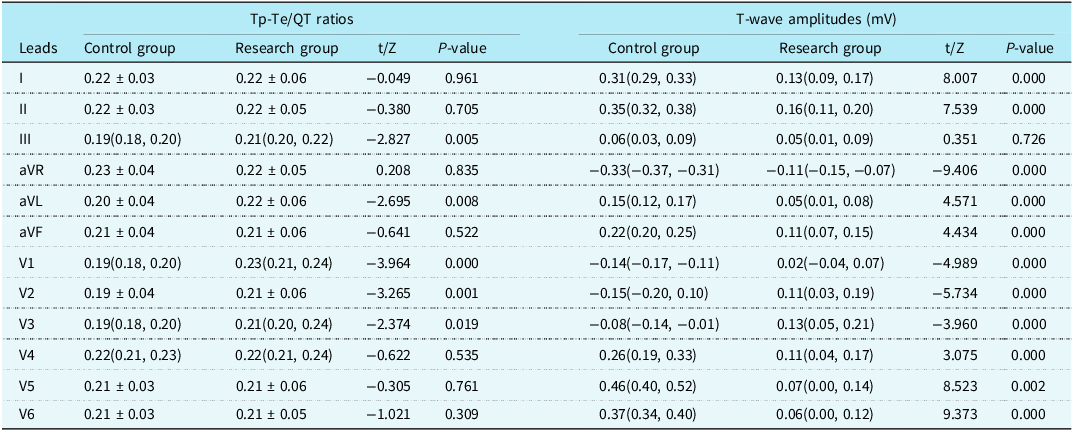
Compared with the control group, the left ventricular ejection fraction and the left ventricular fractional shortening in the research group were decreased (P < 0.05). The diameters of left ventricular, right ventricular, left atrial, and right atrial were increased (P < 0.05). The QTcmax and QTcmin were prolonged (P < 0.05). The Tp-Te/QT ratios in leads III, aVL, V1, V2, and V3 were increased (P < 0.05). The amplitudes of the T wave in leads I, II, aVL, aVF, V4, V5, and V6 were decreased (P < 0.05). (Table 1, 2).
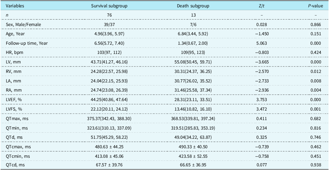
Comparison of general data, echocardiographic and electrocardiographic parameters between the research group and the control group [(
 $\overline{x}$
± s) or M (P25, P75)]

Note: HR = heart rate; LVEF = left ventricular ejection fraction; LVFS = left ventricular short axis shortening rate; LV = left interior; RV = right ventricular; LA = left atrial; RA = right atrial; QTmax = maximum QT interval; QTmin = minimum QT interval; QTd = QT interval dispersion; QTcmax = corrected the maximum QT interval; QTcmin = corrected minimum QT interval; QTcd = corrected QT interval dispersion.
Comparison of 12-lead electrocardiographic tp-te/QT ratios and T-wave amplitudes between research group and control group [(
 $\overline{x}$
± s) or M (P25, P75)]

Comparison between the survival group and the death group
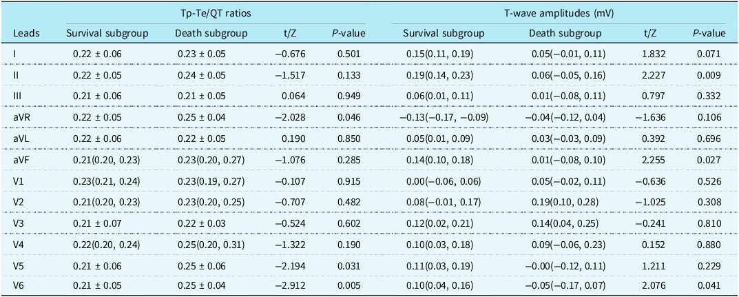
Compared with the survival subgroup, the left ventricular ejection fraction and left ventricular fractional shortening in the death subgroup were decreased, while the diameters of LV, RV, LA, and RA were increased (P < 0.05). There were no statistically significant differences in QTmax, QTmin, QTd, QTcmax, QTcmin, and QTcd between the two subgroups (P > 0.05). The Tp-Te/QT ratios in leads aVR, V5, and V6 were increased (P < 0.05), and the amplitudes of the T wave in leads II, aVF, and V6 were decreased (P < 0.05) in the death subgroup (Table 3, 4).
Comparison of echocardiographic and electrocardiographic parameters between survival subgroup and death subgroup [(
 $\overline{x}$
± s) or M (P25, P75)]

Note: HR = heart rate; LVEF = left ventricular ejection fraction; LVFS = left ventricular short axis shortening rate; LV = left interior; RV = right ventricular; LA = left atrial; RA = right atrial; QTmax = maximum QT interval; QTmin = minimum QT interval; QTd = QT interval dispersion; QTcmax = corrected the maximum QT interval; QTcmin = corrected minimum QT interval; QTcd = corrected QT interval dispersion.
Comparison of 12-lead electrocardiographic tp-te/QT ratios and T-wave amplitudes between survival subgroup and death subgroup [(
 $\overline{x}$
± s) or M (P25, P75)]

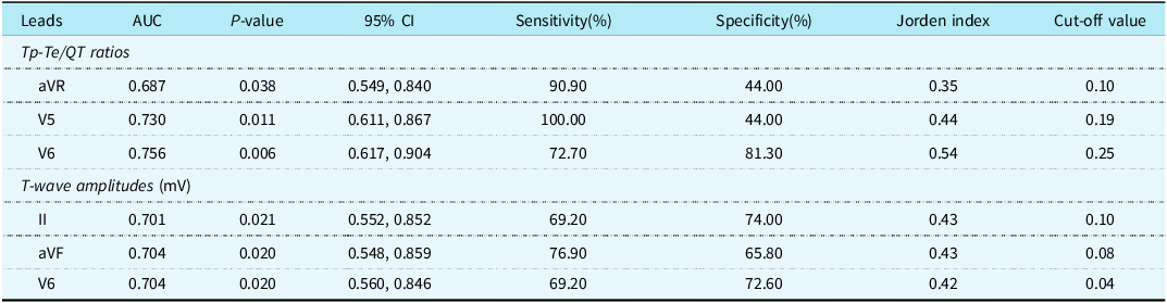
The receiver operating characteristic curve was drawn to evaluate the prognostic estimation value of the Tp-Te/QT ratio for children with dilated cardiomyopathy. The areas under the receiver operating characteristic curves of the Tp-Te/QT ratios in leads aVR, V5, and V6 were 0.687, 0.730, and 0.756, respectively. When the Tp-Te/QT ratios in lead aVR was ≥0.10, that in lead V5 was ≥0.19, and that in lead V6 was ≥0.25, the sensitivity and specificity for predicting the death of children with dilated cardiomyopathy were 90.90% and 44.00%, 100.00% and 44.00%, 72.70% and 81.30%, respectively (Table 5 and Figure 1).
ROC curve of Tp-Te/QT ratios and T-wave amplitudes predicting prognosis of children with dilated cardiomyopathy. (a):Tp-te/QT ratios (b):T-wave amplitudes.

Tp-te/QT ratios and T-wave amplitudes predict the prognosis of children with dilated cardiomyopathy

The receiver operating characteristic curve was drawn to evaluate the prognostic estimation value of the amplitudes of the T wave in leads II, aVF, and V6 for children with dilated cardiomyopathy. The 95% confidence intervals of the amplitudes of the T wave in leads II, aVF, and V6 did not include 0.5, suggesting that they had a relatively good prognostic estimation value for children with dilated cardiomyopathy (P < 0.05). The areas under the receiver operating characteristic curves of the amplitudes of the T wave in leads II, aVF, and V6 were 0.701, 0.704, and 0.704, respectively. The optimal cut-off values were 0.10 mV, 0.08 mV, and 0.04 mV, respectively. The sensitivity and specificity were 69.20% and 74.00%, 76.90% and 65.80%, 69.20% and 72.60%, respectively.
Discussions
Dilated cardiomyopathy is accompanied by abnormal cardiac electrical activities and/or myocardial mechanical dysfunction. Its clinical manifestations include progressive cardiac enlargement, progressive heart failure, and progressive arrhythmia, and the clinical prognosis is poor. The 1-year survival rate is 63–90%, and the 5-year survival rate is 20–80%. It is currently the main cause of heart transplantation in children. Reference Taliercio, Seward, Driscoll, Fisher, Gersh and Tajik18 The main mechanisms for ventricular arrhythmia in children with dilated cardiomyopathy are autonomic nervous system disorders and impaired uniformity of myocardial repolarisation. Reference Türe, Balık, Akın, Bilici and Nergiz5
Electrocardiographic abnormalities can reflect the severity of cardiovascular diseases and provide a simple and useful tool for risk stratification and prognosis prediction. Some electrocardiographic markers are closely related to the prognosis of children with dilated cardiomyopathy. The PtfV1 may be related to increased cardiac load, myocardial hypertrophy, myocardial fibrosis, and conduction delay. Reference Morris, Estes, Whalen, Thompson and Mcintosh19 When myocardial fibrosis and scar repair occur, the activation of ventricular myocardium is delayed, and the heterogeneity of activation in various parts increases significantly, which is manifested as the formation of notches in the QRS wave on the electrocardiogram, resulting in an increase in fragmented QRS. Reference Das, Suradi and Maskoun20 The QTd and QTcd reflect the duration from ventricular depolarisation to the end of repolarisation and the differences in ventricular myocyte repolarisation, and they are the electrophysiological basis for fatal ventricular arrhythmia. Reference Nielsen, Graff and Rasmussen11,Reference Masarone, Valente and Rubino21 An increased rate of change in the ST-T wave and elevation or depression of the ST segment are closely related to arrhythmia and heart failure. Reference Tan, Yi and Chen16 A decrease in the amplitude of the T wave in leads II, V4, V5, and V6 indicates left ventricular myocardial ischaemia and is closely related to the occurrence of heart failure in children with dilated cardiomyopathy. Reference Merlo, Zaffalon and Stolfo22 The interval from the Tp-Te reflects the transmural dispersion of ventricular repolarisation. The Tp-Te and Tp-Te/QT ratios are electrocardiographic indicators for predicting malignant ventricular arrhythmia. Reference Türe, Balık, Akın, Bilici and Nergiz5 Therefore, the T wave represents the process of myocardial repolarisation, and electrocardiographic markers related to the T wave are closely related to the prognosis of patients with cardiomyopathy. Reference Levis23
In this study, 89 children with dilated cardiomyopathy were followed up for a long period, and 13 of them died. There were no statistically significant differences in age, sex, and baseline heart rate between the survival subgroup and the death subgroup. Among the 13 children with dilated cardiomyopathy who died, 8 died within one year after diagnosis. All the 13 deceased children showed manifestations of progressive heart failure. Among them, 2 had malignant arrhythmia manifestations such as severe ventricular fibrillation, Adams–Stokes syndrome, and atrial flutter; 4 had arrhythmia manifestations including supraventricular tachycardia, atrioventricular block, ventricular premature beats, and complete right bundle branch block; and the other 4 cases died after discharge, and it was not recorded whether they had malignant arrhythmia manifestations or not. In the children with dilated cardiomyopathy in this study, the diameters of the LV, RV, LA, and RA were enlarged. At the initial diagnosis, compared with the survival subgroup, the left ventricular ejection fraction and eft ventricular fractional shortening were decreased, and the diameters of the LV, RV, LA, and RA were increased in the death subgroup.
Among the ventricular repolarisation indices, the Tp-Te/QT ratio was increased in children with dilated cardiomyopathy in this study, while the T-wave amplitudes in leads I, II, aVL, aVF, V4, V5, and V6 were decreased, suggesting that there was abnormal ventricular repolarisation in children with dilated cardiomyopathy. In the death subgroup, the Tp-Te/QT ratios were increased in leads aVR, V5, and V6, and the T-wave amplitudes in leads II, aVF, and V6 were decreased, indicating that these T-wave-related electrocardiographic indices were associated with poor prognosis. However, the aetiology of abnormal repolarisation in children with dilated cardiomyopathy is still not completely clear at present, and it may be related to electromechanical coupling and stretch-activated currents. In the mediators of stretch activation of cardiomyocytes, the conduction velocity decreased significantly, and the action potential duration increased. Mechanical deformation affects electrical conduction through feedback on stretch-activated ion channels. Reference Weise and Panfilov24
QTd represents the asynchrony of ventricular muscle repolarisation at different sites and the instability of cardiac electrical activity. An increase in QTd indicates an increased difference in the repolarisation of ventricular myocytes, which is prone to induce malignant arrhythmias or sudden cardiac death. Reference Fauchier, Douglas, Babuty, Cosnay and Fauchier25 Türe et al. Reference Türe, Balık, Akın, Bilici and Nergiz5 reported that the QTd (0.042 ± 0.020 s vs. 0.070 ± 0.0453 s, P < 0.01) and QTcd were prolonged (0.074 ± 0.077 s vs. 0.038 ± 0.027 s, P < 0.05) in children with dilated cardiomyopathy, and their risk of death increased. However, studies on QTd in adult dilated cardiomyopathy patients found no differences among the event-free group, the sudden cardiac death group, and the arrhythmia group (435 ± 42 ms vs. 442 ± 39 ms vs. 440 ± 41 ms, P > 0.05). Reference Fauchier, Douglas, Babuty, Cosnay and Fauchier25 In this study, no differences were found in QTd and QTcd between children with dilated cardiomyopathy and healthy children, or between the survival subgroup and the death subgroup. Therefore, whether QTd and QTcd have predictive value for the prognosis of children with dilated cardiomyopathy still needs further study.
The Tp-Te interval represents the relative refractory period of ventricular repolarisation and reflects the transmural dispersion of ventricular repolarisation, which may be closely related to the occurrence of various cardiovascular diseases that cause sudden cardiac death. Ozdemir et al. Reference Ozdemir, Isguder and Kucuk26 reported that the Tp-Te/QT ratio might be a useful tool for predicting the mortality of patients with sepsis in the paediatric intensive care unit. Türe et al. Reference Türe, Balık, Akın, Bilici and Nergiz5 reported that in 85 children with dilated cardiomyopathy (including 12 deaths), the maximum T-wave peak-to-end time (Tp-Temax) (0.087 ± 0.023 vs. 0.074 ± 0.014, P < 0.05) and T-wave peak-to-end time dispersion (Tp-Ted) (0.050 ± 0.018 vs. 0.037 ± 0.014, P < 0.05) were prolonged in the death group compared with the survival group, suggesting that the prolongation of Tp-Temax and the increase in Tp-Ted were associated with an increased mortality rate in children with dilated cardiomyopathy. However, Porthan et al. Reference Porthan, Viitasalo and Toivonen27 found no association between Tp-Te prolongation and sudden cardiac death (P = 0.231). In this study, it was found that the Tp-Te/QT ratios were increased in leads III, aVL, V1–V3 in children with dilated cardiomyopathy compared with healthy children, and were increased in leads aVR, V5, and V6 in the death subgroup compared with the survival subgroup, suggesting that the Tp-Te/QT ratios in leads aVR, V5, and V6 has predictive value for the prognosis of children with dilated cardiomyopathy.
The T-wave amplitude also reflects changes in ventricular muscle repolarisation. In this study, it was found that the T-wave amplitude was decreased in leads I, II, aVL, aVF, V4, V5, and V6 in children with dilated cardiomyopathy compared with healthy children, and was decreased in the death subgroup compared with the survival subgroup, suggesting abnormal ventricular repolarisation in children with dilated cardiomyopathy. Tan et al. Reference Tan, Yi and Chen16 reported that when the T-wave amplitude was simultaneously ≤0.20 mV in lead II, ≤0.40 mV in lead V4, ≤0.3 mV in lead V5, and ≤0.30 mV in lead V6, it had a good predictive value for left ventricular ejection fraction <50% in children with dilated cardiomyopathy, which might be related to the significant reduction of Ito, Ikr, and Ik1, the decrease in potassium ion efflux, the prolongation of repolarisation time, and the increase in action potential duration in children with dilated cardiomyopathy during heart failure, suggesting that the decrease in T-wave amplitude has predictive value for the decrease in left ventricular ejection fraction. In the follow-up of children with DCM in this study, it was found that when the T-wave amplitude was ≤0.10 mV in lead II, ≤0.08 mV in lead aVF, and ≤0.04 mV in lead V6, it had predictive value for the risk of death in children with dilated cardiomyopathy. This might be related to arrhythmias and heart failure caused by the inhomogeneity of ventricular repolarisation in children with dilated cardiomyopathy. Leads II and aVF correspond to the electrocardiographic activities of the inferior wall of the ventricle, and lead V6 corresponds to the electrocardiographic activities of the lateral wall of the ventricle. Therefore, the decrease in T-wave amplitudes in leads II, aVF, and V6 may reflect myocardial ischaemia in the inferior and lateral walls of the heart in children with dilated cardiomyopathy and can be used as one of the predictive markers for the risk of death in children with DCM.
Limitations
This article is a single-centre retrospective study, and the sample size is limited. The causes of death of children with dilated cardiomyopathy are different, and no classified evaluation has been conducted on cases of death due to different reasons. The electrocardiogram was recorded only once at the first visit, and it might be affected by confounding factors such as the time of electrocardiogram recording and the compliance of children. Electrocardiographic indicators in children have different characteristics as they grow older, and stratified evaluation can be conducted according to different age groups in later studies.
Conclusions
Abnormal ventricular repolarisation parameters were found in children with dilated cardiomyopathy. Increased Tp-Te /QT ratios in aVR, V5, and V6 leads and decreased T-wave amplitudes in II, aVF, and V6 leads were risk factors for predicting mortality in children with dilated cardiomyopathy.
Acknowledgements
We thank the study participants for volunteering to participate in this research.
Financial support
Natural Science Foundation of Hunan Province (2021JJ70093, 2023JJ30812, 2025JJ81175) and Department of Science and Technology of Hunan Province (2023SK4018).
Author contributions
SW and CW conceived the research. MW, TZ, PL, FL, LL, EH, YW, RZ, and YX collected and reviewed subjects’ data. MW performed statistical analysis and SW drafted the manuscript. All authors contributed to its revision.
Competing interests
The authors declare no competing interests.
Ethical standard
Informed consent was signed by the subjects themselves or their legal guardians before HUTT. This research was approved by the Medical Ethics Committee, The Second Xiangya Hospital, Central South University, and conforms to the principles stated in the Declaration of Helsinki [Ethical Audit No. Study 249(2022)].
Data availability
All data generated for this analysis are available from the corresponding author upon reasonable request from qualified researchers.
Declarations
None.