Case report
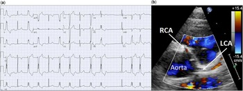
A 16-year-old female evaluated for history of non-exertional, reproducible chest pain was found to have frequent isolated premature ventricular contractions with frequent couplets on resting electrocardiogram as shown in Figure 1. The finding of frequent premature ventricular contractions prompted further cardiac investigation. Holter monitoring detected normal sinus rhythm with premature ventricular contractions comprising 18% of all beats. Transthoracic echocardiogram revealed anomalous origin of the right coronary artery from the left sinus of Valsalva with normal origin of the left main and circumflex coronary arteries as shown in Figure 1. Her cardiac function was normal. A cardiac magnetic resonance scan was performed, confirming the anomalous origin of the right coronary artery with no evidence of ventricular scar on late gadolinium enhancement imaging. Oral verapamil was prescribed in an attempt to decrease the ectopy burden and reduce risk of sustained ventricular arrhythmias. Given the possibility of symptoms and arrhythmia secondary to myocardial ischaemia, the decision was made to proceed with surgical repair of the anomalous right coronary artery. The intramural segment of the right coronary artery was unroofed without complication and the patient was discharged home after a short hospital stay. At follow-up, she was asymptomatic with normal ventricular function on verapamil therapy, although she continued to have high ventricular ectopy, including non-sustained ventricular tachycardia, with premature ventricular contractions comprising 42% of beats on Holter monitoring. The premature ventricular contraction morphology did not change compared with her pre-surgery electrocardiogram (Fig 1).
( a ) Baseline electrocardiogram showing frequent ventricular couplets with inferior axis and left bundle branch morphology. ( b ) Baseline transthoracic echocardiogram, parasternal short-axis view with colour showing anomalous origin of the right coronary artery (RCA) from the left aortic cusp. LCA=left coronary artery.

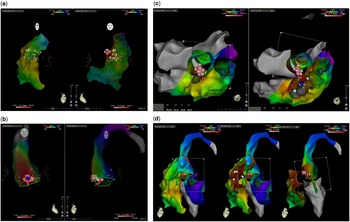
Given the significant burden of ectopy, including non-sustained ventricular tachycardia, the patient underwent an electrophysiology study 18 months following the anomalous right coronary artery unroofing surgery. The baseline surface and intracardiac electrograms confirmed normal sinus rhythm with frequent premature ventricular contractions and couplets. The premature ventricular contractions had left bundle branch block morphology with inferior axis, and therefore the right ventricular outflow tract was predicted as the likely site of origin. The premature ventricular contraction duration was 130 ms. Intracardiac electrogram activation mapping of the right ventricle was performed to identify the site of earliest activation, with initial results suggesting a right ventricular outflow tract focus. Radiofrequency ablation of the right ventricular outflow tract and cryoablation of sites in the right ventricular outflow tract that were close to the His bundle were performed, without alteration in the frequency of ventricular ectopy (Fig 2).
Three-dimensional electroanatomic maps. ( a ) Post-cryoablation followed by radiofrequency ablation of the right ventricular outflow tract. Yellow dots=location of His bundle; red/pink dots=radiofrequency ablation sites. ( b ) After radiofrequency ablation in the region of the aortic cusps. Red/pink dots=ablation sites. ( c ) Composite maps of the right atrium, right ventricle, left ventricle, and aorta in axial projections and ( d ) sagittal projection showing the proximity of the aortic cusp ablation lesions to the radiofrequency ablation sites in the right ventricular outflow tract. Yellow dots=location of His bundle; pink/red dots=radiofrequency ablation sites.

At this point, the mapping and ablation catheter was advanced retrograde from the femoral artery to the left ventricular outflow tract. Premature ventricular contraction onset was measured at −48 ms from the mapping catheter placed in the right coronary cusp, indicating significantly earlier activation than seen within the endocardial ventricular cavity where activation points within the right ventricle mapped at no earlier than 30 ms pre-QRS. Radiofrequency ablation at this site (Fig 2) promptly terminated the premature ventricular contractions within a few seconds of energy application. Verapamil was discontinued, and the patient has remained asymptomatic without any recurrence of ventricular ectopy at the last follow-up.
Discussion
Monomorphic ventricular ectopy with left bundle branch block morphology and inferior axis typically arises from the right ventricular outflow tract. This case report highlights the importance of considering less common origins of ventricular arrhythmia such as the left ventricular outflow tract, aortic cusps, or epicardial sites regardless of the initial prediction. Limited reports indicate that the prevalence of ventricular arrhythmias arising from the right ventricular outflow tract or left ventricular outflow tract that originate in the aortic cups is ~6–21%.Reference Morwood, Triedman and Berul 1 – Reference Yamada, McElderry and Doppalapudi 4 Previous reports of left ventricular outflow tract-origin premature ventricular contractions mimicking right ventricular outflow tract-origin morphology have also described a similar progression during electrophysiology studies from initial right ventricular outflow tract mapping and ablation to left ventricular outflow tract mapping and ablation after the initial approach fails to terminate arrhythmia.Reference Gonzalez y Gonzalez, Will and Tuzcu 5 , Reference Hlivák 6 Various predictive algorithms have been suggested to identify likely left ventricular outflow tract or epicardial origin of ventricular arrhythmias before attempted ablation,Reference Yamada, McElderry and Doppalapudi 4 , Reference Stevenson and Soejima 7 – Reference Strobel 9 including early precordial R wave transition, although these algorithms fail to identify arrhythmia origin correctly in many cases and even in retrospect would not have predicted a right coronary cusp arrhythmia origin in this specific case. The adjacent anatomical location of the ablation sites in the aortic cusp and the right ventricular outflow tract in this case, as shown in Figure 2, illustrate the intrinsic difficulty of precise identification of premature ventricular contraction origin in this region. In cases where the coronary ostia locations are unknown, adjunctive imaging such as coronary angiography, cardiac MRI, or CT merged onto the electroanatomic images and/or transoesophageal echocardiogram may be helpful to prevent inadvertent injury during ablation.
To our knowledge, this is the first report of an anomalous coronary artery and ventricular arrhythmia arising in the same region of the aortic cusps. In previous reports, patients with left ventricular outflow tract and aortic cusp origin of ventricular arrhythmias have had otherwise normal cardiac and aortic anatomy with the arrhythmogenic substrate believed to arise in an extension of the myocardium beyond the aortic annulus.Reference Gami 10 In this case, although probably unrelated and coincidental findings, we speculate that perhaps a common pathological process in embryology with abnormal extension of the myocardium into the aortic cusps resulted in development of both an arrhythmogenic substrate and anomalous origin of the right coronary artery, and we encourage others to report any similar cases.
Although there is general consensus regarding the need for surgical intervention to reduce the risk of sudden cardiac death for symptomatic patients with anomalous origin of the left coronary artery from the right aortic cusp, debate continues for patients with anomalous origin of the right coronary artery from the left aortic cusp. Ventricular arrhythmia associated with an anomalous coronary artery prompted us to consider disruption to normal myocardial perfusion and contributed to the decision to proceed with surgical repair in this case. In retrospect, the location of premature ventricular contraction origin in the left ventricular outflow tract is unlikely to be secondary to inadequate coronary perfusion. Arrhythmia associated with an anomalous coronary artery should therefore be cautiously interpreted with regard to the decision to proceed with surgical repair.
Acknowledgements
None.
Financial Support
This research received no specific grant from any funding agency, commercial or not-for-profit sectors.
Conflicts of Interest
None.
Ethical Standards
The authors assert that all procedures contributing to this work comply with the ethical standards of the relevant guidelines on human experimentation and with the Helsinki Declaration of 1975, as revised in 2008, and has been approved by the institutional committee (Children’s National Medical Center).