Introduction
Interspecies microinsemination assays have been used to study gamete physiology around fertilization; e.g. assays for sperm capacitation using zona-free hamster oocytes (Hanada & Chang, Reference Hanada and Chang1972), for sperm-borne oocyte-activating factor (SOAF) using mouse oocytes (Rybouchkin et al., Reference Rybouchkin, Dozortsev, De Sutter, Quin and Dhont1995) and for the microtubule-organizing centre (MTOC) using rabbit and bovine oocytes (Terada et al., 1993; Kobayashi et al., Reference Kobayashi, Amemiya, Takeuchi, Tsujioka, Tominaga, Hirabayashi, Ishikawa, Fukui and Hochi2006). Ogonuki et al. (Reference Mogoe, Fukui, Ishikawa and Ohsumi2001) investigated the relationship between calcium oscillation-inducing activity and SOAF activity in cynomolgus monkey spermatogenic cells by microinseminating mouse oocytes. The accumulated results of the interspecies and homologous mouse assay (Rybouchkin et al., Reference Rybouchkin, Dozortsev, De Sutter, Quin and Dhont1995; Sousa et al., Reference Sousa, Mendoza, Barros and Tesarik1996; Ogonuki et al., Reference Ogonuki, Sankai, Yagami, Shikano, Oda, Miyazaki and Ogura2001) indicate that the stage at which SOAF first emerges, or becomes biologically active during spermatogenesis, may vary with species. We have recently reported that minke whale spermatogenic cells acquire SOAF activity during spermiogenesis, using the mouse oocyte activation assay (Amemiya et al., Reference Amemiya, Iwanami, Kobayashi, Terao, Fukui, Ishikawa, Ohsumi, Hirabayashi and Hochi2004), as seen in the cases of mouse and rat spermatogenic cells (Kimura et al., Reference Kimura, Yanagimachi, Kuretake, Bortkiewicz, Perry and Yanagimachi1998; Yazawa et al., Reference Yazawa, Yanagida, Katayose, Hayashi and Sato2000).
The aim of the present study was to characterize the haploid spermatogenic cells of minke whales (Balaenoptera bonaerensis), with special reference to Ca2+ oscillations and SOAF activity using interspecies microinsemination of mouse oocytes. The Japanese Whale Research Program under Special Permit in the Antarctic Sea, conducted under Article VIII of the International Convention for the Regulation of Whaling (ICRW), allowed us to use the vasa deferentia and testicular samples from the Antarctic minke whales killed using the most humane method available.
Materials and Methods
Sperm preparation
Spermatozoa from cauda epididymis of a B6D2F1/CrIj male mouse (Charles River Laboratories Japan) were suspended at 4 °C in the Dulbecco's phosphate-buffered saline (PBS) containing 5.6 mM glucose, 5.4 mM sodium lactate and 0.1% polyvinyl pyrrolidone (360 kDa; Sigma-Aldrich), referred to here as GL–PBS. The sperm suspension was centrifuged twice at 200 g for 5 min at 4 °C and cryopreserved in GL–PBS supplemented with 7.5% glycerol (Wako Pure Chemical Industries) and 7.5% fetal bovine serum (FBS; Equitech Bio) in 1 ml cryotubes, according to the method of Ogura – et al. (Reference Ogura, Matsuda, Asano, Suzuki and Yanagimachi1996). Frozen tubes were thawed in a water bath at 20 °C. The warmed samples were washed twice by centrifugation at 200 g for 5 min with GL–PBS.
Spermatozoa from a Japanese Black bull were derived from commercially available semen frozen in 0.5 ml straws. The samples that were thawed in a 37 °C water bath were washed twice by centrifugation at 490 g for 5 min with Brackett and Oliphant solution (Brackett & Oliphant, Reference Brackett and Oliphant1975).
Spermatozoa from an Antarctic minke whale were recovered by squeezing the vasa deferentia downstream and were suspended in 15% egg yolk–Tris buffer/5% glycerol solution in 1.0 ml Eppendorf tubes. The tubes were cooled to 5 °C for 2 h and then to −80 °C overnight and stored in liquid nitrogen, according to the method by Mogoe et al. (Reference Mogoe, Fukui, Ishikawa and Ohsumi1998). Frozen tubes were warmed in a 37 °C water bath and the warmed samples were washed twice by centrifugation at 200 g for 5 min with GL–PBS.
Spermatid preparation
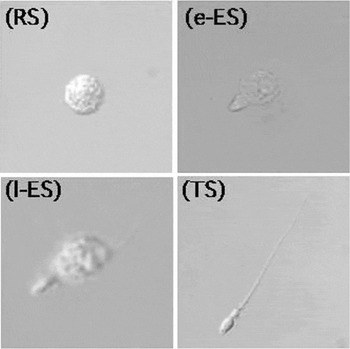
Testicular spermatogenic cells were prepared from mature Antarctic minke whales on the research base ship. Briefly, cubic centimetre testis clods were placed in erythrocyte-lysing buffer (155 mM NH4Cl, 10 mM KHCO3, 2 mM EDTA; pH 7.2). Seminiferous tubules were transferred into GL–PBS at an ambient temperature, cut into small pieces using fine scissors and shaken gently to allow the release of spermatogenic cells into the medium. The cell suspension was filtered through a 40 μm nylon mesh and then centrifuged at 200 g for 5 min. The suspension containing the spermatids and spermatozoa were cryopreserved in 1.0 ml cryotubes in GL–PBS supplemented with 7.5% glycerol and 7.5% FBS, as described above for the mature mouse spermatozoa. The tubes were warmed in a water bath at 20 °C immediately before injection and resuspended with 10-fold volumes of GL–PBS. Whale spermatogenic cells that were included were round spermatids (RS: Gorgi/cap phase; stages 1–7), early-stage elongating spermatids (e-ES: acrosome phase; stages 8–14), late-stage elongating spermatids (1-ES: maturation phase; stages 15–18) and testicular spermatozoa (TS: stage 19), as shown in Fig. 1.
Whale spermiogenic cells under phase-contrast light microscopy (RS, round spermatid; e-ES, early-stage elongating spermatid; l-ES: late-stage elongating spermatid; TS, testicular spermatozoa).

Oocyte preparation
Specific pathogen-free/virus antibody-free B6D2F1/CrIj female mice (7–10 weeks old; Charles River) were superovulated by intraperitoneal injections of 5 i.u. equine chorionic gonadotropin (eCG; Denka Pharmaceuticals) and 5 i.u. human chorionic gonadotropin (hCG; Mochida Pharmaceuticals) at an interval of 48 h. The oocytes were collected from the oviductal ampullae 14 h after hCG injection and were freed from cumulus cells by a 5 min treatment with 0.1% hyaluronidase (Sigma-Aldrich) suspended in CZB medium (Chatot et al., Reference Chatot, Lewis, Torres and Ziomek1990). Denuded oocytes were rinsed and incubated in CZB medium for up to 2 h at 37 °C in an atmosphere of 5% CO2 in air.
Microinsemination
Sperm injection
Prior to the ICSI, 1.7 μl of the sperm suspension was mixed with 8.3 μ1 of CZB medium supplemented with 22 mM HEPES and 12% PVP, defined hereafter to as HEPES–CZB/PVP medium, at an ambient temperature of 23 ± 2 °C. Using a piezo-impact driving unit (PMM-150FU; Prime Tech) with a pulse controller (PMAS-CT150; Prime Tech), the sperm head was dissociated from the tail by a few piezo pulses. Ten denuded oocytes were placed in 10 μ1 of CZB medium supplemented with 22 mM HEPES, called hereafter HEPES–CZB medium. The sperm head was then deposited into an ooplasm with a blunt-ended injection pipette with an inner diameter of 5–6 μm.
Spermatid injection
RS, e-ES and 1-ES samples were each suspended in 10 μ1 of the HEPES–CZB/PVP medium. The procedure for spermatid injection was according to the method described by Hirabayashi et al. (Reference Hirabayashi, Kato, Aoto, Ueda and Hochi2002). Briefly, the spermatid's membrane was broken by shear stress during repeated pipetting. The spermatid nucleus with surrounding cytoplasm was then microinjected into an ooplasm in HEPES–CZB medium.
Kinetics of intracellular Ca2+
Intracellular Ca2+ level was measured by a Ca2+-imaging method described previously (Yazawa et al., Reference Yazawa, Yanagida, Katayose, Hayashi and Sato2000) with some minor changes. Briefly, oocytes were treated with a Ca2+-sensitive fluorescent dye, fluo-3-acetoxymethylester (fluo-3/AM; Molecular Probes), at a concentration of 22 μM in HEPES–CZB medium for 30 min at 37 °C. A number of oocytes (three to five) was microinseminated within 7 min and then was transported within 3 min to another laboratory kept at 30 °C. The kinetics of intracellular Ca2+ level was measured for up to 3 h at 20 s intervals under a confocal laser-scanning microscope (Bio-Rad 2100; Nippon Bio-Rad Laboratories) equipped with an image processor.
Evaluation of oocyte activation
After the 3 h observation of intracellular Ca2+ oscillations at 30 °C, each microinseminated oocyte was placed on a glass slide, mounted with a coverslip and then fixed with acetic acid:ethanol (1:3 v / v) for 20 min. The oocytes stained with 1% aceto-orcein for 5 min were observed under a phase-contrast microscope at ×200 magnification. The oocytes with the anaphase-II or telophase-II plate were defined as having resumed meiosis (activated).
Statistical analysis
The proportions of oocytes exhibiting Ca2+ oscillations and resuming meiosis were compared by the Ryan method (Ryan, Reference Ryan1960). A p-value < 0.05 was chosen as an indication of statistical significance.
Results
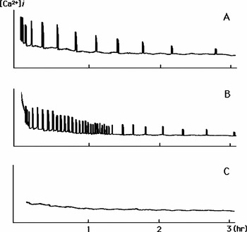
Two oscillation patterns were observed in mouse oocytes. Intracellular Ca2+ concentration increased repetitively with intervals of 10–30 min, lasting for 3 h (pattern A; Fig. 2A) or with intervals of 2–5 min for an initial 1–1.5 h and then intervals of 10–30 min for the subsequent 1.5–2 h (pattern B; Fig. 2B). Otherwise, there was no detectable increase in intracellular Ca2+ concentration during the observation period (Fig. 2C).
Intracellular Ca2+ oscillation patterns. (A) normal; (B) short-cycled; (C) no response.

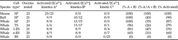
The Ca2+ oscillation patterns induced in microinseminated mouse oocytes and their interaction with oocyte-activating abilities are shown in Table 1. Either pattern A or pattern B oscillation was observed in all mouse oocytes injected with mature spermatozoa. Mouse mature spermatozoa induced the pattern A oscillation in all the 23 mouse oocytes (100%). Bull and whale mature spermatozoa induced not only pattern A oscillation (43 and 35%, respectively), but also pattern B oscillation (57 and 65%, respectively). The proportions of mouse oocytes that exhibited oscillation (pattern A or B) after microinjection of whale RS, e-ES, 1-ES and TS were 0, 65, 81 and 96%, respectively. Pattern A oscillation was observed in 26–47% of these oscillated oocytes. In exact one-to-one correspondence to the occurrence of Ca2+ response across the species and cell type, it was found that most oocytes that exhibited the pattern A oscillation (98%) and the pattern B oscillation (85%) also resumed meiosis.
Pattern of Ca2+ oscillations triggered in mouse oocytes by mature spermatozoa (mouse, bull and whale) and haploid spermiogenic cells (whale) and the one-to-one correspondence to oocyte activation

ae-ES, early-stage elongating spermatids; 1-ES, late-stage elongating spermatids; RS, round spermatids; SP, mature spermatozoa; TS, testicular spermatozoa.
bRefer to Fig. 2.
Discussion
Physiological characteristics of haploid spermatogenic cells in marine mammals, such as dolphins and whales, are poorly understood. Although Antarctic minke whale spermatogenic cells acquire SOAF activity at a relatively early phase of spermiogenesis (Amemiya et al., Reference Amemiya, Iwanami, Kobayashi, Terao, Fukui, Ishikawa, Ohsumi, Hirabayashi and Hochi2004), the relationship to Ca2+ oscillation-inducing capacity of the haploid spermatogenic cells has not been investigated. In the present study, the time at which SOAF becomes biologically active (e-ES) matched well with the time at which spermatid cells became Ca2+ oscillation inducible. In contrast, Ogonuki et al. (Reference Ogonuki, Sankai, Yagami, Shikano, Oda, Miyazaki and Ogura2001) reported that some monkey RS were capable of inducing repetitive Ca2+ oscillations (45%, 10/22) or a single large increase in intracellular Ca2+ concentration (18%, 4/22) in mouse oocytes. In their report, the oocyte activation rate, although not the one-to-one correspondence to occurrence of Ca2+ response, was 67% (8/12). Yazawa et al. (Reference Yazawa, Yanagida, Katayose, Hayashi and Sato2000) reported that mouse and rat elongating spermatids induced activation in 31–89% of injected mouse oocytes, while most of the intracellular Ca2+ concentration responses did not show the normal oscillation pattern. In the present study, observation under exact one-to-one correspondence of oocyte activation to the occurrence of Ca2+ response suggests that intracellular Ca2+ elevation(s), triggered by whale elongating spermatids, is the minimal prerequisite for oocyte activation.
The pattern A oscillation (Fig. 2A) must be a physiologically normal response as a postfertilization event, based on numerous previous reports in mammalian species (Swann, Reference Swann1990; Stricker, Reference Stricker1999; Yazawa et al., Reference Yazawa, Yanagida, Katayose, Hayashi and Sato2000; Ogonuki et al., Reference Ogonuki, Sankai, Yagami, Shikano, Oda, Miyazaki and Ogura2001). Another pattern of the oscillations (Fig. 2B) was induced specifically in interspecies microinsemination (mouse–bull and mouse–whale), probably due to the qualitatively or quantitatively higher SOAF activity in bull and whale sperm or spermatogenic cells, because bull and whale spermatozoa may have to activate homogenous oocytes that are larger than mouse oocytes. Such a short-cycled oscillation pattern, as shown in Fig. 2B, has been reported when a non-physiologically larger volume of porcine sperm extracts was microinjected into mouse oocytes (Gordo et al., Reference Gordo, Wu, He and Fissore2000). The cRNA of human phospholipase C-ζ (PLCζ) injected into mouse oocytes correlated with the interval of Ca2+ pulses in a dose-dependent manner (Cox et al., Reference Cox, Larman, Saunders, Hashimoto, Swann and Lai2002). A single transient increase of intracellular Ca2+ was not observed in the present study. Some chemical or electrical stimuli (e.g. ethanol or direct current pulse) is known to trigger a single increase in intracellular Ca2+ concentration and a non-physiological Ca2+ response induced subsequent activation of oocytes (Cuthbertson et al., Reference Cuthbertson, Whittingham and Cobbold1981; Sun et al., Reference Sun, Hoyland, Huang, Mason and Moor1992).
The stage at which the SOAF first emerges or becomes biologically active during spermatogenesis was at the elongating spermatid stage in whales (Amemiya et al., Reference Amemiya, Iwanami, Kobayashi, Terao, Fukui, Ishikawa, Ohsumi, Hirabayashi and Hochi2004), mice (Kimura et al., Reference Kimura, Yanagimachi, Kuretake, Bortkiewicz, Perry and Yanagimachi1998) and rats (Kimura et al., Reference Kimura, Yanagimachi, Kuretake, Bortkiewicz, Perry and Yanagimachi1998; Yazawa et al., Reference Yazawa, Yanagida, Katayose, Hayashi and Sato2000). In contrast, round spermatids from hamsters (Yazawa et al., Reference Yazawa, Yanagida, Katayose, Hayashi and Sato2000; Haigo et al., Reference Haigo, Yamauchi, Yazama, Yanagimachi and Horiuchi2004), rabbits (Yazawa et al., Reference Yazawa, Yanagida, Katayose, Hayashi and Sato2000; Ogonuki et al., Reference Ogonuki, Inoue, Miki, Mochida, Hatori, Okada, Takeiri, Shimozawa, Nagashima, Sankai and Ogura2005), monkeys (Ogonuki et al., Reference Ogonuki, Sankai, Yagami, Shikano, Oda, Miyazaki and Ogura2001) and humans (Tesarik et al., Reference Tesarik, Mendoza and Testart1995; Yazawa et al., Reference Yazawa, Yanagida, Katayose, Hayashi and Sato2000) have already shown SOAF activity (in primates probably even diploid spermatocytes carry SOAF; Yazawa et al., Reference Yazawa, Yanagida, Katayose, Hayashi and Sato2000; Ogonuki et al., Reference Ogonuki, Sankai, Yagami, Shikano, Oda, Miyazaki and Ogura2001). The mechanism of interaction between Ca2+ oscillation-inducing ability and SOAF activity in whales might be be understood when the identity of SOAF molecules are determined. To date, the PLCζ seems to be the strongest candidate to be the SOAF (Cox et al., Reference Cox, Larman, Saunders, Hashimoto, Swann and Lai2002; Saunders et al., Reference Saunders, Larman, Parrington, Cox, Royse, Blayney, Swann and Lai2002) and evidence is accumulating.
In conclusion, the Antarctic minke whale spermatogenic cells acquire SOAF activity that is closely related to their Ca2+ oscillation-inducing ability at a relatively early stage of spermiogenesis.
Acknowledgements
The authors thank the crew of the research base ship, Nisshin-maru, for their technical assistance. Mr T. Terao (Shinshu University) and Mr T. Fujihira (Obihiro University) were also acknowledged for processing frozen whale spermatid samples. This study was supported in part by a Grant-in-Aid for 21st Century COE Program from the Ministry of Education, Sports, Culture, Science and Technology of Japan and by the Co-operative Study Program of National Institute for Physiological Sciences, Japan.