Introduction
Many of the manual tasks that are performed on a daily basis comprise of a sequence of actions. For instance, to drink water from a cup, we must reach and grasp the cup, lift it to the mouth, then drink. To efficiently complete such actions, one has to prioritise grasping the object in order to end the movement comfortably, which, at times could be at the expense of comfort at the beginning of, and/or during, a movement (Rosenbaum, Vaughan, Barnes, & Jorgensen, Reference Rosenbaum, Vaughan, Barnes and Jorgensen1992). This is known as the ‘end-state-comfort’ (ESC) effect, and is a hallmark of a mature motor system. Indeed, while the tendency to opt for ESC when planning actions becomes stronger during childhood (Wunsch, Henning, Aschersleben, & Weigelt, Reference Wunsch, Henning, Aschersleben and Weigelt2013). Interestingly, little is known about the neural mechanisms that subserve childhood motor planning, nor variations in the tendency to opt for ESC when performing actions. Answering these questions is critical to understanding individual differences in childhood motor planning.
There is compelling evidence from adult neuroimaging studies that suggests areas of the frontoparietal network are central for planning of movements. For instance, in a functional magnetic resonance imaging (fMRI) study, a sample of the healthy population was asked to indicate their grasp selection when a bar had to be transported from its starting position to a specified end position and orientation (Zimmermann, Meulenbroek, & de Lange, Reference Zimmermann, Meulenbroek and de Lange2012). The researchers found that areas of the dorsal and ventral premotor network that include the precentral gyrus (PcG), middle frontal gyrus (MFG) and superior medial gyrus were activated. In addition to the premotor network, areas of the parietal lobe such as the superior parietal lobule (SPL) were also linked to complex motor planning activity. In another study researchers looked at the neural correlates of planning and execution of voluntary inhibition of ongoing actions and found that areas such as the supplementary motor area (SMA), inferior frontal gyrus (IFG) and inferior parietal cortex were involved in both phases (Omata, Ito, Takata, & Ouchi, Reference Omata, Ito, Takata and Ouchi2018).
The efficacy of motor planning is thought to depend heavily on one’s capacity to generate, and engage with, internal modelling systems (Johnson-Frey, McCarty, & Keen, Reference Johnson-Frey, McCarty and Keen2004; Rosenbaum, Herbort, van der Wei, & Weiss, Reference Rosenbaum, Herbort, van der Wei and Weiss2014). Experimentally, one’s capacity to generate (or manipulate) internal models has classically been inferred from tasks of motor imagery (MI), which involve mental simulation of actions without physically executing the movements (Decety & Grezes, Reference Decety and Grezes1999). In support of the view that the internal modelling systems are critical to motor planning, greater efficiency of MI performance (a proxy for internal modelling) was shown to be related to a stronger propensity to opt for ESC during motor planning (Fuelscher, Williams, Wilmut, Enticott, & Hyde, Reference Fuelscher, Williams, Wilmut, Enticott and Hyde2016). Further, the frontoparietal regions implicated in motor planning (e.g., PcG, MFG, SMA and SPL) overlap considerably with those thought to subserve MI performance. In addition to these areas, the supramarginal gyrus (SMG), inferior parietal gyrus (IPG) and precuneus (PreC) were also activated to support MI of upper limb performance (Hétu et al., Reference Hétu, Grégoire, Saimpont, Coll, Eugène, Michon and Jackson2013). Of note, the SPL is connected to the prefrontal and premotor regions via the bidirectional associative white matter pathway, the superior longitudinal fasciculus (SLF) (Floris & Howells, Reference Floris and Howells2018). Evidence from the adult literature has proposed that this white matter tract is critical for complex motor planning of upper limb movements, by facilitating the integration of visuospatial and somatosensory inflow and transmitting visuomotor representations for the hand and reach direction from posterior association regions to frontal motor cortices (Budisavljevic et al., Reference Budisavljevic, Dell’Acqua, Zanatto, Begliomini, Miotto, Motta and Castiello2017; Parlatini et al., Reference Parlatini, Radua, Dell’Acqua, Leslie, Simmons, Murphy and Thiebaut de Schotten2017).
The neural architecture of the SLF is complex, meaning that voxels included in analyses often contain numerous bundles of crossing fibre populations. Since the more commonly adopted tensor model (i.e., DTI) is unable to reconcile more than a single fibre within a given voxel, it has been argued that that extreme caution should be exercised when interpreting findings derived from this approach (Jeurissen, Leemans, Jones, Tournier, & Sijbers, Reference Jeurissen, Leemans, Jones, Tournier and Sijbers2011), particularly in regions such as the SLF where the proportion of voxels containing crossing fibres is high. Indeed, this issue can lead to the premature termination of fibre tracts or incorrectly switching to an adjacent tract resulting in isolation of a fibre pathway that does not exist during tractography (Behrens et al., Reference Beurze, de Lange, Toni and Medendorp2007; Jeurissen et al., Reference Jeurissen, Leemans, Jones, Tournier and Sijbers2011). This crossing fibre issue can also make tensor-derived metrics (such as fractional anisotropy and mean diffusivity) difficult to interpret. As a result, higher-order diffusion approaches, such as constrained spherical deconvolution (CSD), are therefore recommended to address the methodological difficulties with crossing fibres (Jeurissen et al., Reference Jeurissen, Leemans, Jones, Tournier and Sijbers2011; Tournier et al., Reference Tournier, Yeh, Calamante, Cho, Connelly and Lin2008). Via spherical harmonics, CSD is able to detect and model the presence of crossing fibres within a given voxel. This allows for tractography that better reflects the underlying anatomy than that derived from DTI. Further, CSD allows for the generation of metrics such as apparent fibre density (AFD) (Raffelt et al., Reference Raffelt, Tournier, Rose, Ridgway, Henderson, Crozier and Connelly2012), which provide an index of underlying white matter microstructure that are less susceptible to the ‘crossing fibres’ problem than tensor metrics (Farquharson & Tournier, Reference Farquharson and Tournier2016; Jones, Knösche, & Turner, Reference Jones, Knösche and Turner2013; Raffelt et al., Reference Raffelt, Tournier, Rose, Ridgway, Henderson, Crozier and Connelly2012).
Despite evidence in the adult literature suggesting a role for frontodorsal cortical regions in motor planning, as well as microstructural properties of the white matter fibres that facilitate communication within this network, no study has reported on the cortical or microstructural basis of motor planning in children. There is, however, indirect evidence implicating these frontal and parietal regions in childhood action planning, as well as the white matter tracts that connect them (e.g., SLF). Indeed, children with developmental coordination disorder show a decreased tendency to opt for ESC during complex motor planning (Bhoyroo, Hands, Steenbergen, & Wigley, Reference Bhoyroo, Hands, Steenbergen and Wigley2020; Bhoyroo, Hands, Wilmut, Hyde, & Wigley, Reference Bhoyroo, Hands, Wilmut, Hyde and Wigley2018, Reference Bhoyroo, Hands, Wilmut, Hyde and Wigley2019; Fuelscher et al., Reference Fuelscher, Williams, Wilmut, Enticott and Hyde2016; Wilmut & Byrne, Reference Wilmut and Byrne2014a). In addition, they also present with alterations in frontal and parietal areas during tasks of movement, while atypical white matter organization has also been observed within the SLF in these children (Biotteau et al., Reference Biotteau, Peran, Vayssiere, Tallet, Albaret and Chaix2017; Fuelscher et al., Reference Fuelscher, Caeyenberghs, Enticott, Williams, Lum and Hyde2018; Hyde et al., Reference Hyde, Fuelscher, Enticott, Jones, Farquharson, Silk and Caeyenberghs2019; Kashiwagi, Iwaki, Narumi, Tamai, & Suzuki, Reference Kashiwagi, Iwaki, Narumi, Tamai and Suzuki2009; Langevin, Macmaster, Crawford, Lebel, & Dewey, Reference Langevin, Macmaster, Crawford, Lebel and Dewey2014; Langevin, MacMaster, & Dewey, Reference Langevin, MacMaster and Dewey2015; Williams, Kashuk, Wilson, Thorpe, & Egan, Reference Williams, Kashuk, Wilson, Thorpe and Egan2017).
The aim of the study was to explore the associations between complex motor planning and the macrostructural and microstructural properties of the frontoparietal neural structures thought to subserve motor planning to gain an understanding of the involvement of the frontoparietal network in children’s motor planning. These include macrostructural metrics include cortical thickness and cortical surface area of the superior frontal gyrus (SFG), MFG, PcG, SPL, IPG and SMG acquired using MRI, and the white matter organization within the bilateral SLF as expressed by mean AFD obtained from higher order diffusion weighted imaging (DWI). Complex motor planning was measured using the octagon task, a protocol that elicits the ESC effect across the developmental spectrum (Wilmut & Byrne, Reference Wilmut and Byrne2014b), and is effective at distinguishing individual differences in childhood action planning (see Bhoyroo et al., Reference Bhoyroo, Hands, Steenbergen and Wigley2020 for a review). It was hypothesised that, similar to adults, greater cortical thickness in the abovementioned frontoparietal regions would be associated with a greater propensity to adopt ESC on the octagon task. We also expected the latter to be associated with AFD within the SLF.
Methods
Participants
Nineteen right-handed boys aged 8–11 years participated in the study (M age = 9.84, SD = 1.14). Participants were recruited from local schools, advertisements in the local newspaper over 24 months. Given the modest sample size, only boys were included in order to eliminate any potential gender differences. Parents completed the SNAP IV questionnaire and none of the participants reported concerns for attention deficit hyperactive disorder. The children had no prior diagnosis as well as showed no indication of learning difficulties, visual impairments or neurological diagnosis (e.g., cerebral palsy, muscular dystrophy) that could affect movement as reported by their parents. This study was granted ethics approval by the University of Notre Dame Australia Human Resources Ethics Committee in according with the Helsinki Declaration involving human research. Informed consent was obtained from all individual participants and their parents included in the study.
Behavioural Data Acquisition: Motor Planning
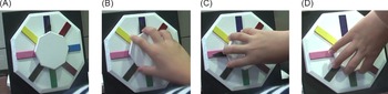
In order to assess motor planning, participants completed the octagon task. This apparatus is similar to that used in previous studies that examined motor planning in children and adults (Bhoyroo et al., Reference Bhoyroo, Hands, Wilmut, Hyde and Wigley2018, Reference Bhoyroo, Hands, Wilmut, Hyde and Wigley2019; Fuelscher et al., Reference Fuelscher, Williams, Wilmut, Enticott and Hyde2016; Wilmut & Byrne, Reference Wilmut and Byrne2014a, Reference Wilmut and Byrne2014b). It consisted of a wooden octagon with each side coloured differently, mounted on a wooden black board (Figure 1). On the bigger octagon, a smaller ‘dial’ octagon was placed. This dial was available in different sizes ranging between 6.5 cm and 12.5 cm, allowing participants to opt for the one that most comfortably fits their grip. The dial consisted of a black pointer placed at 0° (pointing upwards to the purple colour) prior starting any trial. Participants used a whole hand grasp to complete the colour sequence.
A. Set up of Octagon task with pointer facing north (purple colour). B–D. Participant use a whole hand grasp to complete the colour sequence.

Participants were tested individually in a quiet room. They sat comfortably in front of the apparatus at a distance ensuring they could complete the tasks without difficulty and placed their palms on their thighs. This task is usually completed in one, two and three colour sequences. However, a previous study identified that the three colour sequence is too complex for this age group (Bhoyroo et al., Reference Bhoyroo, Hands, Steenbergen and Wigley2020), therefore the one and two colour sequences are reported. All sequences were completed with the self-reported preferred hand. Participants were instructed to grasp the dial in their preferred way, with no more than one finger on a given side of the octagon. They were then required to move the black pointer to the designated colour and hold the position. Participants completed two successful practice trials for each colour sequence before commencing the experimental trials. The colour sequences used for the practice trials were not included in the experimental trials.
Participants completed four experimental trials for each colour sequence. The sequences were presented in a blocked order, starting with the simplest sequence (one colour sequence). Each trial began when the researcher called out the colour or the colour sequence. Participants were free to rotate the dial in a clockwise or anticlockwise direction and any combination could be used for the two colour sequence. For the two sequences, participants were instructed to move to the first colour, pause and then move onto the next colour. Once the movement started participants were not allowed to adjust their grasp, doing so resulted in the trial being restarted. This occurred for a few participants (n = 2). For each colour sequence, the initial position of the thumb, the dial rotation and end colour were noted. The comfort rating scheme reported by Wilmut and Byrne (Reference Wilmut and Byrne2014b) was used and higher scores indicate poorer motor planning. All trials were scored by two independent researchers.
Structural Data Acquisition
All participants completed an MRI familiarisation session at the hospital where they were being scanned. They were introduced to the scanning environment, noise, confined space and explained the importance of remaining still. They were screened for MRI contraindications such as metal implants, claustrophobia and none presented with any concerns. The participants selected a movie to watch during the scan. The researcher ensured that the participants could communicate before, during and after the scan. Two sequences of data were acquired. Firstly, high-resolution T1-weighted 3D fast-field echo images were acquired on a 3T Philips Ingenia CX with participants wearing a 32-channel head coil. The acquisition parameters included 200 sagittal slices with no gap, voxel size of .9 mm3, repetition time (TR) = 8.9 ms, echo time (TE) 4.1 ms, field of view (FOV) = 230 mm, flip angle = 9° and acquisition matrix = 256 × 256 × 180. The scanning session lasted for approximately 5 minutes. Secondly, a higher order DWI sequence was acquired using single-shot echo planar imaging with a twice-refocused spin echo sequence to reduce eddy-current induced distortions. Sixty-four diffusion weighted directions (b-value = 3000 s/mm2) and one nonweighted image without diffusion-weighting (b-value = 0 s/mm2) were acquired (60 contiguous slices; TR = 7286 ms; TE = 86 ms; FOV = 224 × 224 mm2; flip angle = 90°, matrix size = 128 × 128; voxel size = 2.5 × 2.5 × 1.75 mm3). All anatomical scans were checked by a radiologist to ensure the absence of brain lesions or other major structural abnormalities.
Image Processing
MRI
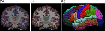
Structural images were analysed using FreeSurfer version 6.0, a software package that can be employed for cortical reconstruction which is documented and is freely available online (http://surfer.nmr.mgh.harvard.edu/). Preprocessing of the structural images is a multistep process including (but not limited to) skull stripping, bias field correction, grey–white matter segmentation and reconstruction of cortical surface models (grey–white boundary surface and pial surface). Results (grey and white boundary and the pial surface) for each participant were visually inspected to ensure accurate cortical surface reconstruction. No abnormalities were observed affecting the frontal and parietal areas therefore reconstruction was not required (Figure 2). Motion artefacts were minimum and did not require exclusion of MRI datasets from the study. Cortical structures were segmented using the Desikan–Killiany atlas (Desikan et al., Reference Desikan, Ségonne, Fischl, Quinn, Dickerson, Blacker and Killiany2006).
Preprocessing of MRI data using FreeSurfer. A–B. Inspection of cortical segmentation and surface reconstruction of images. C. Inspection of atlas parcellation using the Desikan–Killiany atlas.

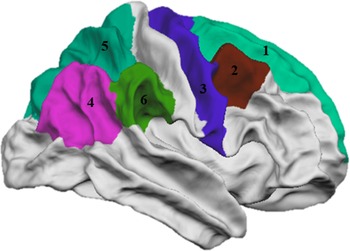
Surface area and cortical thickness were extracted from regions of interest (ROIs) which have been reported to be involved in motor planning, and/or the internal modelling process that is thought to subserve motor planning (viz MI) (Andersen & Cui, Reference Andersen and Cui2009; Hanakawa, Dimyan, & Hallett, Reference Hanakawa, Dimyan and Hallett2008; Zimmermann et al., Reference Zimmermann, Meulenbroek and de Lange2012). These included three regions from the frontal lobe; SFG (1), caudal MFG (2), PcG (3) and three from the parietal lobe; IPG (4), SPL (5) and SMG (6) from each hemisphere (Figure 3). The Desikan–Killiany atlas does not include cortical parcellation of the SMA, another area important for representing motor actions. As this area lies partly in the SFG and MFG, these two cortical regions have been included in the analyses. Further, regions of the temporal lobe parcellated from the Desikan–Killiany atlas were included as a comparison site as this particular region is generally not associated with motor planning.
Lateral view of the left hemisphere representing ROIs using Desikan–Killiany atlas. 1: Superior frontal gyrus, 2: Caudal middle frontal gyrus, 3: Precentral gyrus, 4: Inferior parietal gyrus, 5: Superior parietal lobule, 6: Supramarginal gyrus.

DWI
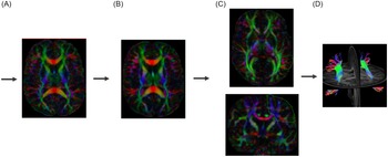
The DWI data were processed using ExploreDTI version 4.8.6. (Leemans, Jeurissen, Sijbers, & Jones, Reference Leemans, Jeurissen, Sijbers and Jones2009). The images were processed in two main steps: (1) evaluation of artefacts and (2) motion correction and quality assessment. During artefacts evaluation phase, images were visually inspected by looping through the images in different orthogonal views (i.e., axial, sagittal and coronal) to evaluate any obvious artefacts. The average residuals per DWI volume and across the DWIs for each voxel and the outlier profiles were also inspected. In the motion correction phase, the DWI datasets were corrected for subject (head) motion, eddy current induced distortions and geometric deformations due to susceptibility artefacts and the REKINDLE approach was taken to maximise accuracy (Figure 4A and 4B) (Tax, Otte, Viergever, Dijkhuizen, & Leemans, Reference Tax, Otte, Viergever, Dijkhuizen and Leemans2015). The same quality assessment procedure as described in the evaluation phase was performed to check the quality of the corrected DWIs. Major motion artefacts were observed in three datasets. The DWI images of these two participants were not included in the study leaving the final sample for the DWI study to 16 participants (M age = 10.01, SD = 1.07).
A. Raw DWI images are evaluated for artefacts and subjected to motion and eddy current induced distortions. B. CSD tractography are performed on all preprocessed images. C. The SLF is reconstructed using ‘AND’ gates. D. 3D representation of bilateral SLF.

Whole brain tractography deterministic CSD was conducted to the preprocessed DWI data in ExploreDTI. The following parameters were used: seedpoint resolution = 2 × 2 × 2 mm, angle threshold = 30°, fibre orientation distribution (FOD) = .05, fibre length range = 10–500 mm.
The protocol for extracting the SLF was based on the work of Wakana et al. (Reference Wakana, Caprihan, Panzenboeck, Fallon, Perry, Gollub and Mori2007). The SLF was manually delineated for all participants by RB. On the coronal slide, a large ‘AND’ gate was drawn gate was drawn at the posterior commissure line to capture trajectories passing through the parietal lobe. A second large ‘AND’ gate was drawn at the middle of the splenium of the corpus callosum identified by a midsagittal view on a coronal slice. These ‘AND’ gates include the core and all branches of the SLF (Figure 4C). To remove any spurious tracts ‘NOT’ gates were applied as required. The reconstructed bilateral SLF is presented in Figure 4D. The diffusion metric, mean AFD, was extracted from the left and right SLF for all participants. Reconstruction of the SLF for two participants was checked by CH.
Statistical Analyses
The tests of normality indicated that not all behavioural data and brain metrics met the assumptions of normality. To maintain consistency across reporting of results, appropriate nonparametric tests were used. To investigate the relationship between the dependent variable, ESC and the macrostructural properties (cortical thickness and surface area) of each of the ROIs, separate nonparametric partial (Spearman’s Rho) correlations were conducted for both colour sequences. Since the current sample consisted of participants aged between 8 and 11 years, age was controlled in these analyses. Similar tests were conducted to look at the associations between the bilateral SLF and ESC for both colour sequences; again age was controlled for. To explore the associations between the grey and white matter, partial correlations were run for significant cortical areas and white matter bundles while controlling for age. In order to ensure specificity of any effects observed when investigating the relationship between putative motor planning structures and complex motor planning performance, the relationship between cortical thickness and surface area of the temporal lobe (a region typically not associated with higher order motor functions such as the planning of complex movement) and the percentage of trials terminating in ESC were conducted bilaterally. All statistical analyses were conducted using SPSS 26. Type 1 error was controlled using the false discovery rate (FDR) using a family-wise error rate approach (Benjamini & Hochberg, Reference Benjamini and Hochberg1995). Using the family-wise error rate approach, the p-values for the left and right hemisphere for each ROI were controlled using the FDR approach for cortical thickness and cortical surface areas separately. Alpha was set at .05 for all correlational analyses and adjusted p-values were reported for significant associations.
Results
ESC
Descriptive analyses on motor planning indicated that overall children (n = 19) had a lower median score on the two colour sequence (median = 5.02, interquartile range = 1.26) compared to their performances on the one colour sequence (median = 3.81, interquartile range = .85).
Associations between Brain Morphology and ESC for One Colour Sequence
No significant correlations were observed between ESC for the one colour sequence and the cortical ROIs when controlling for age. While a negative relationship was observed between cortical surface area of the right SPL and ESC for the one colour sequence (r = −.51, p = .032), the effect did not survive the FDR correction (FDR adjusted p = .064). No significant association was found between cortical thickness of bilateral temporal lobe and ESC for the one colour sequence.
Association between Brain Morphology and ESC for Two Colour Sequence
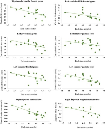
In Table 1, the associations between the cortical thickness and cortical surface area of the ROIs of both hemispheres with ESC for two colour sequence are summarised. Results indicated that, after correcting for multiple comparisons and age, significant associations were observed between the cortical thickness of the left hemisphere for MFG (r = −.553, p = .017), SFG (r = −.542, p = .04), PcG (r = −.562, p = .03), inferior parietal lobe (IPL) (r = −.551, p = .036) and the SPL (r = −.695, p = .002) and ESC for the two colour sequence. The right MFG was also negatively associated with ESC (r = −.557, p = .032) which meant that decreased cortical thickness of the right MFG was associated with a greater end state discomfort. When looking at cortical surface area, significant negative associations were observed for the SPL and ESC for the two colour sequences of the right hemisphere only (r = −.669, p = .004). Cortical surface area of bilateral temporal lobe was not significantly associated with ESC for the two colour sequence. The visual representations are shown in Figure 5.
Correlations between cortical thickness and cortical surface area of ROIs and ESC for the one and two colour sequence

Note. * Adjusted p-values < .05; ** adjusted p-values < .01; FDR corrected.
Scatterplots representing significant associations between end-state-comfort for the two colour sequence and cortical thickness and cortical surface area and the superior longitudinal fasciculus (SLF).

Associations between the SLF and ESC
After correcting for multiple comparison and age, a negative correlation between ESC for the two colour sequence and AFD of the right SLF (r = −.591, p = .020) which suggested that poorer motor planning (as per the end state discomfort rating) associated with greater AFD (Figure 5). No relationship was observed between ESC and the SLF for the one colour sequences (Table 2).
Correlations between the SLF, ESC and cortical measures of grey matter

Note. * Adjusted p-values < .05; FDR corrected.
Association between Significant Cortical Areas and Left SLF
A significant positive association was observed between cortical thickness of the left SFG and the right SLF (r = .530, p = .042) while controlling for age. No other significant correlations were observed between morphological metrics and AFD within the SLF (Table 2).
Discussion
The aim of this study was to probe the neurostructural basis of motor planning in children using a multimodal approach. Overall, children performed poorer on the two colour sequences compared to the one colour sequence. This finding is in line with previous studies that reported on motor planning efficiency using the octagon task in children of similar age group (Bhoyroo et al., Reference Bhoyroo, Hands, Wilmut, Hyde and Wigley2018, Reference Bhoyroo, Hands, Wilmut, Hyde and Wigley2019; Wilmut & Byrne, Reference Wilmut and Byrne2014a, Reference Wilmut and Byrne2014b). Significant associations between motor planning efficiency and several cortical areas of the frontoparietal lobe as well as the right SLF were observed. Cortical thickness of bilateral MFG, left SFG, left PcG, left IPL and left SPL were correlated negatively with motor planning of the two colour sequences. As higher scores on the ESC rating indicate lower motor planning capacity, this suggested that a decrease in motor planning efficiency is associated with cortical thinning of the abovementioned frontoparietal areas. With regards to the white matter tracks, the observed negative relationship between motor planning rating scores of the two colour sequence and AFD suggested that poorer motor planning efficiency was associated with increased AFD in the right SLF. Further, results indicated that an increase in the left cortical thickness of the SFG was associated with increased AFD of the right SLF. As no other correlations were found, including between motor planning efficiency and morphology of the comparison region (the temporal lobe), these effects cannot, at least entirely, be attributed to general neurostructural effects. Taken together, in childhood motor planning, areas of both the frontal and parietal cortex are recruited for complex motor planning. More broadly, these data indicate that morphology within frontodorsal circuity, and the white matter bundles that support communication between them, may be associated with individual differences in childhood motor planning.
The Frontal Lobe and Motor Planning
Cortical thickness of the frontal lobe that include the MFG, SFG and the PcG were related with individual differences on complex motor planning in children. More specifically, greater cortical thickness was associated with a stronger tendency to plan the octagon task. Of note, cortical surface area of none of the implicated regions of the frontal lobe correlated with motor planning. These findings are broadly consistent with neuropsychological accounts of motor planning, and action sequencing based on adult studies. Further, the findings of this cross-sectional study support that of Langevin and colleagues who showed that thicker cortical areas are associated with improved task performances in children and adolescents (Langevin et al., Reference Langevin, MacMaster and Dewey2015). Indeed, the MFG and SFG form part of the dorsolateral prefrontal cortex (DLPFC; BA 6/10/46), and have previously been implicated in the ‘planning phase’ of action in adults (Omata et al., Reference Omata, Ito, Takata and Ouchi2018). The DLPFC is considered to operate as the center of executive function in complex cognitive tasks such as making plans for the future (Gilbert & Burgess, Reference Gilbert and Burgess2008). The DLPFC also plays a critical role in controlling deliberate and intentional behaviour (Fassbender et al., Reference Fassbender, Murphy, Foxe, Wylie, Javitt, Robertson and Garavan2004; Wagner, Maril, Bjork, & Schacter, Reference Wagner, Maril, Bjork and Schacter2001). Indeed, the two colour sequence used in the present study is more complex than the one colour sequence considering that participants have to choose a particular grip point from a eight options so that they can make two rotations (clockwise and/or anticlockwise) to reach the final allocated colour. Given the importance of the DLPFC in controlling behaviour, and organizing complex behaviours (both motor and cognitive), the observed relationships between cortical thickness in the DLPCF and complex motor planning efficiency are consistent with neuropsychological accounts of executive and motor control. Thicker MFG, SFG and PcG may relate to better movement choices resulting appropriate planning of motor actions.
In addition, the PcG which is located in primary motor cortex is involved in hand movements (and limb control more broadly) and is activated more often during performance of complex motor tasks (Chen, Gerloff, Hallett, & Cohen, Reference Chen, Gerloff, Hallett and Cohen1997; Hanakawa et al., Reference Hanakawa, Dimyan and Hallett2008; Verstynen & Ivry, Reference Verstynen and Ivry2011). Completing the two colour sequence involved complex hand movements as the performer might need to sacrifice comfort at the beginning of the task when grasping the dial by turning the pointer to one colour at a time until the end colour is reached. This is in line with involvement of the PcG, an area that is recruited during performance of complex movement tasks. Altogether our results support the concept that motor planning ability is related to alterations with brain morphology of the frontal lobe in children, and supports evidence from adult studies.
The Parietal Lobe and Motor Planning
A strong association was observed between planning of movement sequences and areas of the parietal lobe that include SPL and IPL. Briefly, the SPL and broader posterior parietal cortex (PPC), is critical for generating limb to body reference frames, as well as the visuomotor transformation necessary for planning of upper limb movements (Battaglia-Mayer & Caminiti, Reference Battaglia-Mayer and Caminiti2018). The observed relationship between complex motor planning and brain morphology within the PPC is in line with neurocomputational accounts of motor planning with several functional MRI studies in adults highlight the importance of the parietal regions in motor planning in healthy population (Andersen & Cui, Reference Andersen and Cui2009; Beurze, de Lange, Toni, & Medendorp, Reference Beurze, de Lange, Toni and Medendorp2007; Tunik, Lo, & Adamovich, Reference Tunik, Lo and Adamovich2008; Zimmermann et al., Reference Zimmermann, Meulenbroek and de Lange2012). Interestingly, the left SPL as well as the IPL are known for their involvement in MI (Hétu et al., Reference Hétu, Grégoire, Saimpont, Coll, Eugène, Michon and Jackson2013), a process thought to provide insight into one’s capacity to engage internal models thought to support motor planning (Shadmehr & Krakauer, Reference Shadmehr and Krakauer2008; Wolpert & Flanagan, Reference Wolpert and Flanagan2001). This study is the first to show that similar to healthy adults, children may rely on the parietal lobe during motor planning. In addition, as the SPL is broadly associated with MI, these findings provide support for computational modelling of action planning, and demonstrate that similar to adults, the same effects may be present in children.
The SLF and Movement Planning
The right SLF was associated with motor planning efficiency of complex movement sequences. In typically developing children and adolescents, it has been shown that the right SLF correlates with attention (Urger et al., Reference Urger, De Bellis, Hooper, Woolley, Chen and Provenzale2015). While general cognitive demands likely increase from one to two sequence tasks, task instructions remain relatively simple meaning that demands on peripheral cognitive processes would be expected to be minimal (e.g., working memory). Instead, the principal factor driving the increase in task difficulty reflects the need to accommodate two, rather than one, target location in the upcoming movement and relies more heavily on executive functioning, our findings support the relationship between the right SLF and a complex motor task. The SLF is a major white matter tract that covers extensive areas of frontal and parietal lobe, several regions within which an association between cortical morphology and motor planning efficiency was observed. Thus, our findings support the broader literature that demonstrates the pivotal role of the frontoparietal network for motor control (Budisavljevic et al., Reference Budisavljevic, Dell’Acqua, Zanatto, Begliomini, Miotto, Motta and Castiello2017; Filimon, Reference Filimon2010; Parlatini et al., Reference Parlatini, Radua, Dell’Acqua, Leslie, Simmons, Murphy and Thiebaut de Schotten2017). The SLF connects regions in the frontoparietal cortices (Thiebaut de Schotten et al., Reference Thiebaut de Schotten, Ffytche, Bizzi, Dell’Acqua, Allin, Walshe and Catani2011) and previous tractography studies revealed that the SPL is connected to the ventral premotor network and the primary motor network by bundles of the SLF (Hecht, Gutman, Bradley, Preuss, & Stout, Reference Hecht, Gutman, Bradley, Preuss and Stout2015). The SFG holds the premotor networks. In the present study, the frontal and parietal regions were associated with motor planning and connections between the SLF and these regions are crucial for planning reach-to-grasp tasks (Koch et al., Reference Koch, Cercignani, Pecchioli, Versace, Oliveri, Caltagirone and Bozzali2010). The observed relationship between the SLF and motor planning highlights the importance of considering the broader frontodorsal network, and connectivity therein, in order to understand the structural basis of childhood differences in motor planning efficiency.
Strengths, Limitations and Future Research
Using a multimodal design, we provide evidence of the involvement of the frontoparietal cortical regions as well as the white matter bundles that facilitate transmission of information between them in contributing to individual differences in motor planning efficiency in school-aged children. This approach demonstrates the importance of examining the involvement of the frontoparietal network rather than focusing individually on grey or white matter structures when seeking to understand the brain basis of motor planning. High angular resolution diffusion imaging (HARDI) sequence and CSD tractography were used in this study. These advanced DWI parameters are more robust and less susceptible to ‘crossing’ fibres problems compared to traditional methods such as diffusion tensor imaging when involving white matter tracts such as the SLF (Farquharson et al., Reference Farquharson, Tournier, Calamante, Fabinyi, Schneider-Kolsky, Jackson and Connelly2013; Raffelt et al., Reference Raffelt, Tournier, Rose, Ridgway, Henderson, Crozier and Connelly2012). Another strength of this study was the adoption of manual tracing which increased accuracy during reconstruction compared to using a generic template for automatic tracing of the white matter tract. Several limitations are also acknowledged. Even though, robust statistical techniques were used, findings are preliminary given the modest sample size and should be replicated with a larger sample. Further, the design of the study was correlational, and did not allow one to infer causality. Therefore, it is still unknown whether cortical thickness and/or surface area affects planning ability or vice versa. This may be addressed using longitudinal designs.
Conclusion
This study was the first to probe the neural basis of individual differences in childhood motor planning efficiency using a multimodal approach. Findings support the neurocomputational accounts of motor planning based on adult data, suggesting that morphology within frontodorsal circuits may be associated with planning efficiency, as shown by a greater tendency to plan complex motor tasks. Interestingly, microstructure within the SLF, a white matter bundle that supports communication between frontoparietal regions, is also associated with planning efficiency. The multimodal approach allowed identification of the importance of the broader frontoparietal structural networks in childhood motor efficiency.
Acknowledgments
Heartfelt thanks go to all participants and their families. We would also like to specially thank Dr. Kate Wilmut for her help with the octagon task. Finally, thanks to Dr. Katrina Spilsbury for her statistical input.
Author Contributions
Beth Hands and Karen Caeyenberghs contributed equally to this work.
Financial Support
This research was funded by Collaborative Research Network.
Conflict of Interest
The authors have nothing to disclose.