INTRODUCTION
Schistosomiasis is a chronic parasitic disease which affects more than 200 million people, being endemic in 76 countries around the world and causing approximately 200 000 deaths per year (Chitsulo et al. Reference Chitsulo, Loverde and Engels2004). The pathology characteristic of this disease is a granulomatous reaction around parasite eggs within the liver and other organs (Boros, Reference Boros1989). Currently, chemotherapy is the control strategy used; however, large extension of endemic areas and constant reinfection of individuals together with poor sanitary conditions in the tropical countries make drug treatment inefficient (Bergquist, Reference Bergquist1995). Therefore, a vaccine that induces even a partial reduction in worm burden could considerably reduce pathology and limit parasite transmission (Chitsulo et al. Reference Chitsulo, Loverde and Engels2004), contributing enormously to disease control. Several studies are in progress in this field, testing different antigens of the parasite and different vaccination strategies (Bergquist et al. Reference Bergquist, Al-Sherbiny, Barakat and Olds2002; Wilson and Coulson, Reference Wilson and Coulson2006; Bethony et al. Reference Bethony, Diemert, Oliveira and Loukas2008; Oliveira et al. Reference Oliveira, Fonseca, Cardoso, Farias and Leite2008).
At the beginning of the 1990s, the WHO selected 6 antigens as candidates to compose a subunit vaccine against schistosomiasis. More recently, 2 tegument antigens Sm-TSP2 (Tran et al. Reference Tran, Pearson, Bethony, Smyth, Jones, Duke, Don, McManus, Correa-Oliveira and Loukas2006) and Sm29 (Cardoso et al. Reference Cardoso, Macedo, Gava, Kitten, Mati, De Melo, Caliari, Almeida, Venancio, Verjovski-Almeida and Oliveira2008) induced around 50% protection against experimental infection and became promising antigens to be used as a schistosome vaccine (Hotez et al. Reference Hotez, Bethony, Oliveira, Brindley and Loukas2008; Oliveira et al. Reference Oliveira, Fonseca, Cardoso, Farias and Leite2008). The sequencing of the S. mansoni transcriptome (Verjovski-Almeida et al. Reference Verjovski-Almeida, DeMarco, Martins, Guimaraes, Ojopi, Paquola, Piazza, Nishiyama, Kitajima, Adamson, Ashton, Bonaldo, Coulson, Dillon, Farias, Gregorio, Ho, Leite, Malaquias, Marques, Miyasato, Nascimento, Ohlweiler, Reis, Ribeiro, Sa, Stukart, Soares, Gargioni, Kawano, Rodrigues, Madeira, Wilson, Menck, Setubal, Leite and Dias-Neto2003) and the development of proteomic and microarray technologies have dramatically improved the possibilities for identifying novel vaccine candidates (Liu et al. Reference Liu, Lu, Hu, Wang, Cui, Chi, Yan, Wang, Song, Xu, Wang, Zhang, Zhang, Wang, Xue, Brindley, McManus, Yang, Feng, Chen and Han2006; Verjovski-Almeida and DeMarco, Reference Verjovski-Almeida and DeMarco2008), particularly proteins that are secreted or anchored on the surface of schistosomes. Recently, Braschi et al. (Reference Braschi, Curwen, Ashton, Verjovski-Almeida and Wilson2006) identified and characterized the major proteins of the apical membranes of the Schistosoma mansoni tegument, one of them (Accession number C608350.1) was selected by our group as the focus of this study.
Herein, we have identified in a S. mansoni adult-worm cDNA library a gene encoding a protein of unknown function, which was previously identified on the tegument (membrane anchored) of the parasite through proteomic analysis (Braschi et al. Reference Braschi, Curwen, Ashton, Verjovski-Almeida and Wilson2006). In this present study, its amino acid sequence was analysed by bioinformatics and shown to have 73% similarity with a hypothetical S. japonicum protein that also possesses an immunoglobulin domain. Termed here SmIg, it was produced as a recombinant protein and evaluated as a potential candidate vaccine against S. mansoni infection in a murine model. Mice immunized with rSmIg failed to reduce worm burden but diminished liver pathology.
MATERIALS AND METHODS
Mice and parasites
Female C57BL/6 mice aged 6–8 weeks were purchased from the Federal University of Minas Gerais (UFMG) animal facility. Cercariae of S. mansoni were maintained routinely on Biomphalaria glabrata snails at CPqRR (Centro de Pesquisa René-Rachou-Fiocuz) and prepared by exposing infected snails to light for 1 h to induce shedding. Cercarial numbers and viability were determined using a light microscope prior to infection. All protocols involving animals were approved by the local Ethics Committee on Animal Care (CETEA – UFMG No. 023/05).
SmIg cDNA isolation and amino acid sequence analysis
The SmIg cDNA sequence was obtained from the Minas Gerais Genome Network by searching homologue proteins to the tegumental protein Accession number C608350.1 (http://cancer.lbi.ic.unicamp.br/schisto6/; Braschi et al. Reference Braschi, Curwen, Ashton, Verjovski-Almeida and Wilson2006). Analysis of the SmIg deduced amino acid sequence was performed using ExPASy (Expert Protein Analysis System) computer program (Gasteiger et al. Reference Gasteiger, Gattiker, Hoogland, Ivanyi, Appel and Bairoch2003). The signal peptide was identified using the SignalP 3.0 (Nielsen et al. Reference Nielsen, Engelbrec, Brunak and Von Heijne1997). Transmembrane protein topology prediction was analysed by SOSUI (http://sosui.proteome.bio.tuat.ac.jp/sosuiframe0.html). N-glycosylation and O-glycosylation sites were analysed using the NetNGlyc 1.0 (www.cbs.dtu.dk/services/NetNGlyc/) and YinOYang (www.cbs.dtu.dk/services/YinOYang), respectively. To identify sequences similar to SmIg we used the BLAST network server at the National Center for Biotechnology Information (NCBI). The search for conserved domains was performed using InterPro Scan, which integrates searches in the PROSITE, Pfam and PRINTS (http://www.ebi.ac.uk/InterProScan/) Databases.
Subcloning of the S. mansoni SmIg cDNA
The cDNA coding for SmIg (Accession no. GQ149769) without the signal peptide and the C-terminal transmembrane domain was amplified by PCR from a S. mansoni adult-worm cDNA library using the following primers: 5′-CCGGGATCCAAACTTGAAG TTGACTTTGAT-3′ (sense orientation) and 5′-CGTATTAAAAGCAAATTGGCTATGAATTCGCC-3′ (antisense orientation) which include, respectively, BamHI and EcoR I sites (underlined). The parameters for the PCR reaction were as follows: 95°C, 3 min, 1 cycle; 95°C, 30 s, 56°C, 30 s, 72°C, 2 min, 35 cycles; 72°C, 10 min, 1 cycle. The PCR fragment (650 pb) corresponding to the amplified SmIg cDNA was isolated from agarose gels using the ConcertTM Rapid Gel Extraction System (Gibco BRL) and digested with BamHI and EcoR I according to the manufacturer's recommendations. The digested cDNA was subcloned into pET21a expression vector that had been previously digested with these same restriction enzymes, generating a pET21aSmIg construct. This constructs was transformed into E. coli electrocompetent cells using the gene Pulser SystemTM (Bio-Rad) according to standard procedures (Sambrook et al. Reference Sambrook, Fritsch and Maniatis1989). Escherichia coli transformants harbouring the constructed plasmids were screened on LB agar plates containing ampicillin (100 μg/ml). DNA sequencing was performed to confirm the presence and the correct orientation of the SmIg cDNA.
SmIg transcript analysis in parasite life-stage by real-time PCR
Absolute quantities of SmIg mRNA in various life-cycle stages of S. mansoni were determined using a real-time RT-PCR analysis. Total RNA was prepared from S. mansoni eggs, cercariae, lung-stage schistosomulum, and separated male and female adult worms. Total RNA was isolated using Trizol (Invitrogen) and the cDNA were synthesized using the Superscript III Reverse Transcriptase (Invitrogen) in the presence of oligo(dT), according to the manufacturer's instruction. RT reactions without reverse transcriptase were used as negative controls. Target sequences were amplified from the prepared cDNA templates using Power SYBR green PCR master mix (Applied Biosystems) and the SmIg primer pair (5′TGCATATCAAAGTGCCAACC 3′ and 5′ Ctgatttggctctgggaaac 3′). Real-Time RT-PCR was performed with ABI 7900 Real-Time PCR System (Applied Biosystems), using the following cycling parameters: 60°C for 10 min, 95°C for 10 min, 40 cycles of 95°C for 15 sec and 60°C for 1 min, and a dissociation stage of 95°C for 15 sec, 60°C for 1 min, 95°C for 15 sec, 60°C for 15 sec. Primers were used to amplify a specific 200 bp fragment corresponding to the specific gene. All PCR reactions were carried out in triplicate wells of a 96-well microamp optical plate (Applied Biosystems). A standard curve generated using different concentrations (0·1, 0·01, 0·001 and 0·0001 ng) of SmIg plasmid was used for quantitative determination of SmIg in the samples. The differences in the absolute expression of SmIg among the stages were analysed by Student's t-test where P<0·05 was considered statistically significant.
Expression and purification of recombinant SmIg
Recombinant SmIg (26–243) amino acid fragment was expressed in E. coli with an in-frame six-histidine N-terminal tag using the pET21a expression vector. An E. coli BL21DE03 culture (1L) containing the recombinant plasmid was grown at 37°C to an optical density at 600 nm of 0·5–0·8, and the expression of rSmIg was induced by 1 mM IPTG. After 4 h of induction, the bacterial cells were harvested by centrifugation at 4000 g for 20 min. The pellet was resuspended in 50 ml of 10 mM Na2HPO4, 10 mM NaH2PO4, 0·5 m NaCl and 10 mM imidazole. Subsequently, the cells were submitted to 3 cycles of sonication lasting 30 s each and centrifuged at 5400 g for 20 min. The rSmIg was recovered as inclusion bodies and solubilized in 50 ml of 8 M urea, 10 mM Na2HPO4, 10 mM NaH2PO4, 0·5 M NaCl, and 10 mM imidazole. The protein was purified by affinity chromatography on a Ni-Sepharose column (Hitrap FF crude 5 ml) under denaturing conditions using an AKTAexplorer chromatograph (GE Healthcare), according to the manufacturer's protocol. Fractions containing rSmIg (approximately 400 μg/ml) were dialysed against decreasing concentrations of urea (6 M, 4 M, 3 M, 2 M, 1·5 m, 1·0 M, 0·75 M, 0·5 M, 0·25 M) in PBS buffer containing 10% glycerol, 50 mM glycine, pH 8·0, followed by dialysis against PBS buffer. The dialysis was carried out at 4°C using a Spectra/Por2 membrane (MWCO 6 to 8 kDa; Spectrum Medical Industries, Inc., Laguna Hills, CA, USA). Protein concentration was quantified using the Bradford's method (Bradford, Reference Bradford1976). This recombinant protein was used as antigen for immunization and immunological experiments. The level of LPS contamination was tested using a commercially available LAL Chromogenic Kit. The concentration of LPS found in rSmIg was 1·32 EU/ml.
SDS-PAGE and immunobloting
SDS-PAGE of purified rSmIg was performed (Laemmli, Reference Laemmli1970). The gel was electroblotted onto nitrocellulose membrane (Towbin et al. Reference Towbin, Staehelin and Gordon1979). The membrane was blocked with TBST (0·5 M NaCl−0·02 m Tris (pH 7·5), 0·05% Tween 20) containing 5% dry milk for 16 h at room temperature. Subsequently, the membrane was incubated in a 1:3000 dilution of mouse alkaline phosphatase (AP) conjugated anti-6xHIS antibodies (Invitrogen) in TBST plus 5% dry milk for 3 h at room temperature. After 3 washes using TBST, the membrane was treated with AP reaction-developing buffer containing nitroblue tetrazolium (NBT) and 5-bromo-4-chloro-3-indolyl-1-phosphate (BCIP). After the reaction developed, the membrane was washed using distilled water and dried in filter paper.
Immunization of mice
Six to eight-week-old female C57BL/6 mice were divided into 2 groups of 10 mice each. Mice were subcutaneously injected in the nape of the neck with 25 μg of rSmIg on days 0, 15 and 30. The recombinant protein was formulated diluted in 100 μl of PBS plus 100 μl of Complete Freund's Adjuvant (CFA) in the first immunization and 100 μl of Incomplete Freund's Adjuvant (IFA) in the subsequent immunizations. In the control group, PBS with Freund's adjuvant was administered using the same immunization protocol.
Challenge infection and worm burden recovery
Fifteen days after the last boost, mice were challenged through percutaneous exposure of abdominal skin for 1 h in water containing 100 cercariae (LE strain). Forty-five days after challenge, adult worms were perfused from the portal veins (Fonseca et al. Reference Fonseca, Brito, Alves and Oliveira2004). Two independent experiments were performed to determine protection levels. The protection was calculated by comparing the number of worms recovered from each vaccinated group with its respective control group, using the formula:

where PL=protection level, WRCG=worms recovered from control group, and WREG=worms recovered from experimental group.
Measurement of specific anti-SmIg antibodies
Following immunization, sera of 10 mice from each rSmIg vaccinated or control group were collected at 2-week interval. Measurement of specific anti-SmIg antibodies was performed using indirect ELISA. Maxisorp 96-well microtitre plates (Nunc, Denmark) were coated with 5 μg/ml rSmIg in carbonate-bicarbonate buffer, pH 9·6 for 16 h at 4°C, then blocked for 2 h at room temperature with 200 ml/well PBST (phosphate buffered saline, pH 7·2, with 0·05% Tween-20) plus 10% FBS (fetal bovine serum). Then 100 μl of each serum, diluted 1:100 in PBST, were added per well and incubated for 1 h at room temperature. Plate-bound antibody was detected by peroxidase-conjugated anti-mouse IgG, IgG1 and IgG2a (Sigma) diluted in PBST 1:10000, 1:5000 and 1:2000, respectively. Colour reaction was developed by addition of 100 ml per well of 200 pmol OPD (o-phenylenediamine, Sigma) in citrate buffer, pH 5·0, plus 0·04% H2O2 for 10 min and stopped with 50 ml of 5% sulfuric acid per well. The plates were read at 495 nm in an ELISA plate reader (Bio-Rad, Hercules, CA, USA).
Cytokine analysis
Cytokine experiments were performed using splenocyte cultures from individual mice immunized with rSmIg plus CFA/IFA (n=5 for each group). Splenocytes were isolated from macerated spleen of individual mice 1 week after the third immunization and washed twice with sterile PBS. After washing, the cells were adjusted to 1×10⁶ cells per well for IL-4, IL-10, IFN-γ and TNF-α assays in RPMI 1640 medium (Gibco) supplemented with 10% FBS, 100 U/ml of penicillin G sodium, 100 μg/ml of streptomycin sulfate, 250 ng/ml of amphotericin B. Splenocytes were maintained in culture with medium alone or stimulated with rSmIg (25 μg/ml) or concanavalin A (ConA) (5 μg/ml) as previously described (Fonseca et al. Reference Fonseca, Brito, Alves and Oliveira2004; Pacifico et al. Reference Pacifico, Fonseca, Barsante, Cardoso, Araujo and Oliveira2006). The 96-well plates (Nunc) were maintained in an incubator at 37°C with 5% CO2. For cytokine assays, polymyxin B (30 μg/ml) was added to the cultures and this treatment completely abrogated the cytokine response to LPS as previously described by our group (Cardoso et al. Reference Cardoso, Araujo, Goes, Pacifico, Oliveira and Oliveira2007). Culture supernatants were collected after 24 h of ConA stimulation, 48 h of rSmIg stimulation for IL-4 and TNF-α analysis and 72 h of rSmIg stimulation for IL-10 and IFN-γ. The assays for measurement of IL-4, IL-10, IFN-γ and TNF-α were performed using the Duoset ELISA kit (R&D Diagnostic, Minneapolis, MN, USA) according to the manufacturer's directions.
Histopathological analysis
Following perfusion for the recovery of the schistosomes, liver sections from mice (8/group) of control and experimental groups were collected to evaluate the effect of immunization in granuloma formation. The liver sections removed from the central part of the left lateral lobe were fixed with 10% buffered formaldehyde in PBS. Histological sections were cut using a microtome at a thickness of 4 μm and stained on a slide with haematoxylin-eosin (HE), and Picrosirius. To perform measurements of the granuloma volume and the fibrosis area (collagen deposition), 80 granulomas from each group of 8 mice with a single well-defined egg were randomly chosen at a microscope with 10×objective lens followed by analysis in the KS300 software connected to a Carl Zeiss image analyser. The fibrosis area was quantified using the image segmentation function. All pixels of collagen zones in Picrosirius sections were selected to build a binary image, subsequently calculating the total area occupied by connective tissue in the granuloma, expressed in square micrometers (μm2). Granuloma liver volume was estimated by point-counting stereology analysis in 6 images of each liver section in 100 grids points generated by KS300 software and laid over each image for point counting according to Bartley et al. (Reference Bartley, Ramm, Jones, Ruddel, Li and McManus2006).
Statistical analysis
Statistical analysis was performed with Student's t-test for using the software package GraphPad Prism (La Jolla, CA, USA).
RESULTS
In silico analysis of predicted SmIg amino acid sequence
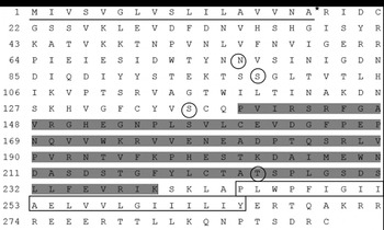
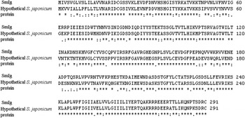
The deduced amino acid sequence for SmIg is 246 residues long composed by a signal peptide of 17 amino acids with the site of cleavage between Ala17 and Arg18 (Fig. 1). Four sites with high probability of glycosylation are present on the Asn76, Ser98, Ser136 and Thr224. Prediction of transmembrane helix using SOSUI revealed a C-terminal sequence between Pro244 and Tyr265 exhibiting high hydrophobicity. The search for conserved motifs showed an immunoglobulin conserved domain at the C-terminus (Fig. 1). Additionally, the search for similarity with sequences deposited in the data-base of the NCBI using Blastp algorithm showed that SmIg has 73% of similarity with a hypothetical Schistosoma japonicum protein with unknown function, which also presents an immunoglobulin domain (Fig. 2).
In silico analysis of amino acids sequences predicted for SmIg. SmIg is composed by a signal peptide (underline) with the signal cleavage site (*), four glycosylation sites (circles), an immunoglobulin domain at C-terminus (grey bar) and a transmembrane helix (box).

Amino acid sequence comparison between SmIg and a hypothetical protein from Schistosoma japonicum with Ig domain [gb|AAP05938.1|]. Identity (*). Strongly similar (:). Weakly similar (.).

SmIg transcript analysis in parasite life-stages
Absolute quantities of SmIg cDNA were determined by real-time RT-PCR. Ct values of standards obtained with SmIg primers and probes were inversely proportional to the amounts of cDNA template tested and reaction efficiency was close to 95% (slope: −3·54; R2: 0·996). The amount of SmIg cDNA present in parasite cDNA samples was then determined using the standard curve. These results, shown in Fig. 3, confirmed that SmIg is expressed in adult worms female and male (0·56 and 0·67 pg of SmIg, respectively), cercariae (0·087 pg of SmIg) and schistosomulum (0·73 pg of SmIg) and at low levels in egg stage (0·00036 pg of SmIg). As SmIg transcripts were detected in the lung-stage cDNA library and the lung seems to be the major site of parasite elimination during the irradiated cercariae model of vaccination (Coulson and Wilson, Reference Coulson and Wilson1997), to test SmIg as a vaccine candidate would be an interesting approach for development of schistosomiasis control measures.
Analysis of SmIg expression in Schistosoma mansoni life-stages by real-time RT-PCR. A bar graph comparing the absolute quantities of SmIg (shown as pg mRNA) in different developmental stages of S. mansoni. The following developmental stages were tested: egg, cercariae, lung-stage schistosomulum, female and male adult worms. Data presented are the mean from 3 similar experiments. Statistically significant difference compared to egg stage is denoted by an asterisk for P<0·05.

Production of recombinant SmIg
Recombinant SmIg was expressed as inclusion bodies and purified under denaturing conditions in 8 M urea by affinity chromatography. The refolding resulted in a great lost of protein in a precipitated form, but yielded sufficient soluble protein to be used for immunization of mice (approximately 1·8 mg prot/l). The expression and purity of SmIg as a 6xHis tag fusion protein were checked by SDS-PAGE and Western blotting analysis (Fig. 4), which revealed a protein with approximately 30 kDa corresponding to the estimated molecular mass for the rSmIg (~28 kDa).
Expression and purification of recombinant SmIg-6xHis fusion protein. (a) Coomasie blue-stained SDS-12%-PAGE profile of E.coli expressing the pET21aSmIg construct. Lanes: 1, lysate of induced culture; 2, lysate of uninduced culture; 3, molecular weight marker (kDa). (b) Coomasie blue-stained SDS-12%-PAGE profile of the purified recombinant SmIg. (c) Western blot analysis of the purified recombinant protein using anti 6xHIS antibody. Arrows indicate recombinant SmIg fusion protein (~28 kDa).

Antibody profile following immunization of mice
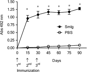
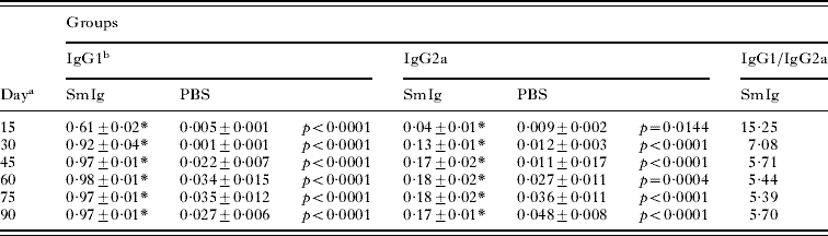
In order to assay for specific IgG, IgG1 and IgG2a antibodies to SmIg, sera from 10 vaccinated animals of each group were tested by ELISA. rSmIg vaccinated mice developed significant levels of specific anti-SmIg antibodies after the first immunization (Fig. 5). Regarding IgG1 and IgG2a, mice immunized with rSmIg showed statistically significant titers of both isotypes compared to control group. However, we detected higher levels of IgG1 compared to IgG2a (Table 1). The IgG1/IgG2a ratio observed in mice immunized with rSmIg decreased over time and it is indicative of a mixed Th1/Th2 type of immune response.
Kinetics of specific anti-SmIg IgG induced in mice immunized with recombinant SmIg. Sera of 10 immunized mice per group were collected at days 15, 30, 45, 60, 75 and 90 after the first immunization and assayed by ELISA. Arrows indicate the timing of vaccination. Results are presented as the mean absorbance measured at 492 nm for each group. Results represent the mean of 2 independent experiments. Statistically significant differences of recombinant SmIg vaccinated mice compared to PBS control group is denoted by one asterisk for (P<0·05).

IgG1 and IgG2a immune profile induced by vaccination with recombinant SmIg
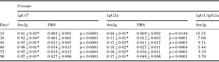
a Days after the first immunization.
b IgG1 and IgG2a numbers represent optical density (O.D.).
* Statistically significant compared to group of animals immunized with PBS with p<0·05.
Cytokine profile
Cytokine production was evaluated in culture supernatants of in vitro-stimulated spleen cells of rSmIg-immunized mice. Statistically significant amounts of IFN-γ, TNF-α, IL-10 and IL-4 were detected in splenocyte culture supernatants of rSmIg-vaccinated mice compared to the control group (Fig. 6). rSmIg immunization induced production of IFN-γ (9217±785·3 pg/ml), IL-10 (3996±202·7 pg/ml), TNF-α (1280±83·4 pg/ml) and IL-4 (59·4±4·6 pg/ml). These results show that immunization with rSmIg induces a Th1/Th2 type of immune response in mouse T cells, characterized by the production of IFN-γ and TNF-α (Th1) and IL-4 (Th2) cytokines.
Cytokine profile of mice immunized with recombinant SmIg. One week after the last immunization, splenocytes were isolated and assayed for IFN-γ, TNF-α, IL-4 and IL-10 production in response to recombinant SmIg (25 μg/ml) or medium alone as a control. The results are presented as mean±S.D. for each group. A statistically significant difference of recombinant SmIg-vaccinated mice compared to control group is denoted by an asterisk for (P<0·05).

Worm burden recovery
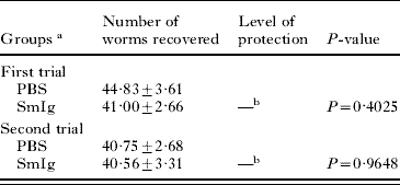
Protective immunity induced by vaccination with recombinant SmIg was evaluated 45 days after challenge with 100 S. mansoni cercarie. Mice vaccinated with rSmIg showed no statistically significant reduction in worm burden recovery compared to control mice (Table 2).
Protection level induced by rSmIg immunization in C57BL/6 mice challenged with 100 S. mansoni cercariae
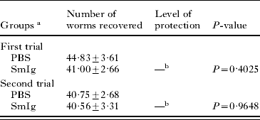
a Results represent the mean of two independent experiments. For each experiment, n=10 mice were used for each vaccinated group.
b Not statistically significant compared to the control group (p<0·05).
rSmIg induces reduction in liver pathology
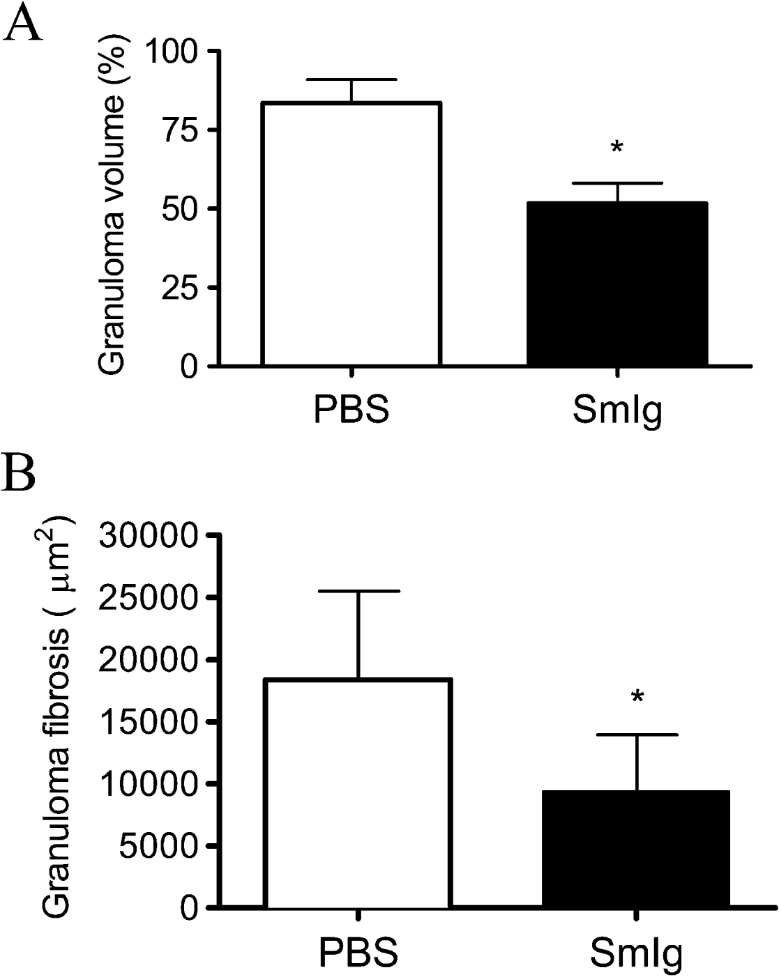
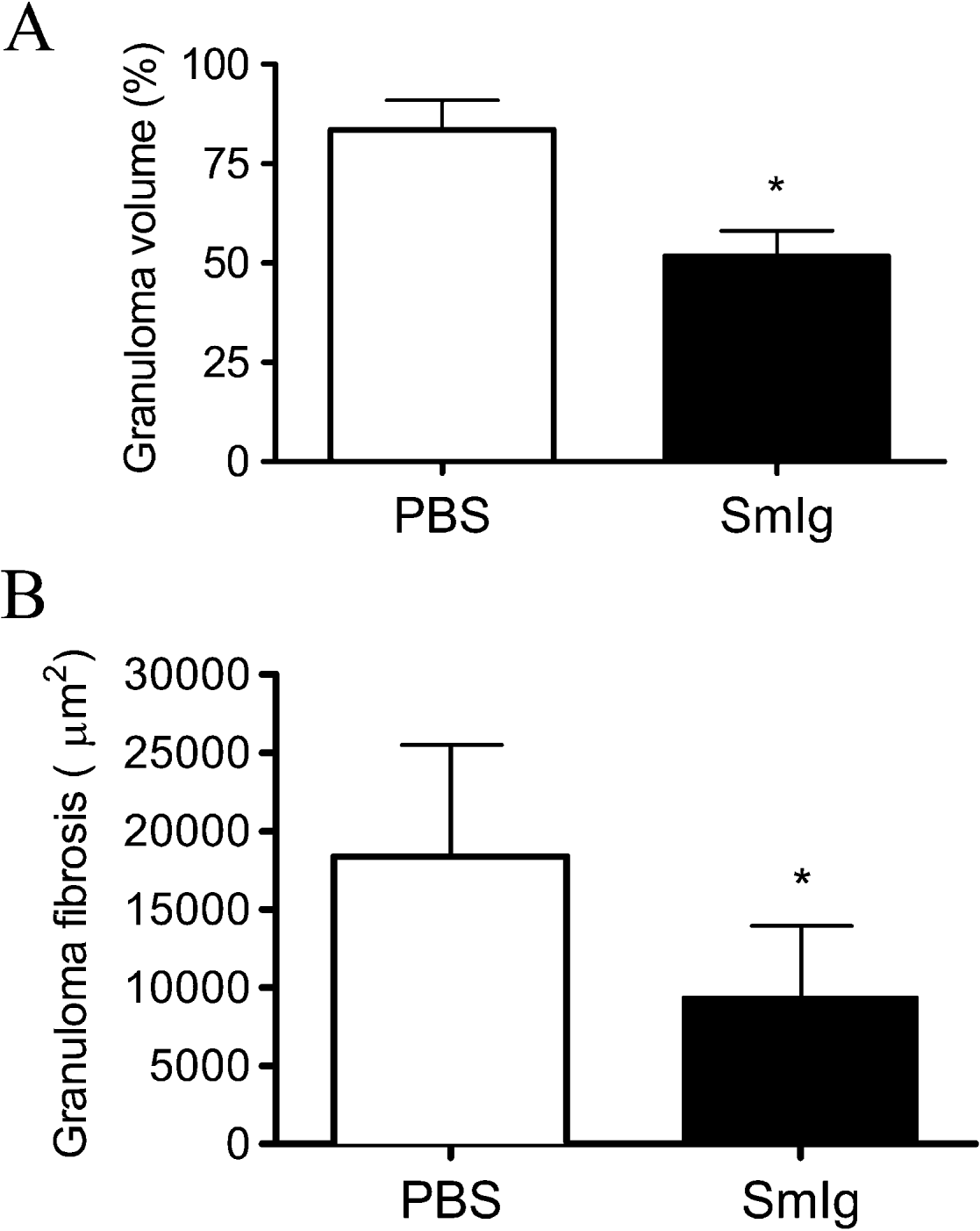
Histological analysis by digital morphometry of haematoxylin-eosin (HE)- and Picrosirius-stained sections obtained from mice immunized with rSmIg showed a reduction of 31·8% in granuloma volume and 49% in collagen deposition compared to mice that received PBS (Fig. 7A and B). Additionally, the micrographs are also shown in Fig. 8, demonstrating reduction in granuloma volume and change in zone of collagen production. These findings demonstrated that SmIg is an important molecule involved in the reduction of liver pathology.
Liver pathology measured in rSmIg vaccinated mice. (A) Hepatic granuloma volume and (B) fibrosis content were measured in rSmIg immunized mice and PBS control group. The granuloma and fibrosis were measured as described in the Materials and Methods section and the results were expressed as % volume for granuloma and square micrometers (μm2) for fibrosis. A statistically significant difference of rSmIg compared to the PBS group is denoted by an asterisk (P<0·05). Results represent data obtained from 2 independent experiments.
Histological analysis of liver tissue from mice immunized with SmIg. Animals were sacrificed and their lungs washed with PBS and stored in formaldehyde until histological procedures. The liver were sliced and stained with Picrosirius. (a) Liver from the PBS control group and (b) liver from SmIg immunized mice. Liver tissue analysis revealed reduced infiltration of inflammatory cells, granuloma volume and fibrosis (red) in SmIg vaccinated group compared to PBS. Magnification, ×40; Scale bars, 50 μm.

DISCUSSION
Tegument proteins are of great importance to schistosome vaccine development since they are a major host–parasite interface (Loukas et al. Reference Loukas, Tran and Pearson2007). Recently, Braschi et al. (Reference Braschi, Curwen, Ashton, Verjovski-Almeida and Wilson2006) identified and characterized the major proteins of the apical membranes of the Schistosoma mansoni tegument. One of these proteins, Accession number C608350.1 (http://cancer.lbi.ic.unicamp.br/schisto6/; Braschi et al. Reference Braschi, Curwen, Ashton, Verjovski-Almeida and Wilson2006), was the focus of our study. Herein, we have isolated a cDNA clone encoding for SmIg and its predicted amino acid sequence was analysed by bioinformatics. In silico analysis revealed interesting characteristics as the presence of a signal peptide, 4 glycosylation sites and a C-terminal transmenbrane region, corroborating the finding of Braschi et al. (Reference Braschi, Curwen, Ashton, Verjovski-Almeida and Wilson2006) as being a membrane-bound protein. Additionally, the search for conserved motifs showed the presence of an immunoglobulin domain, and for this reason this protein has been termed SmIg. Bioinformatic data associated with the result that SmIg transcripts were expressed in the lung-stage schistosomulum encouraged us to test the rSmIg as a vaccine candidate against S. mansoni infection in a murine model. Herein, we investigated murine humoral and cellular immune responses to the rSmIg and evaluated the impact of rSmIg immunization on the worm burden recovery and liver pathology of mice infected with 100 cercariae.
Recent data from double-cytokine knockout mice suggest that the establishment of a robust cellular and humoral response is probably the key to generating maximal immunity to schistosomes (Wynn and Hoffmann, Reference Wynn and Hoffmann2000). In the present study, C57BL/6 mice immunized with rSmIg in the presence of CFA/IFA showed high levels of specific IgG anti-SmIg that appeared after the first immunization. Regarding IgG1 and IgG2a subclasses, specific IgG1 anti-SmIg was predominant. However, the IgG1/IgG2a ratio was reduced from day 30 to day 90 after the first immunization. We confirmed by cytokine analysis that rSmIg immunization elicited a Th1/Th2 immune response characterized by high levels of IFN-γ and TNF-α (Th1) and significant amounts of IL-4 (Th2). Despite high antibody titres vaccination of mice with rSmIg plus Freund's adjuvant failed to provide reduction of parasite burden. Therefore, there was no apparent correlation between the antibody levels generated and protective efficacy in this group. It is unclear what is the relevance of the stimulus of Th1 and Th2 arms of the immune system for the induction of protective immunity. In this study, both arms have been stimulated, but Th2 responses seem to fail to induce protection, while Th1 appears to enhance it. Several S. mansoni antigens tested by our group that had a tendency to induce a Th2 type of immune response failed to engender protection in the mouse model (Pacifico et al. Reference Pacifico, Fonseca, Barsante, Cardoso, Araujo and Oliveira2006; Garcia et al. Reference Garcia, Fonseca, Pacifico, Duraes, Marinho, Penido, Caliari, De Melo, Pinto, Barsante, Cunha-Neto and Oliveira2008). In contrast, Th1 antigens induced partial protection against infection (Fonseca et al. Reference Fonseca, Brito, Alves and Oliveira2004; Cardoso et al. Reference Cardoso, Macedo, Gava, Kitten, Mati, De Melo, Caliari, Almeida, Venancio, Verjovski-Almeida and Oliveira2008; Garcia et al. Reference Garcia, Fonseca, Pacifico, Duraes, Marinho, Penido, Caliari, De Melo, Pinto, Barsante, Cunha-Neto and Oliveira2008). Nevertheless, recent findings by Siles-Lucas et al. (Reference Siles-Lucas, Uribe, Lopez-Aban, Vicente, Orfao, Nogal-Ruiz, Feliciano and Muro2007) demonstrated that protection induced by vaccination with Sb14-3-3zeta antigen was achieved independently of a Th1 response (Siles-Lucas et al. Reference Siles-Lucas, Uribe, Lopez-Aban, Vicente, Orfao, Nogal-Ruiz, Feliciano and Muro2007). Another reason for the lack of protection, could be the fact that SmIg is not accessible to the host immune response in live worms. However, since SmIg was detected on the membrane tegument of schistosomes this might not be the case. Another interesting point is the fact that rSmIg immunization induced high levels of IL-10. The modulation of immunity by schistosomes has been the subject of numerous studies reporting the participation of cellular and molecular mechanisms in the regulation of the granulomatous process (Layland et al. Reference Layland, Wagner and Da Costa2005; Bazzone et al. Reference Bazzone, Smith, Rutitzky, Shainheit, Urban, Setiawan, Blum, Weinstock and Stadecker2008). We speculate that it is the high levels of IL-10 which may have modulated the inflammatory response and granuloma size. Surprisingly, the profile of immune response induced by rSmIg is able to induce significant reduction in the liver pathology, relative to granuloma volume (31·8%) and fibrosis content (49%). Therefore, we speculate that vaccination with rSmIg has an anti-fecundity effect and may have a role in schistosome reproduction. Pathology which results from granuloma formation around the eggs in murine schistosomiasis is characterized by a Th2-type of immune response and the granuloma size can be reduced by neutralization of IL-4 (Wynn and Cheever, Reference Wynn and Cheever1995). Since we detected high production of IL-10 following splenocyte activation with rSmIg, we hypothesize here that this cytokine might be regulating Th2 responses and therefore, reducing liver pathology (Hogg et al. Reference Hogg, Kumkate and Mountford2003; Sadler et al. Reference Sadler, Rutitzky, Stadecker and Wilson2003). In conclusion, SmIg is a new tegument protein from S. mansoni that plays an important role in reducing pathology induced by parasite infection.
ACKNOWLEDGEMENTS
This work was supported by CNPq, FAPEMIG, FAPESP, FINEP/SEBRAE and INCT de Doenças Tropicais/CNPq.