Restrictive cardiomyopathy is a rare form of cardiomyopathy, accounting for less than 5% of all paediatric cases.Reference Nugent, Daubeney and Chondros1 It is characterised by increased stiffness of the myocardium, leading to inappropriately high filling pressures and diastolic dysfunction of the ventricles. Usually, wall thickness is normal, and systolic function is preserved. Sudden death and unfavourable outcomes due to pulmonary vascular disease are common in children with restrictive cardiomyopathy.Reference Rivenes, Kearney, Smith, Towbin and Denfield2, Reference Russo and Webber3 Ultimately, heart transplantation is the only treatment option. Restrictive cardiomyopathy can occur spontaneously or as an autosomal dominant trait; in children, mutations in sarcomeric genes account for one-third of the cases.Reference Kaski, Syrris and Burch4
Case report
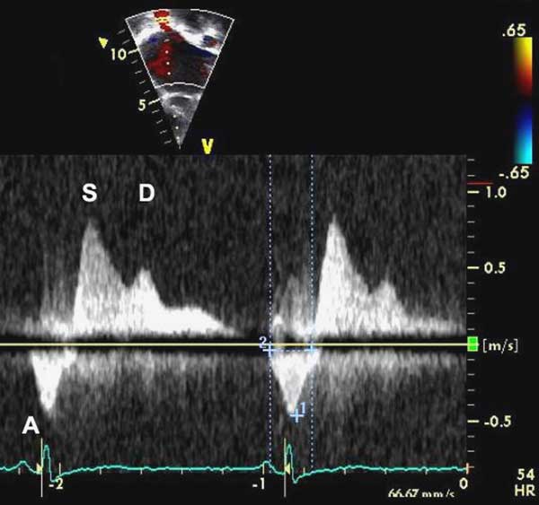
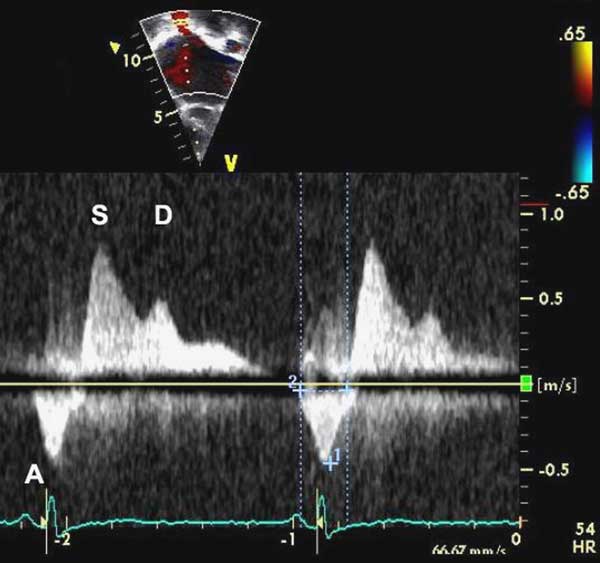
An asymptomatic 4-day-old girl was referred for evaluation of a 2–3/6 systolic heart murmur. She was clinically asymptomatic; family history was unremarkable. Echocardiography showed a restrictive perimembranous ventricular septal defect. Upon follow-up at 18 months, she remained clinically asymptomatic. Echocardiography showed persistence of the ventricular septal defect, new biatrial enlargement, and flow reversal of the pulmonary venous A wave. At the age of 3 years, she underwent cardiac catheterisation, which confirmed the ventricular septal defect and showed an elevated left-ventricular end-diastolic pressure of 16 millimetres of mercury and right-ventricular pressures at 50% of the systemic pressures. We made a diagnosis of restrictive cardiomyopathy in combination with ventricular septal defect.
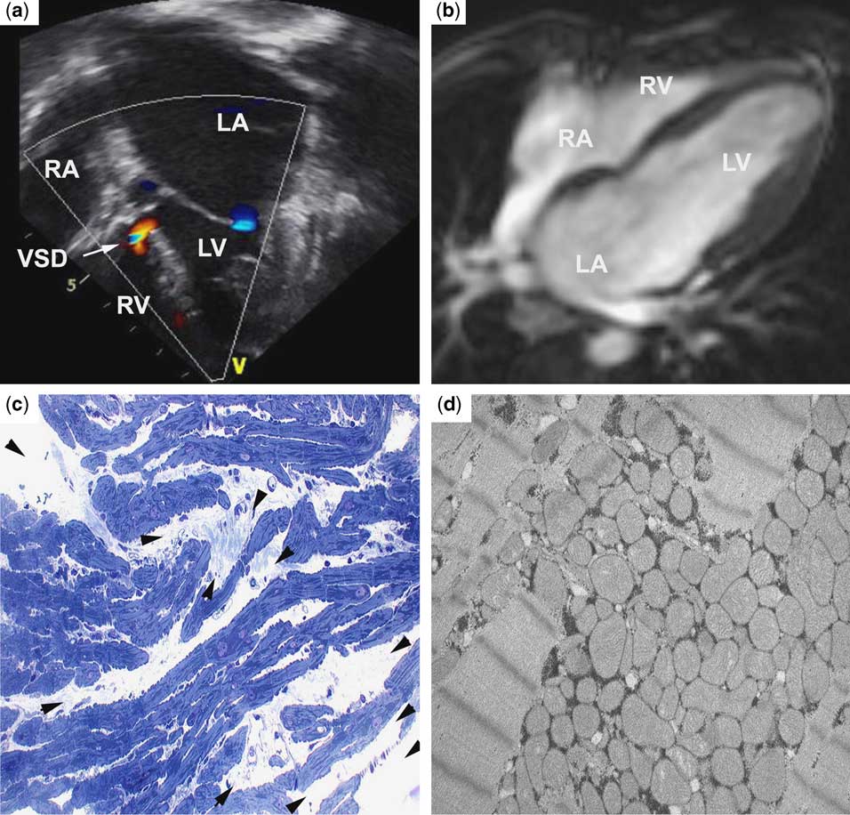
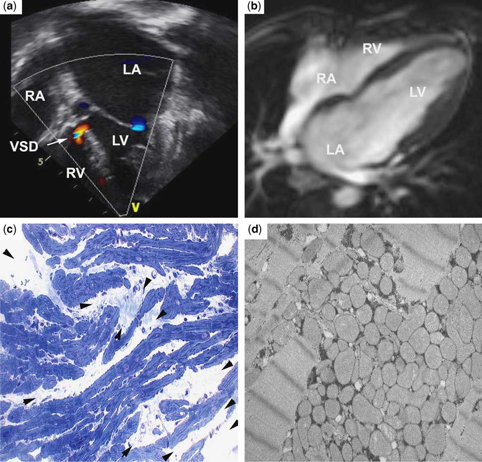
At the age of 8 years, she gradually developed dyspnoea on exertion, with little response to atenolol. Echocardiography showed predominant left atrial enlargement (Fig 1a). Magnetic resonance imaging was performed to better characterise cardiac morphology in this patient and showed predominant left atrial enlargement and thickening of the atrial walls, absence of thrombi, dilation of the pulmonary artery and veins, and no significant ventricular hypertrophy (Fig 1b). The pericardium was normal. Cine magnetic resonance imaging revealed slow, swirling blood flow predominantly in the left atrium (Video 1, supplemental data file; Fig 2).
(a) Apical four-chamber view with biatrial enlargement and a perimembranous ventricular septal defect (VSD, arrow; RA, right atrium; LA, left atrium; RV, right ventricle; LV, left ventricle). (b) Transverse section of magnetic resonance imaging, showing predominant left atrial enlargement with interatrial septum bulging towards the right atrium. (c) Toluidine blue staining of myocardial biopsy indicates massive fibre disarray and interstitial fibrosis (arrowheads; 400×). (d) Electron microscopy with a large stack of irregularly shaped megamitochondria.
Pulsed Doppler flow in the left upper pulmonary vein in apical four-chamber view. S, D, and A: pulmonary venous Doppler waves. Note the largely increased pulmonary venous A wave, and the decreased D wave.
Cardiac catheterisation was repeated in the preparation of the cardiac transplant, and myocardial biopsies displayed extensive anisomorphism and disarray of cardiomyocytes (Fig 1c). Electron microscopy showed large stacks of severely dysmorphic megamitochondria and focal Z-disc streaming (Fig 1d). Owing to the progression of her condition, she was listed for cardiac transplantation.
Genetic analysis identified a heterozygous missense mutation in TNNI3 (R204H), which has been previously identified in both restrictive and hypertrophic cardiomyopathy.Reference Gambarin, Tagliani and Arbustini5, Reference Doolan, Tebo and Ingles6 The patient’s mother tested negative for the mutation; paternal deoxyribonucleic acid was not available for analysis.
Discussion
Ventricular septal defects are a common lesion in children; they occur in 50% of all congenitally malformed hearts, and in 20% as an isolated lesion. Usually, such defects have a high spontaneous closure rate and a benign natural history, which was not the case in our patient. In addition, several recent studies have highlighted a link between congenital cardiac disease and cardiomyopathy caused by sarcomere mutations.Reference Monserrat, Hermida-Prieto and Fernandez7 Importantly, this report is the first one to suggest that those phenotypes may overlap in TNNI3 mutation carriers also.
A wide spectrum of clinical phenotypes can be induced by mutations in TNNI3, including restrictive, hypertrophic, and dilated cardiomyopathies.Reference Kaski, Syrris and Burch4, Reference Mogensen, Kubo and Duque8, Reference Carballo, Robinson and Otway9 It should be noted that mutations in the structural genes, TNNT2 and MYH6, the latter of which codes for the b-myosin heavy chain, have recently been linked to restrictive cardiomyopathy in children.Reference Kaski, Syrris and Burch4, Reference Peddy, Vricella and Crosson10, Reference Ware, Quinn, Ballard, Miller, Uzark and Spicer11 Differential effects of troponin mutations on relaxation have been implicated in the variable expressivity of the phenotypes, with mutations that increase Ca(2+) sensitivity leading to hypertrophic and restrictive cardiomyopathy, and mutations that decrease Ca(2+) sensitivity leading to dilated cardiomyopathy (for a review, see Willott et alReference Willott, Gomes, Chang, Parvatiyar, Pinto and Potter12). However, the mechanism by which sarcomeric mutations may contribute to septal defects remains elusive.
Interestingly, myofibre disarray is much more pronounced than hypertrophy in the histological samples of our patient. A similar finding was described previously in the same TNNI3 R204H mutation in a patient with restrictive cardiomyopathy.Reference Gambarin, Tagliani and Arbustini5 In contrast, the same mutation has been found to cause a predominant hypertrophic cardiomyopathy in an Australian family.Reference Doolan, Tebo and Ingles6 In both cases, the natural history of disease was severe and rapidly progressive, similar to the evolution observed in the patient described here. In conclusion, the TNNI3 mutation, R204H, confers a severe and progressive clinical course, with additional modifying factors in individual patients and families with either predominantly restrictive or hypertrophic phenotypes.
According to the literature, a small subset of patients with persistent small ventricular septal defects in adulthood is prone to developing restrictive cardiomyopathy.Reference Neumayer, Stone and Somerville13 Our case highlights that a high index of suspicion and careful follow-up of patients is mandatory even for seemingly benign congenital cardiac disease such as ventricular septal defects. Knowledge of the underlying genetic basis of the disease can help inform management as well as individual and familial counselling in such cases.
Acknowledgements
The authors gratefully acknowledge grant support of the Canadian Institutes of Health Research (GMHD79045 and Clinician-Scientist program to GA), and of the Nussia et André Aisenstadt Foundation and the Deutsche Herzstiftung (to MPH). The authors have declared no conflict of interest.