Implications
Cecotrophy allows rabbits to reingest microbial products from the cecum as cecotrophs. Cecotrophy has considerable significance for nitrogen (N) metabolism in rabbits. A previous study indicated that as a fermentable sugar, d-mannitol increases N utilization through cecotrophy by deceasing urinary N excretion in rabbits. In the present study, we clarified the possible mechanism by which d-mannitol improves N utilization in growing rabbits. The results indicate that d-mannitol can be used as a cecal microbial stimulator to promote the utilization rate of dietary N in rabbits fed a diet of timothy hay. Moreover, it might aid in reducing the N load passed on to the environment.
Introduction
It is well documented that fermentable carbohydrates can increase urea flux from the blood to the digestive tract and alter fermentation in the large intestine of rats (Rémésy and Demigné, Reference Rémésy and Demigné1989; Younes et al., Reference Younes, Garleb, Behr, Rémésy and Demigné1995b; Henningsson et al., Reference Henningsson, Bjorck, Marfareta and Nyman2002). In rabbits, a considerable portion of endogenous urea is transferred to the alimentary tract (Regoeczi et al., Reference Regoeczi, Irons, Koj and Mcfarlane1965), and the exchange between blood urea nitrogen (N) and cecal ammonia N is well established (Forsythe and Parker, Reference Forsythe and Parker1985a and Reference Forsythe and Parker1985b). However, the exact fate of blood urea in rabbits fed a diet containing fermentable sugar remains unclear.
Urea, as an end product of ammonia and protein metabolism, is synthesized in the liver of mammals and passed into the blood stream (Stewart and Smith, Reference Stewart and Smith2005). Most of the blood urea is eliminated in the urine and the remainder enters the digestive tract (Jackson et al., Reference Jackson, Picou and Landman1984; Lobley et al., Reference Lobley, Bremner and Zuur2000; Stewart and Smith, Reference Stewart and Smith2005), where it can be hydrolyzed to ammonia by the intestinal microflora activity. Ammonia derived from nitrogenous residues (endogenous and undigested food) and urea is the major N source for bacterial growth (Viallard, Reference Viallard1984; Younes et al., Reference Younes, Garleb, Behr, Rémésy and Demigné1995b; Carabaño et al., Reference Carabaño, Villamide, García, Nicodemus, Llorente, Chamorro, Menoyo, García-Rebollar, García-Ruiz and De Blas2009). It has been well documented that the presence of fermentable materials can stimulate bacterial proliferation in the large intestine (Levrat et al., Reference Levrat, Behr, Rémésy and Demigné1991; Djouzi and Andrieux, Reference Djouzi and Andrieux1997; Hanieh and Sakaguchi, Reference Hanieh and Sakaguchi2009). This can then lead to an increase in the transfer of blood urea N to lumen ammonia N, which is then used to synthesize bacterial N in the large intestine of monogastric animals (Rémésy and Demigné, Reference Rémésy and Demigné1989). This results in an increase in bacterial materials (in cecal digesta or feces) and N retention (in cecum and body), concomitantly decreasing plasma urea N and urinary N excretion (Misir and Sauer, Reference Misir and Sauer1982; Kirchgessner et al., Reference Kirchgessner, Kreuzer, Machmüller and Roth-Maier1994; Younes et al., Reference Younes, Demigné, Behr, Garleb and Rémésy1996). The increment in bacterial N in fecal matters is particularly essential for cecotrophic animals like rabbits. Rabbits reingest a significant amount of cecotrophs (or soft feces), which are subsequently digested and absorbed in the stomach and small intestine and make a significant contribution to the total protein requirement (Hörnicke, Reference Hörnicke1981; Belenguer et al., Reference Belenguer, Balcells, Guada, Decoux and Milne2005). When animals consume a low-protein diet, the conversion of blood urea N into bacterial N in the large intestine caused by fermentable carbohydrates is more evident (Levrat et al., Reference Levrat, Rémésy and Demigné1993; Younes et al., Reference Younes, Demigné, Behr, Garleb and Rémésy1996).
In a previous study, we observed that feeding rabbits d-mannitol as an indigestible but fermentable sugar improved N retention due to a significant increase in N in the cecotrophs, and a decrease in urinary N excretion (Xiao et al., Reference Xiao, Xiao, Tsuzuki and Sakaguchi2011). The increase of N shifted from the urine to the cecotrophs may indicate an increase in N transfer from the blood to cecal microbes. To clarify the mechanism by which fermentable sugars such as d-mannitol increase N utilization in rabbits, we administrated 15N-urea to growing rabbits fed a diet containing mannitol to investigate the fate of blood urea N.
Material and methods
Animals and diets
Eight growing male Dutch rabbits aged 100 days were used for this study. The animals were individually housed in metabolic cages with wire-mesh bottoms (diameter, 38 cm; height, 30 cm) and maintained in an air-conditioned room at a constant temperature of 23°C to 25°C and a relative humidity of 50% to 60% with the lights on from 0530 to 1730 h. The rabbits were randomly assigned to one of two treatments and fed a diet of timothy hay (unpelleted timothy hay was chopped to 1 to 2 mm, no addition of vitamins and mineral premix. The timothy hay contains 34.6% of ADF, 11.2% of CP and 1420 kcal/kg of digestible energy; 50 g/kg0.75 per day) with added glucose (80 g/kg; Nacalai Tesque Inc., Kyoto, Japan) or d-mannitol (80 g/kg; Nacalai Tesque Inc.). The animals were allowed free access to water.
The study protocol was approved by the Experimental Animal Care Committee of Okayama University.
Sampling procedures
After 9 days of feeding of the experimental diets, 5 ml of a water solution containing 2 g glucose or d-mannitol was given orally 3 h before killing. A single injection of 20 mg 15N-urea (99.2 atom%, SI Science Co. Ltd, Sugito, Saitama, Japan) in saline was administered under anesthesia (20 mg/kg sodium pentobarbital) via the ear vein 1 h before slaughter. After administration of 15N-urea, urine was collected. All animals were killed between 0900 and 1000 h. A blood sample was taken from the abdominal aorta. Serum was obtained by centrifuging at 3000 r.p.m. for 20 min and stored at −30°C for further analysis. The liver, small intestine, cecum, proximal colon, distal colon and rectum were removed on ice, weighed and stored at −30°C for subsequent analysis.
Analytical procedures
The digesta from each gut segment were weighed and homogenized and the pH was measured immediately with a digital pH meter (Horiba Ltd, Kyoto, Japan). The moisture content of the digesta was determined by drying a sample at 105°C for 24 h. The concentration of short-chain fatty acids (SCFA) in the homogenized cecal contents was determined by HPLC (Column: 2 Shim-pack SCR-102H, Detector: Shimadzu CDD-6A; Shimadzu Corporation, Kyoto, Japan). A liver sample was homogenized in physiological saline (9 g sodium chloride/l). The homogenized liver sample, contents of the small intestine, cecum, proximal colon and distal colon, and the blood serum were quickly precipitated with one-fifth of their volume of trichloroacetic acid (TCA; 500 g/l), centrifuged (12 000 g for 20 min at 4°C) and washed twice with TCA solution (50 g/l; Fürst and Jonsson, Reference Fürst and Jonsson1971). The supernatants of the cecal contents and blood serum were collected. Each sample of cecum, proximal colon and distal colon content was diluted in physiological saline, shaken well and placed in a refrigerator for 24 h at 4°C to dislodge adherent bacteria (Minato and Suto, Reference Minato and Suto1978). Bacteria were retrieved from the solution by differential centrifugation (500 g for 5 min followed by two consecutive centrifugations at 20 000 g for 20 min at 4°C; Belenguer et al., Reference Belenguer, Balcells, Guada, Decoux and Milne2005). The contents of each part of the digestive tract, the urine and the TCA precipitates obtained were analyzed for total N and protein N (TCA precipitates) by the Kjeldahl procedure. The bacteria isolated from the contents of the cecum, proximal colon and distal colon were analyzed for bacterial N also by the Kjeldahl method. Urea in the deproteinized supernatants of the serum and cecal content was converted to ammonia by treatment with urease (EC 3.5.1.5; Nacalai Tesque Inc., Kyoto, Japan) according to the procedure described by Obara et al. (Reference Obara, Fuse, Terada, Shibata, Kawabata, Sutoh, Hodate and Matsumoto1994). The urease-treated samples and non-urease-treated samples were steam distilled with the addition of magnesium oxide or sodium hydroxide and ammonia was trapped in boric acid and titrated with sulfuric acid (0.05 N) for urea N and ammonia N determination (Nolan and Leng, Reference Nolan and Leng1972).
The concentrations of 15N in the total N, protein N, microbial N, urea N and ammonia N of each sample mentioned above were analyzed by a mass spectrometer (MSI-150-MT-600 NC Model, Quadrupole Mass Spectrometry System, J-Science, Kyoto, Japan).
Statistical analysis
Data are presented as means ± standard deviations. The significance of differences between means was analyzed by Student's t-tests. Differences were considered significant for values of P < 0.05.
Results
Feed intake and weight gain
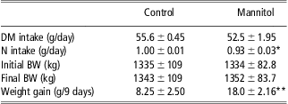
Dry matter intake, N intake and weight gain are shown in Table 1. Daily N intake in rabbits fed the mannitol diet was lower (P < 0.05) than in rabbits fed the control diet, which may be caused by the relatively low dry matter intake in the mannitol group compared with the control group (CG). However, supplementation with d-mannitol increased (P < 0.01) the weight gain over the whole experimental period.
DM intake, N intake and BW change during 9 days feeding period in growing rabbits
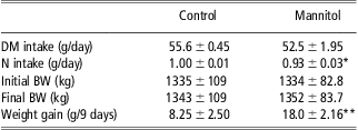
DM = dry matter; N = nitrogen.
*P < 0.05, **P < 0.01, significantly different from control; data are means ± s.d. (n = 4).
Weight of gut
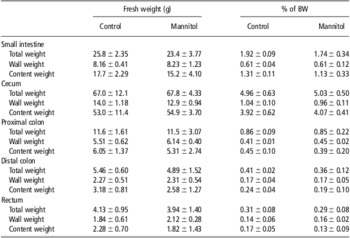
The data on gut development are shown in Table 2. The diet containing d-mannitol did not affect total gut weight, wall weight or content weights of the small (25.8, 8.16 or 17.7 g and 23.4, 8.23 or 15.2 g for CG and mannitol group, respectively) and large (88.2, 23.6 or 64.5 g and 88.1, 23.5 or 64.6 g for CG and mannitol group, respectively) intestines.
Wet weight of gut and content at the end of 9 days feeding period in growing rabbits
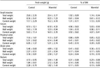
Content weight = total weight − wall weight; data are means ± s.d. (n = 4).
pH and SCFA in the cecal content
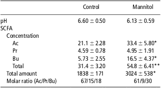
The concentration and total amount of SCFA and the pH in the cecal content are presented in Table 3. d-mannitol feeding increased the concentrations of acetic acid (158% of the CG, P < 0.05) and butyric acid (288% of the CG, P = 0.01), resulting in an increase in the total concentration of SCFA (175% of the CG, P < 0.01). The concentration of propionic acid was not changed (4.59 and 4.95 μmol/g for CG and mannitol group, respectively, P = 0.75). The total amount of SCFA in rabbits fed the mannitol diet was 64% higher than in rabbits fed the control diet. Because of the increase in SCFA, the pH of the cecal content was decreased (93% of the CG), but this was not significant (P = 0.10). The molar ratio of butyric acid was improved (P = 0.04) in the mannitol group compared with the CG.
Luminal pH, concentration (μmol/g contents) and total amount (μmol) of SCFA in cecal content at the end of 9 days feeding period in growing rabbits
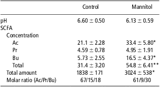
SCFA = short chain fatty acids; Ac = acetic acid; Pr = propionic acid; Bu = butyric acid.
*P < 0.05, **P < 0.01, significantly different from control; data are means ± s.d. (n = 4).
Total N in gut contents and urine
As shown in Table 4, the concentration (114% of the CG, P < 0.05) and amount of total N (127% of the CG, P < 0.05) and total 15N (161% of the CG, P < 0.01) in the cecal content of rabbits fed the mannitol diet were higher than those of rabbits fed the control diet. The concentration of total N and total 15N in the contents of the proximal and distal colon of rabbits fed the mannitol diet were also increased (P < 0.01), whereas the total amount of N (96.8 and 100.8 mg for CG and mannitol group, respectively) and 15N (0.67 and 0.79 mg for CG and mannitol group, respectively) in the colon contents were not different from those of the rabbits fed the control diet. The mannitol diet decreased N excretion in the urine (80% of the CG, P < 0.05). The addition of mannitol to the timothy hay also improved the concentration of total N (112% of the CG, P < 0.05) in the rectum content but not the 15N atom % excess (0.04% and 0.02% for CG and mannitol group, respectively).
Total N and 15N in gut contents and urine after a dose of 15N-urea at the end of 9 days feeding period in growing rabbits

N = nitrogen; DM = dry matter.
Total N means total Kjeldahl N that is the sum of organic N and ammonia.
*P < 0.05, **P < 0.01, ***P < 0.001, significantly different from control; data are means ± s.d. (n = 4).
Protein N in liver and gut contents
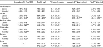
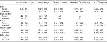
As shown in Table 5, the amount of protein N in the livers of rabbits fed the mannitol diet was lower (74% of the CG, P < 0.05) than that of rabbits fed the control diet, whereas the atom % excess and amount of 15N were greater (P < 0.05) compared with the control animals. The concentration (P < 0.05) and amount of protein N (P < 0.01) and protein 15N (P < 0.001) were increased in the cecum content of rabbits fed the mannitol diet compared with those fed the control diet. The 15N atom % excess of protein N in the proximal colon content of the rabbits fed the mannitol diet was higher (112% of the CG, P < 0.01), but the protein N concentration was not changed (4.59% and 5.36% of DM for CG and mannitol group, respectively, P = 0.10) compared with those of the control animals. Protein N in the contents of the distal colon (24.6 and 29.8 mg for CG and mannitol group, respectively, P = 0.22) and small intestine (180 and 186 mg for CG and mannitol group, respectively, P = 0.24) were not affected by mannitol feeding.
Protein N and 15N in liver and gut contents after a dose of 15N-urea at the end of 9 days feeding period in growing rabbits

N = nitrogen; DM = dry matter.
*P < 0.05, **P < 0.01, ***P < 0.001, significantly different from control; data are means ± s.d. (n = 4).
Bacterial N in cecum and colon
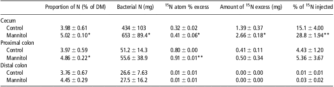
Table 6 shows the concentration and amount of bacterial N and 15N in the contents of the cecum and colon. The bacterial N concentration and 15N atom % excess in the contents of the cecum and proximal colon of rabbits fed the mannitol diet were higher (P < 0.05) than those of rabbits fed the control diet. The amount of bacterial N (150% of the CG, P < 0.05) and 15N (191% of the CG, P < 0.05) was increased only in the cecal content. The addition of mannitol to the diet did not change bacterial N in the distal colon content (26.6 and 27.5 mg for CG and mannitol group, respectively, P = 0.89).
Bacterial N and 15N in large intestine after a dose of 15N-urea at the end of 9 days feeding period in growing rabbits
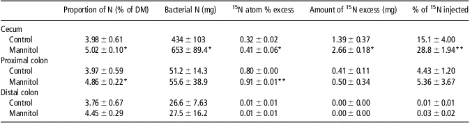
N = nitrogen; DM = dry matter.
*P < 0.05, **P < 0.01, significantly different from control; data are means ± s.d. (n = 4).
Urea N and ammonia N in blood serum and cecal content
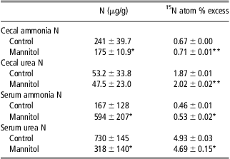
The concentrations of ammonia N and urea N, and 15N atom % excess in wet cecal content and blood serum are presented in Table 7. The concentration of ammonia N in the cecal content was lower (73% of the CG, P < 0.05) in the mannitol group than in the CG, whereas the 15N atom % excess was higher (106% of the CG, P < 0.01) in the mannitol group than in the CG. The concentration of cecal urea N was not affected (53.2 and 47.5 μg/g for CG and mannitol group, respectively, P = 0.82) by mannitol feeding, but the 15N atom % excess in cecal urea N was clearly higher (P < 0.01) in the mannitol group than in the control animals. The concentration of serum ammonia N and 15N atom % excess in the mannitol group was higher (P < 0.05) than those of the CG. In contrast, the concentrations of serum urea N and 15N were lower (P < 0.05) in the mannitol group than in the CG.
Concentration of ammonia N, urea N and 15N in cecal wet content and serum after a dose of 15N-urea at the end of 9 days feeding period in growing rabbits
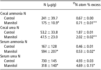
N = nitrogen.
*P < 0.05, **P < 0.01, significant different from control; data are means ± s.d. (n = 4).
Discussion
d-mannitol stimulates bacterial proliferation and increases N content in the large intestine
This study shows that consumption of d-mannitol as a fermentable sugar greatly improved the total N concentration in the rabbit large intestine. The amount of total N, protein N and bacterial N in the cecal content of rabbits fed d-mannitol increased by 27%, 65% and 50%, respectively, compared with the CG. These results indicate that d-mannitol as an indigestible but fermentable sugar can escape degradation in the small intestine and reach the large intestine (Dwivedi, Reference Dwivedi1991), where it stimulates the growth of bacteria. In the present study, the presence of mannitol in the diet stimulates the bacterial proliferation in the cecum of rabbits characterized as higher concentrations of SCFA (P < 0.05), especially of butyric acid (2.9-fold over the CG). The presence of fermentable carbohydrates causes a multiplication of bacteria in the large intestines of rats (Levrat et al., Reference Levrat, Behr, Rémésy and Demigné1991; Younes et al., Reference Younes, Garleb, Behr, Rémésy and Demigné1995b) and humans (Djouzi and Andrieux, Reference Djouzi and Andrieux1997). This causes an increase in demand for N by microorganisms (Hawe et al., Reference Hawe, Walker and Moss1991; Younes et al., Reference Younes, Garleb, Behr, Rémésy and Demigné1995b). The increased N requirement for bacterial growth can result in an increase in diffusion of urea from the blood to the digestive tract (Demigné and Rémésy, Reference Demigné and Rémésy1985), because blood urea is the most readily available N source for bacterial protein synthesis (Levrat et al., Reference Levrat, Rémésy and Demigné1993; Younes et al., Reference Younes, Garleb, Behr, Rémésy and Demigné1995b). In rabbits, blood urea N also represents a significant N source for bacterial growth (Regoeczi et al., Reference Regoeczi, Irons, Koj and Mcfarlane1965). The urea that is transferred from the blood to the large intestine is degraded to ammonia by bacterial urease and subsequently used for bacterial protein synthesis (Demigné and Rémésy, Reference Demigné and Rémésy1979; Viallard, Reference Viallard1984; Sarraseca et al., Reference Sarraseca, Milne, Metcalf and Lobley1998). In the present study, the addition of mannitol to the diet decreased the concentration of cecal ammonia N (P < 0.05), but increased the 15N enrichment of cecal ammonia N (P < 0.01). Together with the increase in 15N enrichment of cecal urea N (P < 0.01), this indicates that the flux of blood urea N towards the cecum was increased and the urea was quickly converted to ammonia by bacterial urease. The ammonia is subsequently used for bacterial protein synthesis and also partially reabsorbed by the cecal vein (Sarraseca et al., Reference Sarraseca, Milne, Metcalf and Lobley1998). A similar decrease in cecal ammonia concentration was observed in rats fed oligosaccharides (Younes et al., Reference Younes, Garleb, Behr, Rémésy and Demigné1995b). This may be due to the stimulating effect of fermentable carbohydrates on cecal bacterial proliferation. Ammonia may be a main N source for bacterial growth since a drop in unionized ammonia concentration has been reported (Mason, Reference Mason1984).
d-mannitol decreases the blood urea N and urinary N excretion
The increase in urea transfer from the blood to the large intestine by fermentable carbohydrates results in a decrease in blood urea N and N excretion in the urine (Younes et al., Reference Younes, Demigné, Behr and Rémésy1995a and Reference Younes, Garleb, Behr, Rémésy and Demigné1995b). In rats, the consumption of indigestible carbohydrates (Arabic gum, fructooligosaccharides and xylooligosaccharides) results in a greater amount of urea N transferred to the cecum, which has been found to increase fecal N excretion 1.5 to 2.0 fold and reduce urinary N excretion by 25% to 30%, causing a 20% to 30% reduction in blood urea (Younes et al., Reference Younes, Garleb, Behr, Rémésy and Demigné1995b). In the present study, the addition of d-mannitol to the diet decreased urinary N excretion by 20% and lowered the blood urea N level by 56%.
d-mannitol increases the flux of blood urea N to cecal microbial N
The amount of total 15N excess in total N in the cecum was 60% higher in the rabbits fed the mannitol diet than in the control animals. This represents as much as 40% of the 15N administrated, compared with 24.8% in the CG. Conversely, the 15N atom % excess of blood urea N (P < 0.01) and urinary N (P < 0.001) was decreased in rabbits fed the mannitol diet compared with those fed the control diet. Furthermore, the 15N atom % excess of cecal bacterial N was higher (P < 0.05) in the mannitol group compared with the CG. These results suggest that the consumption of mannitol increased the transfer of urea from the blood to the large intestine and the microbial proliferation.
The addition of inulin as a fermentable polysaccharide to the diets of rats considerably increases the flux of urea N to the cecum, resulting in a significant increase in fecal N excretion (Levrat et al., Reference Levrat, Rémésy and Demigné1993). This indicates that inulin contributes to the utilization of more blood urea N for cecal bacterial N synthesis. Bacterial N is subsequently excreted as fecal N, representing the major form of fecal N (Viallard, Reference Viallard1984; Mortensen, Reference Mortensen1992). The close relationship between the flux of urea N towards the large intestine, fecal N excretion and decrease in blood urea N has been demonstrated in several studies with rats fed fermentable carbohydrates (Levrat et al., Reference Levrat, Rémésy and Demigné1993; Younes et al., Reference Younes, Garleb, Behr, Rémésy and Demigné1995b and Reference Younes, Alphonse, Abdelkader and Rémésy2001). These reports are in agreement with our present results.
Low-protein diet facilitates the transfer of urea N from blood to large intestine
The effect of fermentable carbohydrates on urea disposal in the large intestine is closely related to dietary protein levels (Levrat et al., Reference Levrat, Rémésy and Demigné1993). Several studies have demonstrated that low-protein diets combined with fermentable carbohydrates were particularly effective in increasing the transfer of blood urea N to the large intestine and subsequently fecal N excretion (Levrat et al., Reference Levrat, Rémésy and Demigné1993; Younes et al., Reference Younes, Garleb, Behr, Rémésy and Demigné1995b and Reference Younes, Demigné, Behr, Garleb and Rémésy1996). To study the mechanism behind the improvement of N utilization by fermentable sugar, we used timothy hay (it contains 34.6% of ADF, 11.2% of CP and 1420 kcal/kg of digestible energy). The ratios of protein and digestible energy of timothy hay to the requirement of the nutrients in growing rabbits are 63% and 59%, respectively, National Research Council (NRC, 1977) as a low-protein diet in the present study. The results showed that the addition of d-mannitol to timothy hay increased the transfer of urea from the blood to the large intestine and then to bacterial N.
The present study provides a clear explanation for the possible mechanism by which d-mannitol improves N utilization in rabbits. In a previous study, the addition of d-mannitol to the diet increased N utilization by increasing the amount of N in the cecotrophs and decreasing urinary N excretion (Xiao et al., Reference Xiao, Xiao, Tsuzuki and Sakaguchi2011). The increase in N in the cecotrophs is likely caused by the increase in bacterial N in the cecal content, because cecotrophs are derived from the cecal content without any great changes (Carabaño and Piquer, Reference Carabaño and Piquer1998). d-mannitol stimulates bacterial proliferation in the cecum, and the microflora uses blood urea N as an N source for their intense growth. The present study demonstrates that d-mannitol enhances the transfer of blood urea to the cecum by lowering the urinary N excretion, since the blood urea N level was depressed.
Conclusion
The experimental results indicate that d-mannitol increases the transfer of blood urea N to the large intestine for bacterial N synthesis. The present study provides a possible mechanism for the increase in N utilization caused by d-mannitol in growing rabbits. This study also shows that, as a fermentable sugar, d-mannitol can be used as a cecal microbial stimulator to promote the utilization rate of dietary N in rabbits fed a diet of timothy hay.
Acknowledgment
This study was supported by Grants-in-Aid for Scientific Research of the Japan Society for the Promotion of Science, Japan (Grant no. 22580309).