Introduction
The advance in genetic studies has been changing the traditional systematic classification of the nematode parasites of vertebrates (Blaxter et al., Reference Blaxter1998; Medal et al., Reference Medal2007; Anderson et al., Reference Anderson, Chabaud and Willmott2009; Hodda, Reference Hodda and Zhang2011; Blaxter and Koutsovoulos, Reference Blaxter and Koutsovoulos2014). The order Strongylida (‘bursate nematodes’), for example, has lost its taxonomic validity, with representatives re-allocated in Rhabditida (as Rhabditomorpha) according to recent molecular phylogenies (Medal et al., Reference Medal2007; Blaxter and Koutsovoulos, Reference Blaxter and Koutsovoulos2014). These new perspectives may complicate the taxonomic limits among lower taxa (i.e. superfamilies, families and subfamilies), which in several cases are complicated based on morphological classification as well.
The ‘clade Strongylida’ is a monophyletic assemblage, representing one of the greatest radiations of parasitic nematodes (Chilton et al., Reference Chilton2006). Despite its high diversity of species and biological traits, several taxa remain poorly studied. In this sense, the family Diaphanocephalidae Travassos, 1920 allocates only two genera, i.e. Diaphanocephalus Diesing, 1851 and Kalicephalus Molin, 1861 (see Lichtenfels, Reference Lichtenfels, Anderson, Chabaud and Willmott1980), in which several species are poorly described. Furthermore, almost no genetic characterization is available for these nematodes, which, along with the lack of morphological data, presents a real challenge to species identification.
Diaphanocephalus galeatus (Rudolphi, 1819) has broad occurrence in South America, parasitizing teiid lizards (Teiidae), more often in Salvator merianae (Duméril & Bibron, 1839) and Tupinambis teguixin (Linnaeus, 1758) (Freitas and Lent, Reference Freitas and Lent1938; Ávila and Silva, Reference Ávila and Silva2010; Vieira et al., Reference Vieira, Fedatto-Bernardon and Müller2016; Teixeira et al., Reference Teixeira2017). However, the morphological descriptions of the species are incomplete and there is no genetic characterization. The same is observed regarding its congeners, D. diesingi Freitas & Lent, 1938 and D. jacuruxi Alho, 1965, which have not been reported since their original descriptions (Ávila and Silva, Reference Ávila and Silva2010).
Given the poor taxonomic knowledge on the nematodes belonging to Diaphanocephalus from reptiles in South America, the present study had the following goals: (1) redescribe D. galeatus using light and scanning electron microscopy (SEM), (2) genetically characterize the species for the first time and perform a genetic comparison with its closely related taxa, and (3) re-evaluate the type material of D. diesingi and D. jacuruxi as well as those nominated as D. galeatus, deposited in the Coleção Helmintológica do Instituto Oswaldo Cruz (CHIOC).
Materials and methods
Collection and examination of nematodes
On 15 May 2017 one specimen of the Paraguay caiman lizard, Dracaena paraguayensis Amaral, 1950 (Sauria: Teiidae) was found freshly dead by researchers of the Universidade Federal de Mato Grosso do Sul (UFMS), on the federal highway BR 262, which crosses the Pantanal wetlands, in the municipality of Corumbá, State of Mato Grosso do Sul, Brazil. Host nomenclature and classification follows Uetz and Hošek (Reference Uetz and Hošek2018). The lizard was kept in an insulated box filled with ice, brought immediately to the laboratory of Animal Parasitology in UFMS and dissected. Nematodes were found alive in the small intestine, washed in saline, fixed in hot 4% formalin and preserved in 70% ethanol. For morphological examination, nematodes were cleared in glycerine. The middle body part of one male specimen was excised and fixed in molecular-grade 96–99% ethanol for genetic studies; the anterior and posterior parts were fixed for morphological identification, i.e. hologenophores (the voucher specimens from which the molecular sample is directly derived; see Astrin et al., Reference Astrin, Zhoun and Misof2013 for more details).
Drawings were made using a drawing tube attached to a microscope (Olympus BX51). Measurements are given in micrometers unless otherwise stated. Specimens used for SEM were dehydrated through a graded ethanol series, dried by evaporation with hexamethyl disilazane, coated with gold and observed using a JEOL JSM 6460-LV microscope, at an accelerating voltage of 15 kV. Systematic classification of the higher taxa of Nematoda and description of the oral structures followed Lichtenfels (Reference Lichtenfels, Anderson, Chabaud and Willmott1980). The numerical system of Chabaud et al. (Reference Chabaud1970) was adopted to describe the caudal structures of males (papillae and bursal rays). Voucher specimens were deposited in the Coleção Zoológica da Universidade Federal de Mato Grosso do Sul (ZUFMS). The host was deposited in the same collection under the accession number ZUFMS-REP02482.
The following type and voucher specimens, deposited in CHIOC, were observed: D. diesingi (CHIOC 9812, 9832), D. galeatus (CHIOC 1935, 2153, 4126, 5373, 5829, 6694, 6743, 7589, 7595, 8191, 8256, 8419, 8552, 9257, 11119, 18337) and D. jacuruxi (CHIOC 29907, 29908).
Genetic procedures
Genomic DNA was isolated using DNeasy Blood & Tissue Kit (QIAGEN, Hilden, Germany), following the manufacturer's instructions. The nuclear large subunit (LSU) of the rRNA gene (28S) was partially amplified in polymerase chain reactions (PCR) (25 μl) consisting of 2.5 μl of 10X PCR buffer minus MgCl2, 1.5 μl of MgCl2 (50 mm), 0.5 μl of dNTPs (10 mm), 0.5 μl of each oligonucleotide primer (10 μm), 0.2 μl of Recombinant Taq DNA polymerase (5 U/μl) (Invitrogen, Carlsbad, California), 1.25 μl of bovine serum albumin (BSA) (10 μg/μl), 16.05 μl of H2O and 2.0 μl of genomic DNA (c. 50 ng). PCR primers were D2A (5’-ACA AGT ACC GTG AGG GAA AGT-3’) + D3B (5’-TGC GAA GGA ACC AGC TAC TA-3’) (Nunn, Reference Nunn1992) that amplified the domain D2–D3 of the gene; cycling conditions were the same as in Pereira et al. (Reference Pereira2015). PCR products were purified through an enzymatic treatment with ExoProStar™ (GE Healthcare) and sent for sequencing at ACTGene (Ludwig Biotec, Rio Grande do Sul, Brazil) with the same PCR primers. Contiguous sequences were assembled in Geneious (Geneious v. 9.1.5 created by Biomatters, available from http://www.geneious.com/) and deposited in the GenBank database under accession number MH134561.
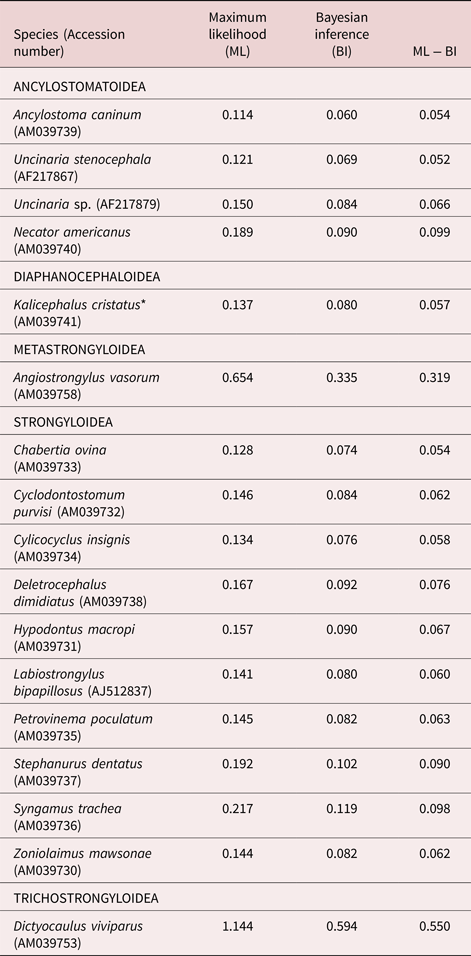
A preliminary BLAST search on the GenBank database was run to confirm the genetic proximity between the present sequence and those of strongylid nematodes. All sequence alignments were performed using M-Cofee (Notredame et al., Reference Notredame, Higgins and Heringa2000) and evaluated by the transitive consistency score, in order to verify the reliability of aligned positions and trim ambiguous sites (Chang et al., Reference Chang, Di Tommaso and Notredame2014). Preliminary phylogenetic reconstructions, including the available sequences of representatives from all superfamilies of the Strongylida (i.e. Ancylostomatoidea, Diaphanocephaloidea, Metastrongyloidea, Strongyloidea and Trichostrongyloidea sensu Anderson et al., Reference Anderson, Chabaud and Willmott2009), were performed to verify the closest taxa to D. galeatus and eliminate those unrelated (see supplementary figs S1 and S2, and table S1 for details). Therefore, Metastrongyloidea and Trichostrongyloidea were largely excluded from the analyses, one representative of each was chosen as outgroup, and sequences of Ancylostomatoidea, Diaphanocephaloidea and Strongyloidea were compared with that of D. galeatus (table 1).
Patristic distances between Diaphanocephalus galeatus and its closely related taxa, estimated from maximum likelihood and Bayesian inference trees, and their differences. Genetic sequences are from the large subunit of the nuclear rDNA (28S) and were retrieved from GenBank. Angiostrongylus vasorum and Dictyocaulus viviparus were considered as outgroups.

*Possible wrong nomination; the correct is probably Kalicephalus costatus indicus Ortlepp, 1923.
The dataset in table 1 was subjected to maximum likelihood (ML) and Bayesian inference (BI), generating trees under the TVM + I+G model of evolution using PHYML (Guindon and Gascuel, Reference Guindon and Gascuel2003) and MrBayes (Huelsenbeck and Ronquist, Reference Huelsenbeck and Ronquist2001), respectively. The model of evolution and its fixed parameters were chosen and estimated under the Akaike information criterion with jModelTest 2 (Guindon and Gascuel, Reference Guindon and Gascuel2003; Darriba et al., Reference Darriba2012). Bayesian posterior probability values were determined after running the Markov chain Monte Carlo (two runs, four chains) for 4 × 10⁶ generations, with sampling frequency every 4 × 10³ generations and discarding the initial ¼ of sampled trees (1 × 10⁶) as bur-in. For ML, bootstrap resampling was performed with 1000 non-parametric replications.
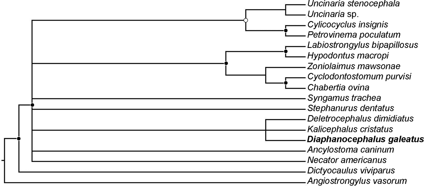
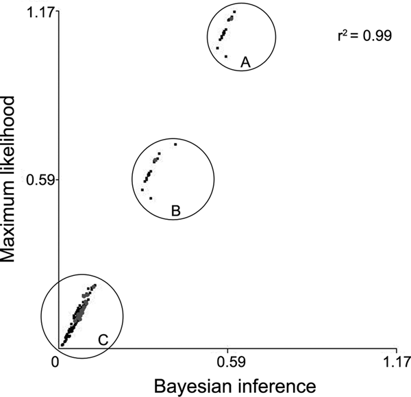
Due to the frequency of low nodal supports and consequent lack of phylogenetic resolution in the assemblage of D. galeatus (see fig. 3 and supplementary fig. S3), the trees were used to estimate the patristic distances among sequences, in the software PATRISTIC (Fourment and Gibbs, Reference Fourment and Gibbs2006). The software estimated the patristic distances from both BI and ML trees and performed a linear regression model with these variables (table 1), generating a plot (fig. 4).
Results
Systematics
Superfamily: Diaphanocephaloidea (Travassos, 1920)
Family: Diaphanocephalidae Travassos, 1920
Genus: Diaphanocephalus Diesing, 1851
Diaphanocephalus galeatus (Rudolphi, 1819)
Description
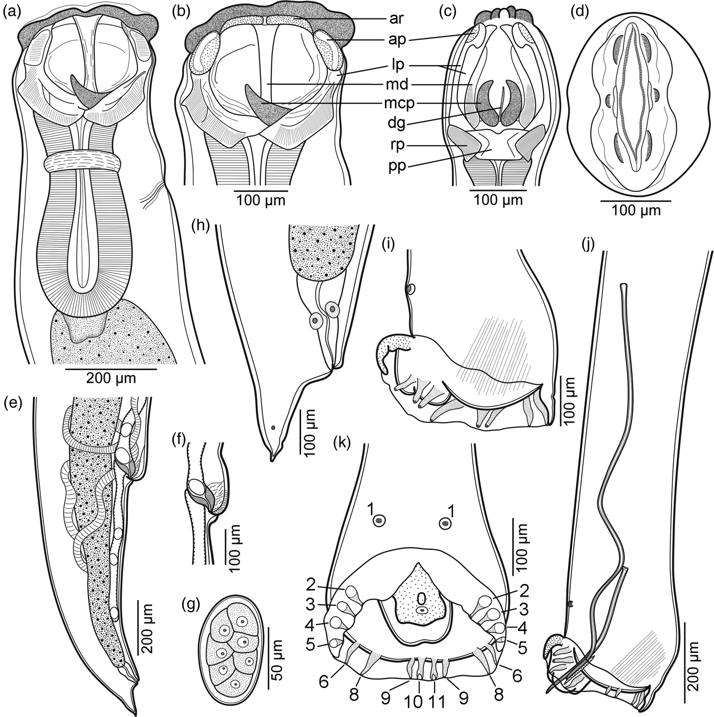
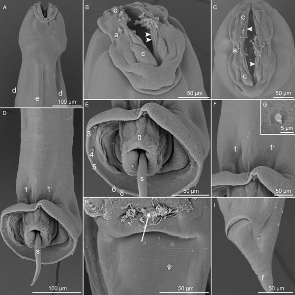
Small, robust, whitish nematodes. Cuticle thick, with fine transverse striations throughout body. Oral aperture dorso-ventrally elongate, inner margin delimited by small membranous denticles (figs 1d and 2B, C). Cephalic end with broad lip-like swelling, bearing four large submedian cephalic papillae and two lateral conspicuous amphids (figs 1a, b, d and 2A–C). Broad, complex, articulated buccal capsule laterally symmetrical (fig. 1a, b), composed of 14 paired strongly sclerotized plates, each side of capsule composed of seven plates, plus a large single plate supporting its base (fig. 1a–c). Anterior ridge of peri-oral groove bearing two articulated narrow plates; ventral and dorsal anterior plates oval, supported by two adjacent and long lateral plates, slightly C-shaped in both lateral and dorso-ventral views (fig. 1a–c). Median lateral plates long and narrow, supporting median region of buccal capsule (fig. 1a–c). Well-developed, horn-shaped, membranous cuticular processes, located at internal base of buccal capsule (fig. 1a–c). Single posterior plate supporting buccal capsule base, ventrally articulated with two slightly rectangular plates on each lateral face (fig. 1c). Small dorsal oesophageal gutter arising between membranous cuticular processes, fused with dorsal wall of buccal capsule (fig. 1c). Oesophagus strongly muscular, club-shaped (fig. 1a). Nerve ring encircling oesophagus on its first third. Excretory pore conspicuous, deirids pointed and small, both located somewhat at same level, slightly posterior to nerve ring (figs 1a and 2A). Sexual dimorphism evident, males with vesicular, well-developed copulatory bursa, females with conical tail (figs 1e, h–k and 2D, E, I).
Diaphanocephalus galeatus (Rudolphi, 1819). (a) Anterior end of male, lateral view; (b, c) female and (d) male cephalic ends, lateral, ventral and apical views, respectively; (e) posterior end of female, lateral view; (f) vulva, lateral view; (g) egg; (h) tail of female, lateral view; (i, k) bursa and (j) posterior end of male, lateral, ventral and lateral views, respectively. Abbreviations (according to Lichtenfels, Reference Lichtenfels, Anderson, Chabaud and Willmott1980): ar, narrow plate of peri-oral anterior ridge groove; ap, anterior oval plates; dg, dorsal oesophageal gutter; lp, C-shaped lateral plates; mcp, horn-shaped membranous cuticular processes; md, median lateral plates; pp, single posterior plate; rp, rectangular plate. Numbering (according to Chabaud et al., Reference Chabaud1970): 0, unpaired papilla; 1, pre-bursal papillae; 2, 3, 4, 5, 6, 8, lateral pairs of bursal rays; 9, outer simple pair of dorsal ray; 10, 11, terminally bifid inner branches of dorsal ray.

SEM micrographs of Diaphanocephalus galeatus (Rudolphi, 1819). (A) Anterior end, ventral view; (B, C) cephalic end, subventral and apical views, respectively (arrowheads indicate denticles); (D) posterior end of male, ventral view; (E) copulatory bursa, ventral view; (F, G) detail of papillae anterior to copulatory bursa, ventral views; (H) vulva, ventral view (arrow indicates vulval aperture); (I) tail of female, sublateral view. Abbreviations: a, amphid; c, cephalic papilla; d, deirid; e, excretory pore; f, phasmid; s, spicule. Numbering (according to Chabaud et al., Reference Chabaud1970): 0, unpaired papilla; 1, pre-bursal papillae; 2, 3, 4, 5, 6, 8, distal ends of lateral pairs of bursal rays.

Male (based on two adult specimens). Body length 4.2–5.0 mm, maximum width 333–389. Buccal capsule 173–176 long, 222–224 wide. Nerve ring, deirids and excretory pore, 302–345, 435–455 and 414–435, respectively, from anterior end. Oesophagus 343–365 long, maximum width 170–175; representing 7.3–8.1% of total body length. Posterior end of body provided with broad, vesicular copulatory bursa, laterally symmetric, supported by six pairs of robust bursal rays along lateral edges (2, 3. 4, 5, 6 and 8) (figs 1i–k and 2D, E), dorsal ray consisting of four segments, outer simple branches (pair 9) and two inner branches (10, 11) with shallow terminal bifurcation (fig. 1k). Thick muscular dorsal bulge anterior to copulatory bursa (fig. 1i, j). Genital cone well developed, covered by membranous process on its ventral surface, 105–123 long, 106–131 wide (figs 1k and 2E). Pair of small ciliary pre-bursal papillae (1), slightly anterior to copulatory bursa, plus unpaired weakly developed papilla (0) near distal edge of genital cone membranous process (figs 1i–k and 2D–G). Spicules without sheath, similar in shape, different in length, proximal end with smooth dilatation, distal end rounded (fig. 1j). Large (left) spicule 970–1090 long; small (right) spicule 322–380 long. Length ratio of spicules 1 : 2.9–3.0. Gubernaculum simple, rod-shaped, 125–126 (fig. 1j).
Female (based on two gravid specimens). Body length 4.7–5.1 mm, maximum width 364–494. Buccal capsule 215–218 long, 255–276 wide. Nerve ring, deirids and excretory pore, 345–353, 404–452 and 381–436, respectively, from anterior end. Oesophagus 403–412 long, maximum width 194–203; representing 8.1–8.6% of total body length. Vulva simple, post-equatorial, guarded by protruded lips (figs 1e, f and 2H), 3.7–4.1 mm from anterior end, at 78.7–80.3% of body length. Ovejector anteriorly directed, 420–460 long, with loose striated musculature and weakly developed sphincter connecting uterine branches. Amphidelphic. Uterus containing few eggs, not reaching oesophageal and anal regions. Thin-shelled ellipsoid eggs in early stages of cleavage, containing large granulose cells (fig. 1g), 81–91 × 44–52 (n = 6). Tail conical, ending in thin point, small phasmidial pores located c. 50 from tail end (figs 1h and 2I).
Taxonomic summary
Host. Dracaena paraguayensis Amaral, 1950 (Sauria: Teiidae).
Site of infection. Small intestine.
Locality. State highway MS 184 crossing Pantanal wetlands, near municipality of Miranda, State of Mato Grosso do Sul, Brazil (19°33′42″S, 57°19′40″W).
Voucher specimens deposited. Two males, two females, one hologenophore ZUFMS-NEM00052.
Remarks
The genus Diaphanocephalus was proposed by Diesing (Reference Diesing1851) to accommodate the species Strongylus galeatus (= D. galeatus), described by Rudolphi (Reference Rudolphi1819), from an unknown lacertid collected in Brazil. According to Lichtenfels (Reference Lichtenfels, Anderson, Chabaud and Willmott1980), updated in Anderson et al. (Reference Anderson, Chabaud and Willmott2009), the genus includes species with peri-oral groove delimited by anterior and posterior ridges, two membranous cuticular processes at the base of the buccal cavity, spicules not alate, a bell-shaped copulatory bursa and a thick muscular dorsal bulge anterior to it. The present specimens have all these features and were assigned to D. galeatus by the overlapping biometric measurements and very similar morphology, which was confirmed after observations of the conspecific specimens deposited in CHIOC (CHIOC 1935, 2153, 4126, 5373, 5829, 6694, 6743, 7589, 7595, 8191, 8256, 8419, 8552, 9257, 11119, 18337).
This is the first time that the following features are observed and described for D. galeatus: lip-like cephalic protrusions, denticles delimiting the inner margin of oral aperture (reminiscent of a rudimentary corona radiata), cephalic papillae, amphids, deirids, phasmids in females and the unpaired papilla on the membranous projection covering the genital cone in males (papilla 0 according to Chabaud et al., Reference Chabaud1970). Furthermore, the structure of the buccal capsule is described in detail, differing from the description of Freitas and Lent (Reference Freitas and Lent1938). However, we observed the same specimens studied by Freitas and Lent (Reference Freitas and Lent1938) (CHIOC 7589, 8191, 8419) and concluded that the authors overlooked some structures and misinterpreted others. After the above-mentioned study, the only additional morphological data on D. galeatus was provided by Vieira et al. (Reference Vieira, Fedatto-Bernardon and Müller2016), who studied specimens from S. merianae in southern Brazil, but without greater detail (e.g. absence of SEM observation).
Two other congeners have been reported parasitizing teiid lizards in South America, namely, D. diesingi and D. jacuruxi, of which this last is a parasite of Dracaena guianensis Daudin, 1802 from northern Brazil (Alho, Reference Alho1965). After observation of the type specimens of D. jacuruxi (CHIOC 29907, 29908), it was possible to confirm its morphological similarity with the present material, i.e. the presence of the papillae pair 1 (pre-bursal pair) and the shape of gubernaculum, which is not “human foot-shaped” as described by Alho (Reference Alho1965), but rod-shaped as in the present specimens. Moreover, D. galeatus and D. jacuruxi have very similar morphometric features. In this sense, D. jacuruxi should be considered a synonym of D. galeatus.
Diaphanocephalus diesingi, a parasite of T. teguixin (reported as T. nigropunctatus) from the Sate of Pará, northern Brazil, differs from D. galeatus based on the smaller sizes of spicules and gubernaculum (Freitas and Lent, Reference Freitas and Lent1938). Unfortunately, observation of the few type specimens of D. diesingi (CHIOC 9812, 9832) was inconclusive, because the material is poorly preserved and the important morphological features were impossible to verify (e.g. presence or absence of pre-bursal papillae); however, the buccal capsule is very similar to that of D. galeatus. Therefore, D. diesingi is tentatively retained as valid until new data become available, but it should be mentioned that indications are strongly in favour of a possible synonym.
Molecular characterization and genetic comparison
The partial sequence of the 28S rDNA was obtained for D. galeatus (595 bp), representing the first genetic characterization of the species. The present sequence was most similar to that of Nicollina cameroni Thomas, 1959 (sequence identity 97.6%), followed by that of Ancylostoma caninum (Ercolani, 1859) (sequence identity 97.5). The sequence named as Kalicephalus cristatus (accession number AM039741; see discussion for details), which represents the only other diaphanocephalid genetically characterized, showed 97.0% of sequence identity with D. galeatus.
In the phylogenetic reconstructions using BI and ML, D. galeatus appeared in the same clade of K. cristatus, forming a sister group of Deletrocephalus dimidiatus (Diesing, 1851) (Deletrocephalidae); however the nodal supports were low (see fig. 3 and supplementary fig. S3). Conversely, high nodal supports along with the patristic distances indicated the genetic proximity of D. galeatus with representatives of Ancylostomatoidea, Strongyloidea and K. cristatus, as well as the separation between the species and the taxa of Metastrongyloidea and Trichostrongyloidea (table 1, figs 3 and 4, and supplementary table S2). The patristic distances estimated from the BI and ML tree were highly correlated (fig. 4; r2 = 0.99).
Tree inferred based on Bayesian inference of sequences of D2–D3 region of the 28S rRNA gene, from representatives of Ancylostomatoidea, Diaphanocephaloidea and Strongyloidea. Filled and unfilled circles represent high and moderate nodal supports, i.e. Bayesian posterior probability >0.96 and 0.90–0.95, respectively (4 × 10⁶ generations, sampling frequency = 4 × 10³, bur-in = 1 × 10⁶). Specimen in bold is from the present study. Angiostrongylus vasorum and Dictyocaulus viviparus were used as outgroups.

Linear regression plot of the differences between the patristic distances estimated from the maximum likelihood and the Bayesian phylogenetic trees. A represents the group of all patristic distances from Angiostrongylus vasorum (Metastrongyloidea); B represents the group of all patristic distances from Dictyocaulus viviparus (Trichostrongyloidea); C represents patristic distances within Ancylostomatoidea, Diaphanocephaloidea and Strongyloidea. Grey circles represent patristic distances from Diaphanocephalus galeatus.

Discussion
Diaphanocephalus galeatus has a broad occurrence in South America, from northern to southern Brazil, Bolivia and Argentina (Ávila and Silva, Reference Ávila and Silva2010; Vieira et al., Reference Vieira, Fedatto-Bernardon and Müller2016; Teixeira et al., Reference Teixeira2017). The species is considered specific of S. merianae by some authors (Freitas and Lent, Reference Freitas and Lent1938; Ramalho et al., Reference Ramalho2009), but it has been reported in other hosts, e.g. S. rufescens (Günther, 1871) and T. teguixin (Ávila and Silva, Reference Ávila and Silva2010). Indeed, the systematic classification of these host species is complicated (Harvey et al., Reference Harvey, Ugueto and Gutberlet2012), which may confuse the discussion on host specificity of D. galeatus. However, the present results indicate that the parasite is not specific of S. merianae, as its infection was confirmed in D. paraguayensis and D. guianiensis. These findings also reinforce the suspected synonymy of D. diesingi with D. galeatus (see remarks).
The present specimens showed all the morphological features from the generic diagnosis of Diaphanocephalidae, except for the weakly developed dorsal gutter (see Lichtenfels, Reference Lichtenfels, Anderson, Chabaud and Willmott1980). However, individually, this is not a strong character within the family, and Diaphanocephalidae is well established based on morphological traits.
It should be mentioned that the sequence AM039741, isolated from a parasite of the lowland copperhead Austrelaps superbus (Günther, 1858) (Serpentes: Elapidae) in Australia, is named as ‘K. cristatus’ in the GenBank database. Following extensive searches of the revisions on the genus Kalicephalus and the most recent descriptions of congeners (Ortlepp, Reference Ortlepp1923; Schad, Reference Schad1962; Baker, Reference Baker1987; Zhang et al., Reference Zhang, Zhang and Zhang2011; Kuzmin et al., Reference Kuzmin2013) we found no taxonomic references to this species, except in Chilton et al. (Reference Chilton2006). It is probably a case of confusion and the correct name would be K. costatus indicus Ortlepp, 1923, which has been reported from snakes in Australia (sensu Schad, Reference Schad1962).
The nodal supports observed in the phylogenetic trees used for patristic distance estimation were generally low (see fig. 3 and supplementary fig. S3). Even though these support values would be improved with addition of other genetic regions, the present results agree with those in Chilton et al. (Reference Chilton2006), in which Diaphanocephaloidea, and consequently D. galeatus, are closer to Ancylostomatoidea and Strongyloidea compared with any lineage of Trichostrongyloidea and Metastrongyloidea. It is also interesting that D. dimidiatus, a parasite of birds from the genus Rhea Brisson, 1760, in its own family Deletrocephalidae, clustered with diaphanocephalids. However, it is important to highlight that evaluating the phylogenetic position of D. galeatus was not the main goal here.
The patristic distances indicated more proximity of D. galeatus with A. caninum and Uncinaria stenocephala Railliet, 1884 than with ‘K. cristatus’. On one hand, Durette-Desset and Chabaud (Reference Durette-Desset and Chabaud1993) proposed the inclusion of Ancylostomatoidea and Diaphanocephaloidea in the erected suborder Ancylostomatina, suggesting some relationship between these higher taxa. On the other hand, the relatedness among the lower taxa (i.e. families) has been unsolved and subject of discussion (see Durette-Desset et al., Reference Durette-Desset, Beveridge and Spratt1994; Chilton et al., Reference Chilton2006). Hence, we cannot infer any comment on how exactly these taxa are related.
With the present genetic and morphological characterization, the validity of D. galeatus was confirmed. These data may serve for further comparative approaches on different populations of the parasite, from different hosts in different geographical areas, mitigating taxonomic confusions.
The close relatedness between strongylid nematodes and some lineages of rhabditids is undeniable (Medal et al., Reference Medal2007; Blaxter and Koutsovoulos, Reference Blaxter and Koutsovoulos2014). It is clear when their larval development is compared (Anderson, Reference Anderson2000). However, we believe it is premature to accept the suppression of the Strongylida, as observed in recent phylogenies (e.g. Blaxter and Koutsovoulos, Reference Blaxter and Koutsovoulos2014). A strong argument is that Strongylida represents a well-supported monophyletic lineage (Chilton et al., Reference Chilton2006), with the copulatory bursa as a clear synapomorphy (Durette-Desset et al., Reference Durette-Desset, Beveridge and Spratt1994; Anderson et al., Reference Anderson, Chabaud and Willmott2009). Furthermore, the loss of ordinal status may complicate the already confused taxonomy of the bursate nematodes.
Supplementary material
To view supplementary material for this article, please visit https://doi.org/10.1017/S0022149X18000597
Acknowledgements
The authors would like to thank Dr Marcelo Knoff and Dr Daniela A. Lopes from the Coleção Helmintológica do Instituto Oswaldo Cruz (CHIOC) for loan of specimens.
Financial support
F.B.P. was supported by a post-doctoral fellowship PNPD-CAPES (Programa Nacional de Pós-Doutorado-Coordenação de Aperfeiçoamento de Pessoal de Nível Superior-CAPES, Brazil).
Conflict of interest
None.