1. Introduction
Vascular plants underwent a major radiation during Devonian time in both taxonomic diversity and morphological complexity, with the appearance of several groups of basal euphyllophytes such as the Iridopteridales, Pseudosporochnales, Rhacophytales, progymnosperms and many others with uncertain higher-rank assignments (e.g. Skog & Banks, Reference Skog and Banks1973; Cornet, Phillips & Andrews, Reference Cornet, Phillips and Andrews1976; Stein, Reference Stein1982; Stein, Wight & Beck, Reference Stein, Wight and Beck1984; Berry & Stein, Reference Berry and Stein2000; Meyer-Berthaud, Soria & Young, Reference Meyer-Berthaud, Soria and Young2007; Hao & Xue, Reference Hao and Xue2013 a,b; Wang et al. Reference Wang, Xu, Xue, Wang and Liu2015; Xue et al. Reference Xue, Huang, Wang, Wang and Hao2015). These groups are of significance in that they bear directly on the investigation of the origin, early diversification and evolution of the ferns, sphenopsids and lignophytes; however, the systematics and taxonomy of these basal plants have been the subject of much debate.
The Rhacophytales, represented by the Late Devonian Rhacophyton Crépin, was compared to the progymnosperms or to the zygopterid ferns (Leclercq, Reference Leclercq1951; Andrews & Phillips, Reference Andrews and Phillips1968; Cornet, Phillips & Andrews, Reference Cornet, Phillips and Andrews1976; Taylor, Taylor & Krings, Reference Taylor, Taylor and Krings2009). The Iridopteridales and Pseudosporochnales were grouped into the Cladoxylopsida by Berry & Stein (Reference Berry and Stein2000), but others have emphasized the distinction between the Iridopteridales and Cladoxylopsida and raised doubts about the delimitation and monophyly of these groups (Cordi & Stein, Reference Cordi and Stein2005; Meyer-Berthaud, Soria & Young, Reference Meyer-Berthaud, Soria and Young2007; Xue, Hao & Basinger, Reference Xue, Hao and Basinger2010). The phylogenetic analysis conducted by Rothwell (Reference Rothwell1999) showed that the Cladoxylopsida, Rhacophyton and zygopterids form a monophyletic clade sister to the sphenopsids, and that they, as well as the sphenopsids, constitute a sister group to lignophytes stemmed by progymnosperms. A contrary phylogenetic hypothesis holds that the Cladoxylopsida and Rhacophyton were stem groups within the Moniliformopses (the sphenopsid-fern clade), a group characterized by the presence of ‘peripheral permanent protoxylem’ (Beck & Stein, Reference Beck and Stein1993; Kenrick & Crane, Reference Kenrick and Crane1997). These issues can only be resolved by further investigations on the morphology and anatomy of basal euphyllophytes.
Largely unexplored regions of the Canadian Arctic Archipelago have been investigated for remains of early land plants by field parties led by J. F. Basinger of the University of Saskatchewan. Particularly significant discoveries have been made in the Upper Devonian of Ellesmere Island (Hill, Scheckler & Basinger, Reference Hill, Scheckler and Basinger1997) and the Upper Silurian and Lower Devonian of Bathurst Island (Basinger, Kotyk & Gensel, Reference Basinger, Kotyk and Gensel1996; Kotyk & Basinger, Reference Kotyk and Basinger2000; Gensel, Kotyk & Basinger, Reference Gensel, Kotyk, Basinger, Gensel and Edwards2001; Kotyk et al. Reference Kotyk, Basinger, Gensel and de Freitas2002). During Devonian time, the Canadian Arctic Archipelago was distant from other known sources of fossil plants in Asia and Appalachian Laurentia (Europe and eastern North America), and probably occupied a different climatic zone (Scotese, Reference Scotese2001). The plants reported from these islands, such as Bathurstia Hueber and Ellesmeris Hill, Scheckler & Basinger, provide new data on the evolutionary history and palaeophytogeographic zonation of Devonian plants (Hill, Scheckler & Basinger, Reference Hill, Scheckler and Basinger1997; Kotyk & Basinger, Reference Kotyk and Basinger2000).
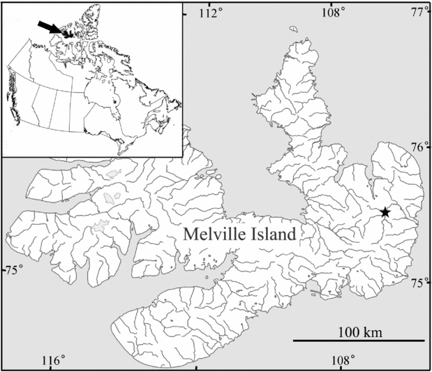
Harrison's (Reference Harrison1995) thorough survey and detailed description of the bedrock geology and the structural and tectonic evolution of Melville Island, Arctic Canada (Fig. 1) revealed extensive exposure of Middle and Upper Devonian non-marine deposits. While palynomorph assemblages were obtained for biostratigraphic control, and the occurrence of coals in several formations was noted (Harrison, Reference Harrison1995), Devonian megafossil plants were not reported from the island. During a palaeobotanical survey led by J. F. Basinger and S. E. Scheckler in 1998, rare occurrences of Late Devonian plants were located, with one locality yielding abundant remains of a new species of fern-like plant with the quadriseriate branching pattern typical of Rhacophytales. This plant provides an opportunity to assess the distribution of quadriseriate branching and the evolutionary relationship among some key basal euphyllophyte plants.
Map of Melville Island, Canadian Arctic Archipelago, Nunavut, Canada, showing the fossil locality (black star).

2. Stratigraphy
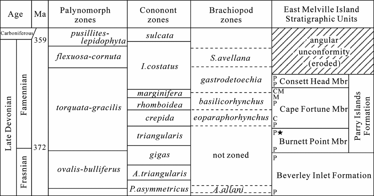
The plant fossils were collected from the west bank of the Baldwin River of eastern Melville Island, Canadian Arctic Archipelago, Nunavut, Canada (75°32.75′N and 106°32.76′W; Fig. 1). Upper Devonian deposits of this area are subdivided into the Beverley Inlet Formation and the overlying Parry Islands Formation (Fig. 2). Dominant rock types of these two formations include quartz sandstone, siltstone, mudrock and minor coals (Harrison, Reference Harrison1995). Three members are recognized within the Parry Islands Formation: in ascending order the Burnett Point, Cape Fortune and Consett Head members (Embry & Klovan, Reference Embry and Klovan1976) (Fig. 2), corresponding respectively to the units DPI1, DPI2 and DPI3–DPI5 of Harrison (Reference Harrison1995). Our fossils were derived from a c. 1m thick bed of dark grey mudrock in the upper part of the Burnett Point Member (DPI1), c. 30m below the base of the overlying Cape Fortune Member (DPI2). The Burnett Point Member, with a thickness of 202–310m in the study area, composed of sandstone, siltstone and minor shale, has been interpreted to be a deltaic facies, while the Cape Fortune Member has been interpreted to be a shallow marine facies on the basis of conodonts and other marine fossils (Harrison, Reference Harrison1995).
Correlation chart of the Upper Devonian strata of eastern Melville Island (modified from figure 38 of Harrison, Reference Harrison1995). Ma – millions of years ago; numerical ages based on Cohen et al. (Reference Cohen, Finney, Gibbard and Fan2013); black star – plant locality; C – conodont collection; M – shelly macrofauna; P – palynomorphs.

Age determination for these formations was presented by Harrison (Reference Harrison1995, appendix 4), and relevant data is summarized here and in Figure 2. The macrofauna contained in the Beverley Inlet Formation indicate a latest Givetian to early Frasnian age, while the palynomorph assemblages are early Frasnian through late Frasnian, spanning the basal to upper ovalis–bulliferus Zone. The Parry Islands Formation was dated on the basis of palynomorph assemblages as late Frasnian through middle Famennian or early late Famennian. The Burnett Point Member contains a palynomorph assemblage that spans the uppermost ovalis–bulliferus Zone (Frasnian) to lower torquata–gracilis Zone (Frasnian–Famennian) (Richardson & McGregor, Reference Richardson and McGregor1986). In the type section of the Cape Fortune Member near Beverley Inlet, conodont assemblages in the lower part of the section represent the crepida Zone of the early Famennian. As the beds bearing our fossil plants occur in the upper part of the Burnett Point Member, and c. 30m below the base of the Cape Fortune Member, we interpret the age as early Famennian.
3. Material and methods
Plant remains were recovered from a single bed c. 1m thick, and represent a monotypic assemblage. Numerous specimens were collected, with c. 90 specimens selected as the basis for describing the main axes, first- and second-order branches, and sterile and fertile ultimate appendages. More than 30 specimens have fertile appendages preserved. The fossils are preserved as impressions and compressions, without any indication of permineralization. Compressions are heavily coalified, with a thick layer of preserved carbon which can be easily broken off and lost. Careful dégagement with steel needles revealed the morphological details of branching and the architecture of fertile appendages. Maceration failed to yield any spores or cellular cuticles. Photographs of specimens were taken with a Lumenera Infinity 4 digital camera. All specimens are housed in the University of Saskatchewan Palaeobotanical Collection, Department of Geological Sciences, University of Saskatchewan, Canada.
4. Systematic palaeobotany
Subdivision EUPHYLLOPHYTINA Kenrick & Crane, Reference Kenrick and Crane1997
Order RHACOPHYTALES sensu Taylor, Taylor & Krings, Reference Taylor, Taylor and Krings2009
Genus Melvillipteris gen. nov.
Generic diagnosis. Plant monopodial, with zigzag to straight, prostrate to upright main axes. Dense roots attached on some internodes of main axes. First-order branches inserted on main axes in alternate pairs, showing a quadriseriate arrangement. First-order branches, on the proximal portions, bearing sterile ultimate appendages composed of dichotomous branching systems. Second-order branches borne alternately on first-order branches, bearing alternate ultimate appendages which may be fertile or sterile. Sterile ultimate appendages of second-order branches with one or two successive dichotomies and paired tips curving oppositely. Each fertile appendage with an initial dichotomy forming two sister branches, each of which dichotomizes three or four times to form a small sporangial cluster. Sporangia with pointed tips and longitudinal dehiscence.
Type species. Melvillipteris quadriseriata sp. nov.
Melvillipteris quadriseriata sp. nov.
Holotype designated here. Specimen US734-9156a (Fig. 3e).
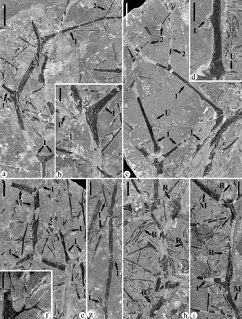
Sterile branches of Melvillipteris quadriseriata gen. et sp. nov. In all Figures 3–7: M with arrow – main axis; 1 with arrow – first-order branch; 2 with arrow – second-order branch; U with arrow – sterile ultimate appendage; R with arrow – roots. (a) Main axis. The proximal node shows a pair of first-order branches; at the middle node, one member of the paired first-order branches has been broken off; while at the distal node, one first-order branch extends to the upper right and the second of the pair of first-order branches is probably buried in the rock matrix. See interpretative drawing in Figure 5a. US734-9135. Scale bar = 15mm. (b) Main axis showing a pair of first-order branches. US734-9146. Scale bar = 5mm. (c) Main axis at lower left and at right, both with paired first-order branches attached. First-order branches bear sterile ultimate appendages proximally, and then second-order branches distally. White arrow points to the part enlarged in (d). See interpretative drawing for the left axis in Figure 5b. US734-9136. Scale bar = 15mm. (d) Enlarged from (c), showing two sterile ultimate appendages, one of which is attached at the swollen base of the first-order branch. Scale bar = 5mm. (e) Holotype. Main axis, showing a zigzag appearance. The proximal node, at right, has a pair of first-order branches. Arrow U points to the part enlarged in (f). See interpretative drawing in Figure 5c. US734-9156a. Scale bar = 10mm. (f) Enlargement of a sterile ultimate appendage in (e), showing dichotomous branching. Scale bar = 3mm. (g) Main axis, showing an upright appearance, with three nodes bearing first-order branches. See interpretative drawing in Figure 5d. US734-9141. Scale bar = 15mm. (h) Main axis, showing attached roots. Note that multiple roots form a group, attached to only one side of each internode. US734-9144. Scale bar = 10mm. (i) Main axis, showing clusters of attached roots to one side of internodes. US734-9145. Scale bar = 10mm.

Paratypes. Specimens US734-9112, -9126, -9133, -9136, -9144, -9177 (Figs 3c, 3h, 4k, 6a, 7a, 7f, 9a).
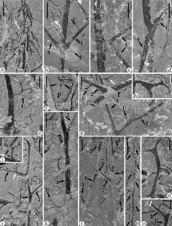
Sterile branches of Melvillipteris quadriseriata gen. et sp. nov. (a) A small axis with irregularly attached roots, or possibly a primary root with secondary roots. US734-9166a. Scale bar = 5mm. (b) Main axis with a pair of first-order branches. Note two sterile ultimate appendages found on the proximal portions of the first-order branch at right. See interpretative drawing in Figure 5e. US734-9140. Scale bar = 10mm. (c) Main axis, showing paired first-order branches. US734-9142. Scale bar = 10mm. (d) Main axis, showing paired first-order branches. US734-9139. Scale bar = 10mm. (e) Proximal portion of a first-order branch, showing the swollen base (bottom) and a sterile ultimate appendage. US734-9133. Scale bar = 5mm. (f) Two first-order branches, with a fragment of a sterile ultimate appendage attached to the base of the right branch. US734-9161a. Scale bar = 5mm. (g) First- and second-order branches, showing attachment of sterile ultimate appendages. White arrow points to the part enlarged in (h). US734-9146. Scale bar = 4mm. (h) Enlargement of a sterile ultimate appendages shown in (g). Scale bar = 1mm. (i) First-order branch, showing alternately arranged second-order branches. White arrow points to the part enlarged in (j). US734-9144. Scale bar = 8mm. (j) Enlarged from (i), showing a sterile ultimate appendage borne on a second-order branch. Scale bar = 2mm. (k) First-order branch, showing a proximal sterile ultimate appendage and then distally a second-order branch. US734-9133. Scale bar = 5mm. (l) Two first-order branches, showing the alternate arrangement of second-order branches. US734-9131. Scale bar = 8mm. (m) Distal portion of a second-order branch, showing alternate arrangement of sterile ultimate appendages (arrows). See interpretative drawing in Figure 5f. US734-9144. Scale bar = 10mm. (n) Enlargement of the top part of (m), showing the distal tip of the second-order branch. Scale bar = 2mm. (o) First- and second-order branches, with the latter bearing sterile ultimate appendages. White arrow points to a sterile ultimate appendage enlarged in Figure 6b. US734-9144. Scale bar = 5mm.

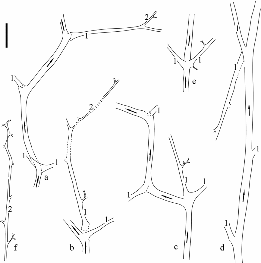
Interpretative drawings of main axes, first-order branches and second-order branches of Melvillipteris quadriseriata gen. et sp. nov. (a–e) Specimens shown in: (a) = Figure 3a; (b) = Figure 3c; (c) = Figure 3e; (d) = Figure 3g; and (e) = Figure 4b. Scale bar = 15mm. (f) Specimen shown in Figure 4m. Note that scale bar = 10mm for (f) only. Numeral 1 indicates first-order branch; numeral 2, second-order branch; and arrows, the growth direction of main axes.

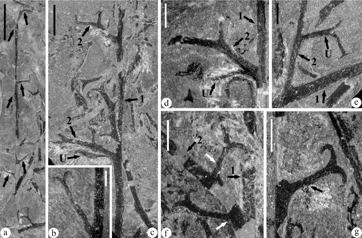
Sterile branches and ultimate appendages of Melvillipteris quadriseriata gen. et sp. nov. (a) Second-order branch, showing regular arrangement of sterile ultimate appendages (arrows). US734-9126. Scale bar = 10mm. (b) Sterile ultimate appendage with only one dichotomy, enlarged from Figure 4o. US734-9144. Scale bar = 1mm. (c) Distal portion of first-order branch. Note the ultimate appendage (arrow U) at the base of a second-order branch. US734-9201a. Scale bar = 4mm. (d) Base of second-order branch with attached ultimate appendage. US734-9128. Scale bar = 2mm. (e) First-order branch, showing a second-order branch with a proximal sterile ultimate appendage. US734-9184. Scale bar = 2mm. (f) First-order branch and a second-order branch, the latter of which bears a complete sterile ultimate appendage at the base (lower white arrow) and a second one at upper white arrow. US734-9198. Scale bar = 2mm. (g) Sterile ultimate appendage of second-order branch. Arrow indicates a dichotomy, with one of the sister branches being buried in the rock matrix. US734-9199. Scale bar = 2mm.

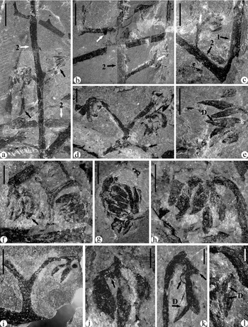
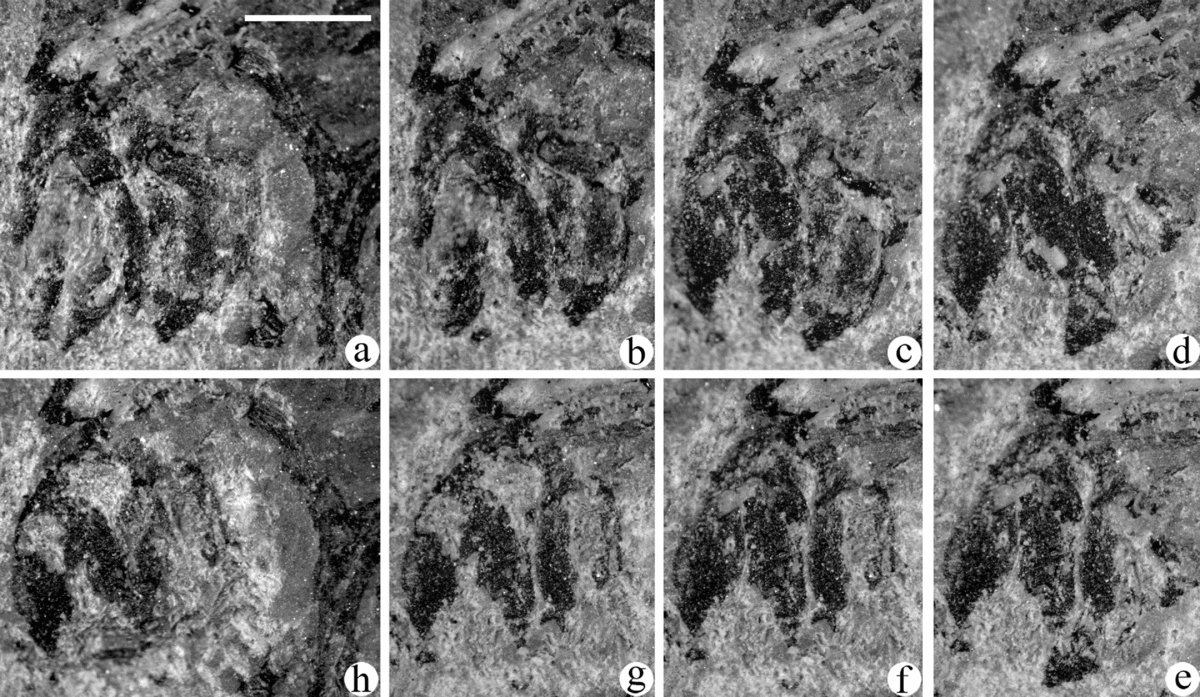
Fertile specimens of Melvillipteris quadriseriata gen. et sp. nov. (a) Two axes interpreted as fertile second-order branches (arrow 2), with fertile appendages attached (black arrows). Upper black arrow points to the fertile appendage enlarged in (e). See line drawing for one axis in Figure 8a. US734-9112. Scale bar = 3mm. (b) Fertile second-order branch, showing an alternate arrangement of fertile appendages. White arrows indicate the initial dichotomy of the fertile appendages. See line drawing in Figure 8b. US734-9112. Scale bar = 3mm. (c) Fertile first- and second-order branches. White arrow indicates the initial dichotomy of the fertile appendage. See line drawing in Figure 8c. US734-9183. Scale bar = 3mm. (d) Fertile appendage, showing an initial dichotomy and characteristic two clusters of sporangia. Arrow indicates an axis which appeared to be attached to the fertile appendage, but close observation shows it is an accident of preservation. US734-9195a. Scale bar = 1mm. (e) Enlarged from (a), showing a sporangial dehiscence line (arrow D). Scale bar = 1mm. (f) Fertile appendage, showing a two-clustered appearance. Eight stages of serial dégagement of the left cluster are shown in Figure 9. US734-9177. Scale bar = 1mm. (g) Sporangial cluster. US734-9190a. Scale bar = 1mm. (h) Sporangial cluster. US734-9171. Scale bar = 1mm. (i) Fertile appendage, showing an initial dichotomy and one pair of sporangia from the cluster. See line drawing in Figure 8d. US734-9169. Scale bar = 2mm. (j) Isolated sporangial truss, showing three sporangia and the base of the fourth (arrow). See line drawing in Figure 8e. US734-9125. Scale bar = 0.5mm. (k) Sporangial truss, showing two sporangia and the bases of two other sporangia (upper arrows). Arrow D indicates a longitudinal dehiscence. US734-9115. Scale bar = 0.5mm. (l) Isolated sporangium, showing a longitudinal dehiscence line (arrows D). US734-9173. Scale bar = 0.5mm.


Repository. Department of Geological Sciences, University of Saskatchewan, Canada.
Type locality. West bank of Baldwin River of eastern Melville Island, Canadian Arctic Archipelago, 75°32.75′N and 106°32.76′W.
Stratigraphy. Burnett Point Member of the Parry Islands Formation, Upper Devonian (Famennian).
Derivation of generic name. ‘Melvillipteris’, from Melville Island, where the plant was collected.
Derivation of species epithet. ‘quadriseriata’, refers to the quadriseriate branching pattern of the first-order branches.
Species diagnosis. Characters as in generic diagnosis. Main axes 3–6mm wide and at least 160mm long, with internode length 20–60mm. First-order branches 0.9–2.5mm wide and up to at least 125mm long. Second-order branches about 0.7mm wide and up to at least 68mm long, borne alternately with internode length 12–22mm. Sterile ultimate appendages inserted at right angles on axes; those on first-order branches restricted to proximal region, 7–11mm long; those on second-order branches 4–5.3mm long. Fertile appendages with axes 0.3–0.7mm wide at base and extending 1.7–4.0mm to point of initial dichotomy to form two sister branches; each branch recurved distally, dividing dichotomously three or four times to support at least 12 sporangia, and with an overall length of 1.8–4.2mm; penultimate divisions of fertile appendages each bearing two to four small fusiform sporangia that are grouped into a truss. Sporangia 1.0–1.4mm long and 0.3–0.5mm wide.
5. Description
5.a. Main axes
Different orders of branching are distinguished on the basis of size, derivation, arrangement of lateral organs and morphology. The main axes are the largest, with a diameter ranging from 3.0mm to 6.0mm, and are the only order of axis that bears roots and paired branches. The longest preserved main axis reaches 16cm without tapering, and in our large collection of over 90 specimens there is no evidence that these axes were borne on a more proximal order of axis; therefore, they are interpreted as the primary stems of the plant. No examples show bifurcations, indicating that they seldom or never branched in this way. Two types of appearance of the main axes were found: straight and zigzag. Those with a zigzag pattern are bent at each node where first-order branches depart (Figs 3a, e, 5a, c); and while some are flexed only slightly (Figs 3a, c, i, 5a, b), others flex strongly, with a bending angle of c. 110° (Figs 3e, 5c). Some axes, on the other hand, are straight in appearance (Figs 3g, 4b–d, 5d, e). The internode between successive pairs of first-order branches on zigzag main axes varies from 20mm to 52mm, while on straight main axes this interval is longer, from 55mm to 60mm. The width of the two types of main axes is similar, and as noted the width remains constant along the preserved length (Fig. 3a, e, g), although a slight swelling occurs below the attachment point of first-order branches to a maximum diameter of about 6.0mm.
Some main axes have adventitious roots attached to the internodes (Fig. 3h, i, arrows R; Fig. 4a). In most examples, more than 15 individual roots are attached as a cluster to only one side of each internode; typically only a portion of the internode, about 17–23mm in length, bears roots. It appears that attachment of roots alternates below and above the node of attachment of first-order branches, so that the roots fan out first to one side, then the other (Fig. 3h, i, arrows R). In the specimen shown in Figure 3h, the proximal internode bears roots on the left side, and the roots of the middle internode are on the right side, and those of the distal internode on the left. The roots of main axes are 0.5–0.9mm wide, with the longest preserved length reaching 27mm. In one specimen, some thinner roots, 0.4–0.5mm wide, extend irregularly from all sides of a small central axis of c. 1.2mm wide (Fig. 4a). This relatively small axis may represent a subterranean part of an axis, or perhaps a primary root, and then the lateral roots would be secondaries.
5.b. First- and second-order branches
Dégagement of many specimens demonstrated that first-order branches diverge from the main axes in distichous pairs, in a quadriseriate arrangement. This paired arrangement is considered typical (Figs 3a–c, e, g, 4b–d, arrows 1; Fig. 5a–e, numerals 1), and where first-order branches appear to be borne singly, this is interpreted as a result of preservational limitation, such that one of the paired branches would have broken off during preservation or is buried in the matrix. In the specimen shown in Figure 3a, the proximal node has a pair of first-order branches; the middle node has only one branch, but thick coalified material marks the base of a second branch, which would have extended towards to the observer; and the distal node also has only one branch visible, with the second branch buried in the matrix. The pairs of first-order branches share a common base at the node, and appear to be alternately arranged along the main axes (Fig. 3a, e, g, arrows 1; Fig. 5a, c, d, numerals 1). The angle between the first-order branches of each pair ranges from 60° to 120° (Fig. 3a–c, e). It is estimated that the first-order branches depart from the main axes at a similar angle, 60° to 115°.
First-order branches have a wide, swollen base (Figs 3b–d, 4b), but quickly taper to a width of 2.0–2.5mm, and then gently taper distally. Distal-most portions of first-order branches are only 0.9–1.5mm wide (Figs 4i, l, 6c, arrows 1). The longest preserved first-order branch approaches 12.5cm in length with only slight tapering.
Second-order branches are borne singly and are alternately arranged on the first-order branches (Fig. 4i, l, arrows 2), with an interval between the bases of two successive branches ranging from 12mm to 22mm. The second-order branches are slender, with an average diameter of c. 0.7mm, and the longest example of a second-order branch reaches 68mm. Besides the difference in size between first- and second-order branches, the second-order branches bear only ultimate appendages, allowing them to be distinguished even from the narrow distal portions of first-order branches (Figs 4g, i, m, o, 5f, 6a, c–f). The length of second-order branches decreases distally on first-order branches.
5.c. Sterile ultimate appendages of first-order branch
One or two sterile ultimate appendages are attached at right angles to the proximal portions of the first-order branches (Figs 3c–f, 4b, e, f, k, arrows U). Where two occur, the first ultimate appendage appears to be borne at the swollen base (Figs 3c, d, 4b, f), but preservation is too poor to generalize; the second ultimate appendage is alternate to the first (Figs 3d, 4b, 5e). Nevertheless, preservation is too poor in many cases to determine whether ultimate appendages are borne at the base of all first-order branches (Fig. 3a, b, i). Distal to these sterile ultimate appendages, second-order branches are borne, with the first attached c. 40–45mm from the base of the first-order branch (Figs 3c, 4k).
Sterile ultimate appendages on first-order branches are dichotomously branching systems, with at least three successive dichotomies (Figs 3f, 4e, k). The branching angles of the dichotomies vary from 55° to 110°. These ultimate appendages appear to be terete and three-dimensional. They have a maximum width of 0.6mm, decreasing distally to a tapering tip. The overall length of an ultimate appendage borne on a first-order branch varies from 7mm to 11mm.
5.d. Sterile ultimate appendages of second-order branch
Ultimate appendages are alternately arranged along the second-order branches. At the base of some second-order branches, the first ultimate appendage can be found (Fig. 6c, d, arrows U; Fig. 6f, lower white arrow), but only one specimen has been found complete. This first ultimate appendage, averaging 0.35mm in width, dichotomously divides only once at c. 3.4mm above the base, and the two daughter divisions, c. 1.0mm in length, terminate in oppositely recurved tips (Fig. 6f, lower white arrow). The next ultimate appendage occurs about 3.5–8.5mm from the base of the second-order branch (Figs 4g, i, j, o, 6e, f). The internodes between successive ultimate appendages decrease in length distally, from c. 9.8mm at the base to c. 3.1mm at the tip (Fig. 4m, arrows; Fig. 4n). Rarely, second-order branches show a regular pattern of alternation between longer (c. 16.6mm) and shorter (c. 3.4mm) internodal lengths (Fig. 6a).
Sterile ultimate appendages on second-order branches are also dichotomously branching systems, but are smaller than those of first-order branches, 4.0mm to 5.3mm in overall length, with a maximum diameter of c. 0.5mm. These ultimate appendages typically dichotomize twice (Figs 4h, j, 6e, g), but may fork only once (Fig. 6b). The ultimate division occurs at right angles to the penultimate division, with the tips sharply recurved, short (0.7–1.9mm) and tapering.
5.e. Fertile axes
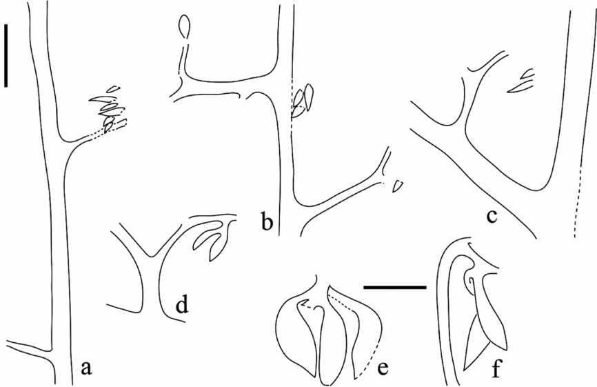
The fertile and sterile axes have not been found in connection. However, there is little doubt that they belong to the same plant. Fertile and sterile axes co-occur on bedding planes, with similar orientation, morphology and surface features. No other plants were found amongst the numerous fossils recovered from the locality. The fertile appendages, comprising complex branching systems and sporangia, are attached alternately at angles of 62–96° on second-order branches of fertile axes (Figs 7a, b, 8a, b); this pattern is consistent with that of sterile ultimate appendages of second-order branches (compare Fig. 7a, b to Figs 4m, 6a, and Fig. 7c to Figs 4g, i, o, 6e). Fertile first-order branches are c. 1.2mm in width, similar in size to the distal portions of sterile first-order branches. Fertile second-order branches are 0.8–1.2mm in width, with the longest preserved length of 28mm. On the proximal portions of fertile second-order branches, the first fertile appendage occurs at c. 6.0mm distal from the base (Figs 7c, 8c). The internode between fertile appendages is 6–10mm in length. Owing to limited material, it remains undetermined whether the fertile branches bear any sterile ultimate appendages, nor how many fertile appendages are attached on an individual fertile branch.
5.f. Fertile appendages
Most fertile appendages are found unattached to axes (Fig. 7d, f–h), and in some cases are compressed together as if they have been lost or shed from their axes and have accumulated as debris. Those fertile appendages found directly attached to axes have been found to occur only on fertile second-order branches, with no evidence of fertile and sterile ultimate appendages borne on the same branch (Figs 7a–c, 8a–c). Fertile appendages are dichotomously branched. After departing from the branch, each fertile appendage extends 1.7–4.0mm to form a short straight stalk, which then divides once to form an initial dichotomy with an angle that ranges from 80° to 100° (Fig. 7b, c, white arrows; Fig. 7d, f, i). The daughter branches following this initial dichotomy are typically 1.8–4.2mm in length and much longer than subsequent branches, making this initial dichotomy more obvious, so that the fertile appendage appears to consist of two major sporangial clusters. Each cluster appears recurved such that sporangia tend to point back towards the base of the fertile appendage (Fig. 7d, e, f). The ultimate divisions of the fertile appendages terminate in sporangia, and generally two to four sporangia are grouped as a small truss supported by a penultimate division (Figs 7i–k, 8d–f). The dichotomizing internal branches of the fertile appendages are 0.3–0.7mm wide, decreasing in width with each dichotomy.
The sporangia are fusiform in shape, 1.0–1.4mm long (including the ultimate divisions) and 0.3–0.5mm in maximum diameter, and with a pointed tip. A longitudinal dehiscence line is seen to extend from the tip to the base on some sporangia (Fig. 7e, k, l, arrows D). No in situ spores were found.
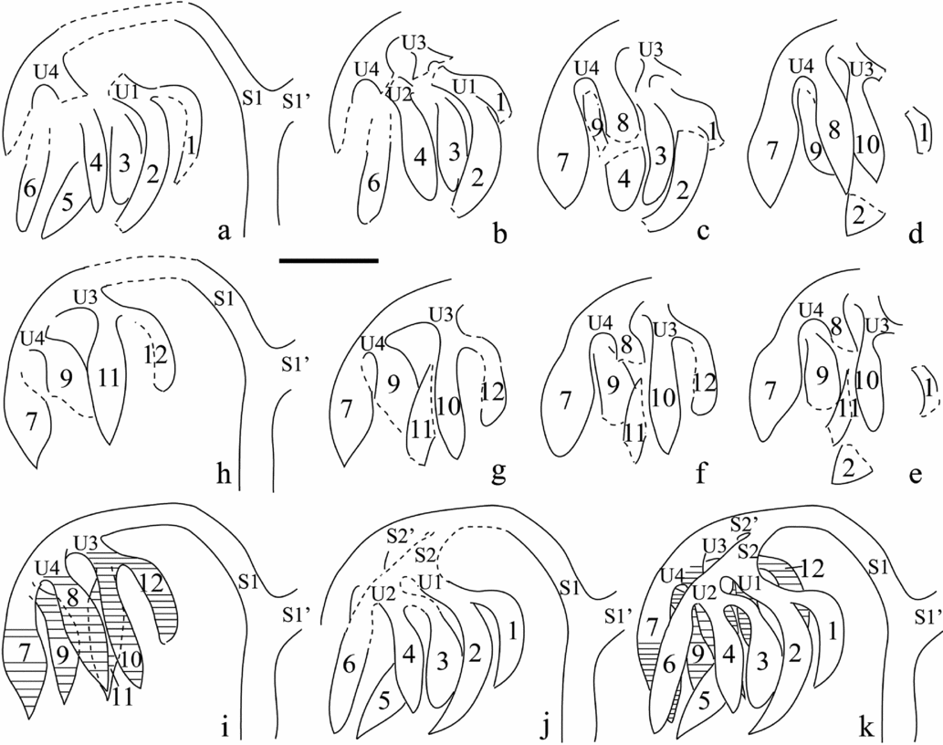
Specimen US734-9177 reveals details of fertile appendage morphology (Figs 7f, 9a–h, 10a–h). After the initial dichotomy, two clusters of sporangia are visible, each supported by a long, recurved branch (Figs 7f, 9a, h; Fig. 10a, h–k, branches S1 and S1'). Multiple sporangia are compressed together, but serial dégagement shows that one cluster has at least 12 sporangia (Fig. 10a–h, sporangia 1–12). The attachment of sporangia 7–12 has been well preserved (Fig. 10c–i), with each sporangium terminating a very short ultimate division, and grouped as small fertile units of three sporangia each, borne on penultimate branches (Fig. 10c–i; U3 with sporangia 10–12; U4 with sporangia 7–9). Although not well preserved, we suggest that sporangia 1 and 2 also form a unit (Fig. 10a, b, j, U1), and sporangia 3, 4 and 6 form another unit (Fig. 10j, U2), for a total of four fertile units in the cluster. Sporangium 5 is the uppermost, and may have been attached to either unit U1 or U2. Branch S1 appears to divide once to produce two sister branches, S2 and S2', which then bifurcate once to produce the penultimate divisions that bear the four fertile units (Fig. 10i, j, U1–U4). A suggested schematic reconstruction of one sporangial cluster of a fertile appendage is shown in Figure 10k, based on the description given here.
Interpretative line drawings of fertile appendage of Melvillipteris quadriseriata gen. et sp. nov. (a–h) Serial dégagment of one sporangial cluster of the fertile appendage shown in Figure 7f (arrow), as shown in Figure 9a–h. Each fertile appendage has an initial dichotomy, producing two sister branches S1 and S1', each of which supports a sporangial cluster. (a) Six sporangia (1–6) of the upper layer are visible. Sporangia 1 and 2 form a fertile unit (U1). (b) Sporangium 5 has been removed. The bases of the fertile units U1 and U2 are visible, but the attachments are not preserved. (c) Sporangium 6 has been removed. Sporangia 7 and 8 are grouped as unit U4. (d) Sporangium 3 has been removed. Sporangium 10 and the base of unit U3 are visible. (e–g) Sporangia 1, 2 and 8 have been removed. Sporangium 9 is attached to the base of U4. Sporangia 10 and 12 form unit U3. (h) Sporangium 10 has been removed. Sporangium 11 is attached to the base of U3. (i) Reconstruction showing attachment of sporangia 7–12 in two fertile units (U3 and U4). (j) Deduced attachment pattern of sporangia 1 and 2 to U1 and sporangia 3, 4 and 6 to U2, based on the pattern of sporangia 7–12 shown in (i). A dichotomy of branch S1 is probable, which produces branches S2 and S2' to support U1, U2 and U3, U4, respectively. Sporangium 5, of the most upper layer, may be attached to unit U1 or U2. (k) Schematic reconstruction of one sporangial cluster of the fertile appendage. Fertile units U1–U4, each comprising two to four terminating sporangia, are supported by the bifurcations of S2 and S2'. (a–k) Scale bar = 1mm.

This pattern is also demonstrated by other sporangial trusses (Figs 7i–k, 8d–f), so it is possible to generalize and describe each fertile appendage as dichotomizing once to produce two sporangial clusters, each cluster dichotomizing twice to produce four fertile units per cluster, and each fertile unit consisting of two to four terminal sporangia.
6. Comparisons with related plants
6.a. Plants with quadriseriate branching
6.a.1. Rhacophyton Crépin
The genus Rhacophyton is characterized by a quadriseriate branching system in certain branching orders, fertile appendages typically comprised of dense masses of sporangia (Leclercq, Reference Leclercq1951; Andrews & Phillips, Reference Andrews and Phillips1968; Cornet, Phillips & Andrews, Reference Cornet, Phillips and Andrews1976; Schultka, Reference Schultka1978) and by a clepsydroid primary xylem strand surrounded by what is interpreted to be secondary xylem (Dittrich, Matten & Phillips, Reference Dittrich, Matten and Phillips1983). Rhacophyton ceratangium Andrews & Phillips, R. zygopteroides Leclercq and R. condrusorum Crépin are included in this genus (Leclercq, Reference Leclercq1951; Andrews & Phillips Reference Andrews and Phillips1968; Cornet, Phillips & Andrews, Reference Cornet, Phillips and Andrews1976; Schultka, Reference Schultka1978), the first two being relatively well known. To facilitate comparison with Melvillipteris, the terms ‘primary pinnae’ and ‘secondary pinnae’ used in the original literature on Rhacophyton are replaced here by the terms first-order and second-order branches, respectively.
Melvillipteris is similar to Rhacophyton in having adventitious roots on the main axes (stems). The roots of Melvillipteris and Rhacophyton ceratangium appear to have occurred in groups that radiate in a similar manner. With few exceptions, the root groups of Melvillipteris are attached on only one side of the main axis internode, which is similar to the condition of R. ceratangium (Cornet, Phillips & Andrews, Reference Cornet, Phillips and Andrews1976; Scheckler, Reference Scheckler1986), and may indicate a similar growth habit, i.e. a scrambling shrub as suggested for Rhacophyton by Scheckler (Reference Scheckler1986). Single individual roots (or root-like structures) have been found on the main axes of R. zygopteroides (Leclercq, Reference Leclercq1951, her pl. II, fig. 2) or at the base of the paired first-order branches (fronds) of R. ceratangium (Cornet, Phillips & Andrews, Reference Cornet, Phillips and Andrews1976, their text-fig. 3), but no such roots have been found in Melvillipteris.
Quadriseriate branching patterns occur in different branching orders in Rhacophyton species. Pairs of first-order branches (primary pinnae) sharing a common base are attached alternately on opposite sides of the main axes of Rhacophyton ceratangium (Cornet, Phillips & Andrews, Reference Cornet, Phillips and Andrews1976), which is similar to the pattern of Melvillipteris, with its main axes bearing paired first-order branches. The arrangement of second-order branches (secondary pinnae) of R. ceratangium is more complex than that of Melvillipteris. Some second-order branches of R. ceratangium show a biseriate arrangement, while others show a transition from a proximal biseriate to a distal quadriseriate pattern. The second-order branches of Melvillipteris show only an alternate, biseriate arrangement. In contrast to R. ceratangium and Melvillipteris, Rhacophyton zygopteroides shows helically arranged first-order branches, which then bear alternate pairs of second-order branches in a quadriseriate arrangement (Leclercq, Reference Leclercq1951).
The attachment and branching of the fertile appendages of Melvillipteris and Rhacophyton are significantly different. In Rhacophyton, a pair of fertile appendages (fertile catadromic aphlebiae) is typically associated with a pair of sterile branches, and together they are attached to the first-order branch as a nodal unit (Leclercq, Reference Leclercq1951; Andrews & Phillips, Reference Andrews and Phillips1968; Cornet, Phillips & Andrews, Reference Cornet, Phillips and Andrews1976). Although fertile appendages of both Melvillipteris and Rhacophyton have a basal dichotomy and a dichotomous branching system with terminating sporangia, those of Rhacophyton are far more complex, more densely branched and bear many more sporangia.
A pair of aphlebia-like appendages is borne at the base of the first-order branches (fronds) of Rhacophyton (Leclercq, Reference Leclercq1951; Andrews & Phillips, Reference Andrews and Phillips1968; Cornet, Phillips & Andrews, Reference Cornet, Phillips and Andrews1976). In Melvillipteris, no comparable structures were found. Although rare sterile ultimate appendages found at the base of some first-order branches to a limited extent resemble the aphlebia-like appendages of Rhacophyton, poor preservation prevents further comparison.
The sterile ultimate appendages of Melvillipteris are similar to those of Rhacophyton in their alternate arrangement, dichotomous branching and recurved tips (Leclercq, Reference Leclercq1951; Andrews & Phillips, Reference Andrews and Phillips1968; Cornet, Phillips & Andrews, Reference Cornet, Phillips and Andrews1976). Nevertheless, the sterile ultimate appendages of Melvillipteris differ from those of Rhacophyton in having a simpler morphology, with only one or two dichotomies. Some of the sterile ultimate appendages of Rhacophyton are markedly planate and flattened (Cornet, Phillips & Andrews, Reference Cornet, Phillips and Andrews1976), while those of Melvillipteris appear terete and three-dimensional.
6.a.2. Cephalopteris Nathorst
Cephalopteris mirabilis Nathorst (originally published as Cephalotheca mirabilis Nathorst but renamed as Cephalopteris mirabilis Nathorst (Nathorst, Reference Nathorst1902; Bureau, Reference Bureau1914, p. 22)) shows close morphological similarities to the genus Rhacophyton and was formally transferred to Rhacophyton by Leclercq (Reference Leclercq1951) as Rhacophyton mirabilis (Nathorst) Leclercq. The shared characters of Rhacophyton and Cephalopteris include quadriseriate branching and the presence of sterile and fertile catadromic aphlebiae (Leclercq, Reference Leclercq1951, her pl. V, fig. 12, 13), the latter not found in Melvillipteris. However, Schweitzer (Reference Schweitzer1968) showed that some of the fertile appendages of Cephalopteris are directly attached along the branch, which is unlike those of Rhacophyton species but similar to those of Melvillipteris. The sterile ultimate appendages of Cephalopteris are laminated (Schweitzer, Reference Schweitzer1968; Hill, Scheckler & Basinger, Reference Hill, Scheckler and Basinger1997; Hilton, Reference Hilton1999), unlike those of Rhacophyton or Melvillipteris. Cephalopteris and Rhacophyton are here considered as distinct genera, and Melvillipteris is considered distinct from both.
6.a.3. Protocephalopteris Ananiev
Protocephalopteris praecox (Høeg) Ananiev is a plant described from the Middle Devonian of Spitsbergen, Siberia, and possibly Belgium and South Africa (Ananiev, Reference Ananiev1960; Schweitzer, Reference Schweitzer1968). Main axes, first-order branches and sterile and fertile ultimate appendages have been recognized for this plant. As in Melvillipteris, the main axes of Protocephalopteris show a zigzag appearance, bending at each successive node. The first-order branches of both plants are borne in alternate pairs arranged on opposite sides of the main axes in a quadriseriate pattern. In Protocephalopteris, two aphlebiae are oppositely arranged at the base of the paired first-order branches, while such structures have not been found in Melvillipteris. The first-order branches of Protocephalopteris bear alternately arranged sterile and fertile ultimate appendages. Schweitzer (Reference Schweitzer1968) considered that these ultimate appendages were also borne in pairs, in a manner similar to the first-order branches. The first-order branches of Melvillipteris, however, bear only one or two sterile ultimate appendages at the base, and distally bear alternate second-order branches. The sterile ultimate appendages of Melvillipteris and Protocephalopteris are similar in gross morphology, dichotomous branching (typically twice dichotomized) and sharply recurved tips.
The fertile appendages of Protocephalopteris were borne at the base of the first-order branches (Ananiev, Reference Ananiev1960, his fig. 2, pl. IV, 1, 1a; Schweitzer, Reference Schweitzer1968, his abb. 9), as are those of Rhacophyton and Cephalopteris mirabilis, although in Protocephalopteris there are fewer dichotomies and fewer sporangia. This differs from the morphology and position of the fertile appendages of Melvillipteris. Simple fertile appendages consisting of two successive dichotomies and four terminal sporangia have been reported to occur along branches of Protocephalopteris (Ananiev, Reference Ananiev1960, his pl. III, 4; Schweitzer, Reference Schweitzer1968, his abb. 8, 9), but these have been poorly preserved.
6.a.4. Ellesmeris Hill, Scheckler & Basinger
Ellesmeris was described from the Frasnian (Upper Devonian) of Ellesmere Island, Canadian Arctic Archipelago, and includes only the type species, Ellesmeris sphenopteroides Hill, Scheckler & Basinger (Reference Hill, Scheckler and Basinger1997). This plant has a pinnate vegetative system, with three orders of branches and laminate sphenopteroid pinnules. The primary pinnae (first-order branches) of Ellesmeris diverge from the main axis in a quadriseriate pattern, similar to those of Melvillipteris. The pinnules (homologous to sterile ultimate appendages) of Ellesmeris borne on the secondary pinnae (second-order branches) are laminated, commonly with three main lobes, while the sterile ultimate appendages of Melvillipteris are dichotomously branched and appear to have been terete and three-dimensional, and neither planate nor laminate. Further comparisons are not possible, as Melvillipteris lacks anatomical information, and sporangia are not known for Ellesmeris.
6.a.5. Stauropteridales
Paired lateral branches are known in some species of the Stauropteridales, including Gillespiea randolphensis Erwin & Rothwell and Stauropteris oldhamia Binney (Erwin & Rothwell, Reference Erwin and Rothwell1989; Taylor, Taylor & Krings, Reference Taylor, Taylor and Krings2009). In G. randolphensis, the largest axes bear alternating pairs of smaller lateral axes, and this pattern is repeated in at least three orders of branching, while in Melvillipteris, only first-order branches are borne in pairs. At least six orders of branches have been reported for S. oldhamia (Farahimanesh et al. Reference Farahimanesh, Gerrienne, Galtier and Prestianni2014), and paired branches occur in multiple orders (Taylor, Taylor & Krings, Reference Taylor, Taylor and Krings2009), also dissimilar to the condition in Melvillipteris. Furthermore, the sporangia of the Stauropteridales are borne in pairs or singly on small axes, unlike the fertile appendages of Melvillipteris with its multiple clustered sporangia.
6.b. Protopteridophyton Li & Hsü
Protopteridophyton devonicum Li & Hsü is a plant with an uncertain affinity described from Givetian–Frasnian strata of Hubei, China (Li & Hsü, Reference Li and Hsü1987). The main axes of Protopteridophyton are similar to those of Melvillipteris in bearing alternate pairs of first-order branches in a quadriseriate arrangement. The sterile ultimate appendages of Protopteridophyton are filiform and divide isotomously up to six times, and have a quadriseriate arrangement on both first- and second-order branches, which are thus unlike Melvillipteris in both arrangement and complexity. The fertile appendages of Protopteridophyton are dichotomously branched and similar to its sterile ultimate appendages, and like the sterile appendages are typically borne in pairs at one insertion point on the axis, again differing from the alternate arrangement of fertile appendages in Melvillipteris. The sporangia of Protopteridophyton occur in pairs, terminating the recurved tips of the terminal dichotomies of the fertile appendages. Unlike Protopteridophyton, the fertile appendages of Melvillipteris show an apparently two-clustered appearance; two to four sporangia form a group (a unit) and several groups are gathered to form each sporangial cluster.
6.c. Chlidanophyton Gensel
The monotypic Chlidanophyton dublinensis Gensel has been reported from the Upper Devonian of Avon Gorge, England and the Lower Mississippian of North America (Gensel, Reference Gensel1973; Hilton, Reference Hilton1999). Unlike the quadriseriate arrangement of first-order branches and the alternate arrangement of second-order branches in Melvillipteris, the branching systems of Chlidanophyton are helically arranged, with the first order bearing the second order and the second in turn bearing a third order. In Chlidanophyton, a pair of non-laminate, dichotomous appendages occurs on opposite sides of the base of each branch (Gensel, Reference Gensel1973; Hilton, Reference Hilton1999), while in Melvillipteris, some lateral branches show only one poorly preserved sterile ultimate appendage, but others appear to have none at their bases. The sterile ultimate appendages of Chlidanophyton dichotomously divide 5–8 times, while most sterile ultimate appendages of Melvillipteris have three or fewer successive dichotomies. The fertile appendages of Chlidanophyton are similar to those of Melvillipteris in their alternate arrangement and their dichotomous branching, with only a few sequential dichotomies, and in both, the initial dichotomy is conspicuous and gives the fertile appendage a two-clustered appearance. The sporangia of both are terminal, but those of Chlidanophyton are ovoid in shape, while those of Melvillipteris are fusiform, with a pointed tip.
6.d. Cladoxylopsida
The Cladoxylopsida is an important plant group spanning the Middle Devonian to Early Carboniferous and has a complex taxonomic history (see Berry & Stein, Reference Berry and Stein2000; Cordi & Stein, Reference Cordi and Stein2005; Meyer-Berthaud, Soria & Young, Reference Meyer-Berthaud, Soria and Young2007; Wang, Reference Wang2008; Xue, Hao & Basinger, Reference Xue, Hao and Basinger2010). We compare Melvillipteris with the morphologically preserved members of the Cladoxylopsida, including the iridopteridaleans, pseudosporochnaleans and others.
The morphologically known iridopteridaleans, including Anapaulia Berry & Edwards, Compsocradus Berry & Stein and Ibyka Skog & Banks (Skog & Banks, Reference Skog and Banks1973; Berry & Edwards, Reference Berry and Edwards1996; Berry & Stein, Reference Berry and Stein2000; Berry, Reference Berry2005; Wang, Reference Wang2008; Fu et al. Reference Fu, Wang, Berry and Xu2011), are quite different from Melvillipteris in the following aspects: (1) the lateral axes and ultimate appendages of these iridopteridaleans are attached in whorls or compressed helices, while the first-order branches of Melvillipteris are quadriseriate and the second-order branches, alternate; (2) the three-dimensional sterile ultimate appendages of the iridopteridaleans have multiple dichotomies (always more than three), and are much larger than those of Melvillipteris (with typically only two dichotomies); and (3) the fertile ultimate appendages of the iridopteridaleans are similar to the sterile ultimate appendages of the same plant, with sporangia replacing the recurved tips, while in Melvillipteris the fertile ultimate appendages, with their sporangial clusters, are more complex than the sterile ultimate appendages.
Pseudosporochnaleans differ from Melvillipteris in having a digitate branching pattern, and their larger axes have a characteristic speckled surface pattern resulting from the ‘nests’ of sclerenchymatous cells in the cortex (Leclercq & Banks, Reference Leclercq and Banks1962; Schweitzer, Reference Schweitzer1973; Berry, Reference Berry2000; Berry & Fairon-Demaret, Reference Berry and Fairon-Demaret2002; Stein et al. Reference Stein, Mannolini, Hernick, Landing and Berry2007). Unlike the alternate sterile ultimate appendages of Melvillipteris with their recurved tips, those of the pseudosporochnaleans have straight tips and a subopposite (in Pseudosporochnus) to whorled (in Wattieza) arrangement (Berry & Fairon-Demaret, Reference Berry and Fairon-Demaret1997; Berry, Reference Berry2000).
Denglongia Xue & Hao, Metacladophyton Wang & Geng, Polypetalophyton Geng, Panxia Wang & Berry and Rhipidophyton Berry & Wang are cladoxylopsid plants described from the Middle to Upper Devonian of South China (Wang & Geng, Reference Wang and Geng1997; Hilton, Geng & Kenrick, Reference Hilton, Geng and Kenrick2003; Berry & Wang, Reference Berry and Wang2006; Wang & Berry, Reference Wang and Berry2006; Wang & Lin, Reference Wang and Lin2007; Xue & Hao, Reference Xue and Hao2008). None of these plants shows the quadriseriate branching pattern found in Melvillipteris. First- and second-order branches of Denglongia are whorled (Xue & Hao Reference Xue and Hao2008), branches of Polypetalophyton are attached helically (Hilton Geng & Kenrick, Reference Hilton, Geng and Kenrick2003), sterile lateral branches of Rhipidophyton show a digitate-like branching pattern (Berry & Wang, Reference Berry and Wang2006) and branches of Panxia are inserted in an opposite to alternate pattern (Wang & Berry, Reference Wang and Berry2006). The first-order branches of Metacladophyton tetraxylum are whorled or nearly whorled and the second-order branches are opposite or subopposite (Wang & Geng, Reference Wang and Geng1997). The second-order branches of Metacladophyton ziguinum occur in pairs, as do the first-order branches of Melvillipteris, but they are attached oppositely and decussately on the first-order branches to form four vertical rows (Wang & Lin, Reference Wang and Lin2007). Unlike the terete and three-dimensional sterile ultimate appendages of Melvillipteris, those of Denglongia have no recurved tips and are planate in some specimens, and those of Polypetalophyton are planate and of the Sphenopteridium type. The fertile appendages of these cladoxylopsid plants are diverse in morphology, but quite different from those of Melvillipteris.
7. Discussion
7.a. Growth habit of Melvillipteris
Melvillipteris is interpreted as a herbaceous plant with thin main axes, 3.0–6.0mm in width, and a procumbent (e.g. Fig. 3e) and/or upright habit (e.g. Fig. 3g). For the zigzag main axis shown in Figure 3e, it seems unlikely that it could have supported the weight of the paired first-order branches. Additionally, with few exceptions, clusters of roots are generally borne on only one side of the main axes, suggesting that the main axes were in direct contact with a moist substrate. Similar dense roots have been reported in Rhacophyton (Cornet, Phillips & Andrews, Reference Cornet, Phillips and Andrews1976) and the cladoxylopsids Metacladophyton and Denglongia (Wang & Geng, Reference Wang and Geng1997; Xue & Hao, Reference Xue and Hao2008). Wang & Geng (Reference Wang and Geng1997) suggested that the root-bearing axes of Metacladophyton are in fact rhizomes. Rhacophyton and Ellesmeris, two plants also with quadriseriate branching, have been interpreted as scrambling shrubs (but see Andrews & Phillips, Reference Andrews and Phillips1968) and their ‘fronds’ (lateral branches) appear to arise from ground-level stems (axially stunted or rhizomatous) (Scheckler, Reference Scheckler1986; Hill, Scheckler & Basinger, Reference Hill, Scheckler and Basinger1997). Melvillipteris may have had a similar scrambling habit, and the main axes may have served as rhizomes, sending adventitious roots into the substrate.
7.b. Phylogenetic position of Melvillipteris
Main axes and two orders of lateral branches have been recognized in Melvillipteris. The first-order branches of Melvillipteris diverge from the main axes in distichous pairs, producing a quadriseriate branching pattern similar to that of Rhacophyton, Cephalopteris, Protocephalopteris, Ellesmeris, Protopteridophyton and some members of the Carboniferous Zygopteridales (Ananiev, Reference Ananiev1960; Schweitzer, Reference Schweitzer1968; Cornet, Phillips & Andrews, Reference Cornet, Phillips and Andrews1976; Li & Hsü, Reference Li and Hsü1987; Hill, Scheckler & Basinger, Reference Hill, Scheckler and Basinger1997; Phillips & Galtier, Reference Phillips and Galtier2005). Rhacophyton is the best-known genus in terms of both morphology and anatomy (Leclercq, Reference Leclercq1951; Andrews & Phillips, Reference Andrews and Phillips1968; Cornet, Phillips & Andrews, Reference Cornet, Phillips and Andrews1976; Dittrich, Matten & Phillips, Reference Dittrich, Matten and Phillips1983). Cephalopteris and Protocephalopteris are based on incomplete materials and lack anatomy (Ananiev, Reference Ananiev1960; Schweitzer, Reference Schweitzer1968). Ellesmeris, although older in age (Frasnian), had already evolved planate and laminate foliage and was originally assigned to the Zygopteridales as the earliest member of this order (Hill, Scheckler & Basinger, Reference Hill, Scheckler and Basinger1997), although Phillips & Galtier (Reference Phillips and Galtier2005) considered this to be a member of the Rhacophytales because there is lack of clear distinction between stem and leaf. The morphology of Protopteridophyton is well known, but its anatomy has been only partially preserved (Li & Hsü, Reference Li and Hsü1987). These five plant genera (Rhacophyton, Cephalopteris, Protocephalopteris, Ellesmeris, Protopteridophyton) are assigned to the Rhacophytales (sensu Taylor, Taylor & Krings, Reference Taylor, Taylor and Krings2009), with an underlying hypothesis that the quadriseriate branching in these taxa may be a shared derived character (synapomorphy). It would follow that Melvillipteris should be considered a new member of the Rhacophytales. Quadriseriate branching is also the basis for the proposed relationship between the Rhacophytales and Zygopteridales (Leclercq, Reference Leclercq1951; Rothwell, Reference Rothwell1999; Xue, Hao & Basinger, Reference Xue, Hao and Basinger2010), the zygopterids having the derived conditions of laminate foliage and well-differentiated stem/leaf organography (thus distinct from Melvillipteris) and being considered early ferns (Phillips & Galtier, Reference Phillips and Galtier2005).
While Melvillipteris shares a quadriseriate branching pattern with the Rhacophytales, assignment must be qualified by the lack of anatomy for Melvillipteris. Rhacophyton has a typical clepsydroid-shaped strand of primary xylem surrounded by secondary xylem. As well, the catadromic fertile appendages of Rhacophyton are borne at the base of lateral branches and occupy a position similar to sterile aphlebiae (Leclercq, Reference Leclercq1951; Andrews & Phillips, Reference Andrews and Phillips1968), and some fertile appendages of Cephalopteris and Protocephalopteris are borne at a similar position (Ananiev, Reference Ananiev1960; Schweitzer, Reference Schweitzer1968). This contrasts with the position of the fertile appendages of Melvillipteris (alternately arranged along the second-order branches). Protocephalopteris, however, also bears another type of fertile appendage with simpler morphology along the branches (Ananiev, Reference Ananiev1960; Schweitzer, Reference Schweitzer1968) that resembles the arrangement in Melvillipteris, although preservation of this second type of fertile appendage in Protocephalopteris is poor and possibly incomplete (e.g. Schweitzer, Reference Schweitzer1968, his abb. 8). Finally, in Melvillipteris, a poorly preserved sterile ultimate appendage can be found at the base of some first-order branches (e.g. Figs 3c, d, 4b), and may be interpreted as: (1) a possible equivalent to the aphlebiae found in Rhacophyton, Cephalopteris, Protocephalopteris and Ellesmeris (Schweitzer, Reference Schweitzer1968; Cornet, Phillips & Andrews, Reference Cornet, Phillips and Andrews1976; Hill, Scheckler & Basinger, Reference Hill, Scheckler and Basinger1997); or, (2) a proximal ultimate appendage equivalent to other ultimate appendages of the same branch. Because many first-order branches of Melvillipteris do not appear to bear such basal ultimate appendages (Fig. 3a, b), the second interpretation is more likely.
Xue, Hao & Basinger (Reference Xue, Hao and Basinger2010) suggested a tentative ‘iridopteridalean-based group’, within which some cladoxylopsids (Denglongia, Metacladophyton and Polypetalophyton), Rhacophyton and Zygopteridales form a subclade (Xue, Hao & Basinger, Reference Xue, Hao and Basinger2010, their fig. 7b and fig. 8, C). This phylogenetic hypothesis is supported by the following three factors. (1) Metacladophyton has both whorled and paired lateral branches (Wang & Geng, Reference Wang and Geng1997; Wang & Lin, Reference Wang and Lin2007). The second-order branches of Metacladophyton ziguinum are in pairs (Wang & Lin, Reference Wang and Lin2007), sharing a common base, similar in a sense to the condition of Melvillipteris and rhacophytaleans, although the bases of these branches in M. ziguinum are opposite and decussate, forming four rows and a structure more complex than the quadriseriate branching of Melvillipteris and rhacophytaleans. In Denglongia, branches are arranged in whorls, but within the whorls adjacent branches may be so close as to resemble pairs (Xue & Hao, Reference Xue and Hao2008, their fig. 2a, e, f, i); thus, a scenario for transformation from whorled (as in Denglongia and Metacladophyton) to quadriseriate branching (as in Melvillipteris and Rhacophyton) is possible. (2) The fertile ultimate appendages of Melvillipteris and Metacladophyton are very similar in that they are arranged alternately along the branches and each fertile appendage shows a two-clustered appearance, with each sporangial cluster consisting of a few dichotomous divisions and terminal sporangia. (3) Metacladophyton and Rhacophyton have substantial secondary xylem, and a similar manner of trace departure (by the formation of a lacuna within the protoxylem and subsequent detachment of a crescent-shaped trace, as noted in Wang & Geng, Reference Wang and Geng1997, Berry & Wang, Reference Berry and Wang2006 and Wang & Lin, Reference Wang and Lin2007).
If the suggestion by Xue, Hao & Basinger (Reference Xue, Hao and Basinger2010) of an ‘iridopteridalean-based group’ is correct, then Melvillipteris would nest within this clade, and remain closest to those taxa typically assigned to the Rhacophytales. But in a broader context, the phylogenetic positions of the Rhacophytales and Cladoxylopsida are highly problematic. Rhacophyton, the Pseudosporochnales and the Iridopteridales have been considered the stem groups of the sphenopsid-fern clade (Moniliformopses), supported by a mesarch protoxylem organization and the presence of ‘peripheral permanent protoxylem’ (Kenrick & Crane, Reference Kenrick and Crane1997). In an alternative hypothesis, Cladoxylon (Cladoxylales), Rhacophyton and Zygopteridales were grouped in a monophyletic group sister to the equisetophytes by Rothwell (Reference Rothwell1999). The grouping of Rhacophyton and Zygopteridales is supported by the quadriseriate branching and some anatomical characters that have not been preserved in Melvillipteris (pith in stem, clepsydroid petiole trace and lateral position of protoxylem in rachis/axis). The current state of phylogenetic knowledge, however, suffers from two sources of criticism. On one side, interpretation of homology is problematic for some characters, including the relationship between the lacunae formed by disintegration of protoxylem tracheids (as in the iridopteridaleans) and the peripheral loops of Early Carboniferous Zygopteridales (Scheckler, Reference Scheckler1974; Stein, Reference Stein1981, Reference Stein1982; Berry & Stein, Reference Berry and Stein2000; Cordi & Stein, Reference Cordi and Stein2005). On the other hand, only a few of the best-known representatives were selected for these phylogenetic analyses, such as Rhacophyton, Pseudosporochnus and Ibyka, while many other plants showing diverse morphologies and character variations, and which are probably related to the Cladoxylopsida and Rhacophytales (e.g. Chlidanophyton, Ellesmeris, Panxia, Polypetalophyton, Protopteridophyton and Rhipidophyton), were not included. Melvillipteris is a new addition to this list. To refine the taxonomic framework for these diverse groups of plants will remain a challenge for future work.
8. Conclusions
Melvillipteris is a new plant that shows a novel combination of characters, including quadriseriate first-order branching, simple sterile ultimate appendages and fertile ultimate appendages on second-order branches. Melvillipteris, with the genera Cephalopteris, Protocephalopteris, Ellesmeris and Protopteridophyton, shares features with the well-known genus Rhacophyton, and thus is assigned to the Rhacophytales. Nevertheless, caution must be exercised, as characters such as quadriseriate branching may have multiple independent origins, and these issues are complicated by incomplete preservation of most of these plants. The comparison of Melvillipteris to Rhacophyton on one hand, and the cladoxylopsids (e.g. Metacladophyton) on the other, is suggestive of a possible relationship among them, together nesting within the ‘iridopteridalean-based group’ (sensu Xue, Hao & Basinger, Reference Xue, Hao and Basinger2010), an unresolved plexus and stem groups of sphenopsids and/or ferns.
Acknowledgements
Professor Stephen E. Scheckler (Virginia Polytechnic Institute and State University) is thanked for his contribution during the field work. The authors are grateful for the help of professors Shougang Hao and Deming Wang (Peking University) and Dr Chris Berry (Cardiff University) for their discussions about the plant. Professor Susan Kaminskyj (University of Saskatchewan) is specially thanked for her support during the laboratory studies of the plant. Collection of the fossils was supported by the Natural Sciences and Engineering Research Council of Canada (DG 1334 to JFB). J.-Z. Xue's travel and visit to the Department of Geological Sciences, University of Saskatchewan was supported by the University of Saskatchewan (Research Grant to JFB). The National Natural Science Foundation of China (grant 41272018) is also thanked. Logistic support for arctic fieldwork was provided by the Polar Continental Shelf Project of Natural Resources Canada.