


INTRODUCTION
Throughout the Quaternary period, Africa experienced climatic and vegetation changes that had strong influences on the evolution, adaptation, and dispersal of faunas and humans in and out of Africa (e.g., DeMenocal, Reference DeMenocal1995, Reference DeMenocal2004; Carto et al., Reference Carto, Weaver, Hetherington, Lam and Wiebe2009; Trauth et al., Reference Trauth, Larrasoana and Mudelsee2009; Compton, Reference Compton2011; Abbate and Sagri, Reference Abbate and Sagri2012; Whiting Blome et al., Reference Whiting Blome, Cohen, Tryon, Brooks and Russel2012; DeMenocal and Stringer, Reference DeMenocal and Stringer2016; Lazagabaster et al., Reference Lazagabaster, Rovelli, Fabre, Porat, Ullman, Davidovich and Lavi2021). Morocco, Algeria, and Tunisia, located in northwestern-most Africa, may have alternately played the role of “dead-end,” refuge area or crossroads for faunas and humans between western Europe, the Near East, and sub-Saharan Africa. During wetter periods, the development of vegetation, rivers, and lakes in the Sahara may have favored the passage of faunas and humans (e.g., Osborne et al., Reference Osborne, Vance, Rohling, Barton, Rogerson and Fello2008; Castañeda et al., Reference Castañeda, Mulitza, Schefuß, Lopes dos Santos, Sinninghe Damsté and Schouten2009; Balter, Reference Balter2011; Garcea, Reference Garcea2012; Coulthard et al., Reference Coulthard, Ramirez, Barton, Rogerson and Brücher2013; Drake et al., Reference Drake, Breeze and Parker2013; Scerri, Reference Scerri2017). These events can be documented through the study of fossil faunas, and especially micromammals such as rodents, found in archaeological sites. Indeed, rodents are recognized as good paleoenvironmental proxies (e.g., Chaline, Reference Chaline1972, Reference Chaline1973; Avery, Reference Avery1982; Andrews, Reference Andrews1990; Cuenca-Bescos et al., Reference Cuenca-Bescos, Melero-Rubio, Rofes, Martínez, Arsuaga, Blain, Lopez-Garcia, Carbonell and Bermudez de Castro2011; Stoetzel, Reference Stoetzel2017), and can help us to better understand human population movements and their effect on the environment (e.g., Abdel Rahman et al., Reference Abdel Rahman, Ducroz, Mitchell, Lamb, Contrafatto, Denys, Lecompte and Taylor2008; Jones et al., Reference Jones, Eager, Gabriel, Johannesdottir and Searle2013; Stoetzel, Reference Stoetzel2017; Weissbrod et al., Reference Weissbrod, Marshall, Valla, Khalaily, Bar-Oz, Auffray, Vigne and Cucchi2017; Cucchi et al., Reference Cucchi, Papayianni, Cersoy, Aznar-Cormano, Zazzo, Debruyne and Berthon2020; Lazagabaster et al., Reference Lazagabaster, Rovelli, Fabre, Porat, Ullman, Davidovich and Lavi2021).
We investigate here fossil and extant specimens of the Grass Rat Arvicanthis, which is a good example to address issues related to dispersal events and relationships to environmental changes and human populations through time in Africa. The genus Arvicanthis most probably originated in eastern Africa during the Mio-Pliocene transition (Winkler, Reference Winkler2002; Manthi, Reference Manthi2007) and is today represented by (at least) seven species: A. abyssinicus, A. ansorgei, A. blicki, A. nairobae, A. neumanni, A. niloticus, and A. rufinus (Musser and Carleton, Reference Musser, Carleton, Wilson and Reeder2005; Monadjem et al., Reference Monadjem, Taylor, Denys and Cotterill2015; Denys et al., Reference Denys, Taylor, Aplin, Wilson, Lacher and Mittermeier2017). However, the taxonomic status of some taxa is still debated, notably in western (Happold, Reference Happold2013) and eastern (Castiglia et al., Reference Castiglia, Bekele, Makundi, Oguge and Corti2006; Bryja et al., Reference Bryja, Colangelo, Lavrenchenko, Meheretu, Sumbera, Bryjova, Verheyen, Leirs and Castiglia2019) Africa, where several cryptic species seem to exist, and relationships between most species are not yet resolved. The data also suggest a rapid radiation, probably during the Early–Middle Pleistocene (Bryja et al., Reference Bryja, Colangelo, Lavrenchenko, Meheretu, Sumbera, Bryjova, Verheyen, Leirs and Castiglia2019). Now restricted to sub-Saharan savannas and the Nile Valley, its past distribution during the Pleistocene and the Early Holocene included parts of the Middle East and northern Africa (Tchernov, Reference Tchernov1968, Reference Tchernov, Prakash and Ghosh1975; Jaeger, Reference Jaeger1975; Stoetzel, Reference Stoetzel2013, Reference Stoetzel2017).
Genetic studies on modern populations of A. niloticus showed a phylogeographic pattern that suggests a strong link to climatic and vegetation changes through the Quaternary (Abdel Rahman et al., Reference Abdel Rahman, Ducroz, Mitchell, Lamb, Contrafatto, Denys, Lecompte and Taylor2008; Dobigny et al., Reference Dobigny, Tatard, Gauthier, Ba, Duplantier, Granjon and Kergoat2013; Bryja et al., Reference Bryja, Colangelo, Lavrenchenko, Meheretu, Sumbera, Bryjova, Verheyen, Leirs and Castiglia2019). According to these studies, in sub-Saharan Africa, allopatric genetic divergence within savanna refugia occurred during humid periods (development of forests, reduction and fragmentation of savannas), while dispersals occurred during arid periods (reduction of forests and expansion of savannas). But it is likely that the development of grassland habitats in the Sahara also allowed expansion across this area during humid periods. Moreover, this rodent has undergone a major population expansion at the Middle-Late Pleistocene boundary, corresponding in time with the expansion of early Homo sapiens within and out of Africa (Abdel Rahman et al., Reference Abdel Rahman, Ducroz, Mitchell, Lamb, Contrafatto, Denys, Lecompte and Taylor2008; Weissbrod and Weinstein-Evron, Reference Weissbrod and Weinstein-Evron2020).
To date, there have been no comprehensive studies of Arvicanthis populations in northern Africa, either to try to clarify specific attributions or to better understand how these savanna rodents adapted to steppe and Mediterranean environments in regions farther north than their current distribution. A local endemic species, A. arambourgi, has been described in the Early–Middle Pleistocene site of Tighennif in Algeria (Jaeger, Reference Jaeger1975), and another one, A. ectos, has been described in the Levant for the Middle Pleistocene (e.g., Tchernov, Reference Tchernov1968, Reference Tchernov, Prakash and Ghosh1975). But most of the remains coming from the other sites North of the Sahara were assigned to Arvicanthis sp. or A. niloticus, without thorough analysis.
The aim of the present study is not to solve the current taxonomic issues related to the genus Arvicanthis, but to find a method to efficiently discriminate both extant and extinct species on the basis of molar shape that will facilitate future analyses of Arvicanthis fossil remains. In archaeological and paleontological contexts, complete crania and mandibles are quite rare, while molars (either isolated or in situ) are generally numerous and well preserved, facilitating the use of Geometric Morphometric methods. We also highlight some aspects of the diet and ecological specificities of selected Arvicanthis species using molar microwear analyses, with application to the fossil population from Tighennif.
MATERIAL AND METHODS
Modern specimens
The current reference specimens used for Geometric Morphometrics (GM) and microwear analyses come from the rodent collections of the Muséum national d'Histoire naturelle (MNHN) of Paris. As far as possible, we used the same specimens for both GM and microwear analyses, but more specimens were considered for GM in order to enhance the significance of discrimination.
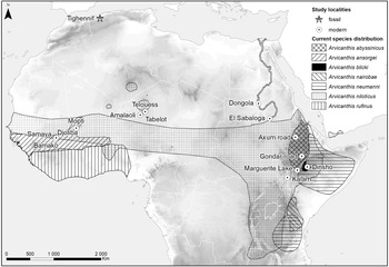
Five species of Arvicanthis from different localities in Africa were considered for this study (Table 1; Figs. 1, 2): A. abyssinicus and A. blicki (both endemics of Ethiopia), A. neumanni (eastern Africa), A. ansorgei (western Africa), and four populations of the widespread A. niloticus. No specimen was available or usable for A. nairobae (eastern Africa) or A. rufinus (western Africa). Data concerning the distribution and ecology of extant species mainly come from the International Union for Conservation of Nature (IUCN) database (www.iucnredlist.org) and Monadjem et al. (Reference Monadjem, Taylor, Denys and Cotterill2015), as well as more specific references when cited.
Current distribution of the modern Arvicanthis species and localities from which the modern and fossil Arvicanthis specimens used in the present study originate.

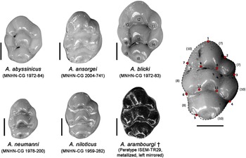
Examples of right first upper molars of each studied modern and fossil Arvicanthis species, nomenclature of cusps (t1–t9), and positions of the landmarks (LM, red points) and sliding semi-landmarks (SLM, dashed lines, numbers in parentheses) used in this study. CG = collection number of the MNHN specimens; TR = collection number of the ISEM specimen. Scale = 1 mm.

Modern and fossil Arvicanthis specimens used in the present study. N. sp. = number of specimens, GM = geometric morphometrics, Mw = microwear analyses. Detailed list of specimens in Appendix (Supplementary Table 1).

Fossil specimens
Various paleontological and archaeological northern African sites have yielded Arvicanthis remains (Stoetzel, Reference Stoetzel2013, Reference Stoetzel2017), but generally in low numbers, preventing any analysis at a populational level. One of the possible explanations for the scarcity of Arvicanthis remains may be the diurnal habits of this rodent, which meant they were likely not often hunted by owls, the major accumulators of small mammal assemblages. However, in other geographical areas, Arvicanthis remains are well represented in fossil assemblages produced by owls (e.g., Stoetzel et al., Reference Stoetzel, Sime, Pleurdeau, Asrat, Assefa, Desclaux and Denys2018). Another explanation would be that this sub-Saharan rodent, which has only made occasional incursions into the region, would have been much less abundant in the environment than the local rodent species, notably Gerbillinae. It is also less likely that remains of Arvicanthis would be fossilized and preserved if the incursion of this taxon in the region was brief.
Only two Middle Pleistocene sites from northwestern Africa have yielded large numbers of Arvicanthis remains: Tighennif in Algeria (N = 457) and Irhoud Derbala Virage in Morocco (N = 67), both studied by Jaeger (Reference Jaeger1975). Both collections were supposed to be stored at the Institut des Sciences de l'Evolution de Montpellier (ISEM, France), but only the Tighennif collection could be analyzed for the present study (Table 1), while the Arvicanthis remains from Irhoud Derbala Virage currently are not accessible.
Geometric morphometrics
Geometric morphometric (GM) methods represent useful tools to precisely quantify and describe anatomical shapes of biological objects (e.g., Zelditch et al., Reference Zelditch, Swiderski and Sheets2012; Adams et al., Reference Adams, Rohlf and Slice2013). Several studies in classical and geometric morphometrics have been carried out on Arvicanthis (Bekele et al., Reference Bekele, Capanna, Corti, Marcus and Schlitter1993; Corti and Fadda, Reference Corti and Fadda1996; Fadda and Corti, Reference Fadda and Corti2001), but they have focused mainly on the skull. These studies are unlikely to be applicable in archaeological or paleontological contexts, because complete skulls are extremely rare, and the best preserved and most abundant identifiable remains are usually isolated molars or in situ molars embedded in fragmented mandibles and maxillae. Moreover, some authors have shown that in murine rodents the upper molars are more diagnostic and show a faster evolution than the lower molars (e.g., Renaud et al., Reference Renaud, Benammi and Jaeger1999, Reference Renaud, Pantalacci and Auffray2011), which is why we primarily focused on the first upper molars (M1).
In the present study, we analyze both the outline and the relative position of cusps on M1, by combining anatomical landmarks (LM) and sliding semi-landmarks (SLM) (Gunz and Mitteroecker, Reference Gunz and Mitteroecker2013), a method that has proven effective in several studies on modern and fossil murid rodents (e.g., Macholan, Reference Macholan2006; Kimura et al., Reference Kimura, Jacobs and Flynn2013; Stoetzel et al., Reference Stoetzel, Cornette, Lalis, Nicolas, Cucchi and Denys2017; Hulme-Beaman et al., Reference Hulme-Beaman, Claude, Chaval, Evin, Morand, Vigne, Dobney and Cucchi2019; Cucchi et al., Reference Cucchi, Papayianni, Cersoy, Aznar-Cormano, Zazzo, Debruyne and Berthon2020). The position of LM was based partially on Janzekovic and Krystufek (Reference Janzekovic and Krystufek2004), Siahsarvie and Darvish (Reference Siahsarvie and Darvish2008), and Matthews and Stynder (Reference Matthews and Stynder2011).
The 11 LM are located on the local maximum curvature of the outline of the Arvicanthis M1 associated with cusps and valleys between cusps (Macholan, Reference Macholan2006; Kimura et al., Reference Kimura, Jacobs and Flynn2013) as follows (Fig. 2):
1. Apex = most anterior point of the prelobe.
2. Maximum curvature of the valley between the prelobe and the cusp t1.
3. Maximum curvature of the valley between cusps t1 and t4 = most posterior point of the cusp t1 on the outline of the tooth.
4. Maximum curvature of the valley between cusps t4 and t8 = most posterior point of the cusp t4 on the outline of the tooth.
5. Maximum curvature of the valley between cusps t8 and t9 = junction between t8 and t9.
6. Most posterior point of the cusp t6 on the outline of the tooth.
7. Maximum curvature of the valley between cusps t6 and t3 = most posterior point of the cusp t3 on the outline of the tooth.
8. Junction between cusps t3 and t2.
9. Junction between cusps t2 and t1.
10. Junction between cusps t6 and t5.
11. Junction between cusps t5 and t4.
In addition, a series of 60 SLM was placed between the LM along the outline of the tooth (Fig. 2), which makes a total of 71 points.
After verification of their state of preservation, the fossil and recent Arvicanthis molars were photographed following a precise protocol allowing GM analyses. The photographs were all taken by the same person (E.S.) and with the same equipment (Nikon D750FX camera with a macro lens 85mm 1:3.5 G positioned on a stand with double side lighting or in a lightbox). A picture of the same ruler was taken at each session in order to check the scale. Each tooth was positioned with the most horizontal occlusal surface possible. Only the right side was considered, except for modern specimens in which the right M1 was absent or damaged, and fossils, for which all molars have been considered. In these cases, the picture of the left side was mirrored before positioning LM and SLM. To avoid any bias related to the age of the individuals, only sub-adults and adults were considered (thus excluding juveniles and old individuals). Age classes were based upon tooth-wear stages (juvenile: distinct cusps and incipient wear, m3/M3 unerupted or partially erupted; sub-adult: little wear beginning to unite cusps into laminae, exposed dentine of the cusps not yet fused; adult: intermediate wear, cusps united transversely but still discernible, dentine-bridge between the cusps becomes progressively wider as the cusps wear down; old: advanced wear, cusps no longer discernible, different rows/laminae become confluent).
The image set was assembled using TpsUtil (Rohlf, Reference Rohlf2019a), and LM and SLM were positioned with TpsDig2 (Rohlf, Reference Rohlf2017). To remove information on position, scale, and orientation from the Cartesian coordinate configurations, we performed a generalized Procrustes analysis (Rohlf and Slice, Reference Rohlf and Slice1990) using TpsRelw (Rohlf, Reference Rohlf2019b) to produce a new set of shape coordinates. The semi-landmarks were allowed to slide, minimizing the bending energy between each specimen and the mean conformation of all specimens (Bookstein, Reference Bookstein1997; Gunz and Mitteroecker, Reference Gunz and Mitteroecker2013). The resulting Procrustes shape coordinates from this superimposition were used as shape variables for subsequent statistical analyses. The centroid size was measured using the square root of the sum of the squared distances between each point and the centroid of the configuration.
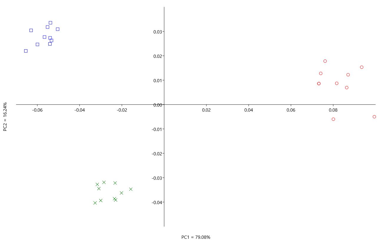
A standard preliminary repetition procedure of orienting-digitizing and landmark positioning was performed on molars. Three specimens of the same species and from the same locality (A. niloticus from Dongola, Sudan) were photographed 10 times, and the LM and SLM were positioned manually on each of the 30 pictures. A Principal Component Analysis (PCA) revealed that the intra-specimen measurement variability, corresponding to the repetition, was significantly lower than the inter-specimen variability (Supplementary Figure 1).
Size differences between groups were investigated using ANOVA with pairwise t-tests using Bonferroni correction. Patterns of shape variation were investigated using a PCA and shape differences between groups were tested using a MANOVA and pairwise comparisons using t-tests with Bonferroni correction. To compare shape differences between modern and fossil specimens or between modern populations, we performed a Canonical Variate Analysis (CVA) on the n axes of the PCA, representing 95% of the total variance (Baylac and Friess, Reference Baylac, Friess and Slice2005)—in our case, the first 14 axes of the PCA. Because of the potential presence of several clades/species among the A. niloticus group (Bryja et al., Reference Bryja, Colangelo, Lavrenchenko, Meheretu, Sumbera, Bryjova, Verheyen, Leirs and Castiglia2019), the four localities were considered separately, and specific analyses on this group were performed to explore possible population differences. The k-Nearest Neighbors (KNN) algorithm was used to compute the cross-validated correct classification percentages. This method allows us to classify a particular object in a group, taking into account its KNN by Euclidian distance after a leave-one-out cross validation. This method is less sensitive to small sample size than linear discriminant analysis (e.g., Hanot et al., Reference Hanot, Gerber, Guintard, Herrel, Verslype and Cornette2020). In order to increase the efficiency of the discriminations, size was included in the KNN analysis. Statistical analyses and graphical representations were performed using PAST, MorphoJ, and R software programs.
Microwear analyses
Dental microwear patterns reflect abrasion resulting from contact between tooth enamel and food items consumed by an animal in the last few days prior to its death (e.g., Walker et al., Reference Walker, Hoeck and Perez1978; Teaford and Oyen, Reference Teaford and Oyen1989). The protocol of dental microwear analyses was adapted to muroid rodents in order to infer the dietary habits of extinct or extant taxa (Gomes Rodrigues et al., Reference Gomes Rodrigues, Merceron and Viriot2009, Reference Gomes Rodrigues, Marivaux and Vianey-Liaud2012, Reference Gomes Rodrigues, Renaud, Charles, Le Poul, Solé, Aguilar and Michaux2013; Renaud et al., Reference Renaud, Gomes Rodrigues, Ledevin, Pisanu, Chapuis and Hardouin2015; Kerr et al., Reference Kerr, Cornette, Gomes Rodrigues, Renaud, Chevret, Tresset and Herrel2017). Casts of first upper molars (M1) were analyzed using transmitted light stereomicroscopy (LEICA M205C). Three different variables were quantified using ImageJ: the number of fine scratches (Nfs, more numerous in grass-dominated feeders, GDF), wide scratches (Nws, more numerous in fruit-dominated feeders, FDF), and large pits (Nlp, more numerous in animal-dominated feeders, ADF).
A PCA was performed using Statistica on the three microwear variables (Nfs, Nws, and Nlp) to infer the main dietary components of Arvicanthis species when compared to the reference database (MUROIDEA, Gomes Rodrigues et al., Reference Gomes Rodrigues, Merceron and Viriot2009, Reference Gomes Rodrigues, Marivaux and Vianey-Liaud2012, from which two populations of Arvicanthis niloticus from Sudan, previously belonging to GDF, have been removed). MANOVA and post-hoc tests, namely multiple means comparison tests, Fisher's least significant difference (LSD), and the more conservative Tukey's honestly significant difference (HSD), were then realized on rank-transformed data (to meet parametric assumptions, see Conover and Iman, Reference Conover and Iman1981) to test for significant differences with dietary categories and between species.
RESULTS
Geometric morphometrics
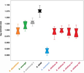
The ANOVA performed on the log of the centroid size shows significant differences between groups (p = 4.85 × 10−61). Arvicanthis blicki is the largest species, while A. neumanni is the smallest (Fig. 3; Table 2). Arvicanthis abyssinicus and all the studied populations of A. niloticus display a similar mean size, while A. ansorgei is a bit larger (but not statistically significant). The fossil species A. arambourgi displays a size close to the upper range of A. ansorgei.
Box plot representation of the size differences (log of the centroid size) between the considered modern and fossil (†) Arvicanthis groups. M = Mali, N = Niger, S1 = Sudan (El Sabaloga), S2 = Sudan (Dongola).

Results of the ANOVA with pairwise t-tests comparisons on the log of the centroid size of the considered modern and fossil Arvicanthis groups. Significant p values (<0.05) are represented in bold.

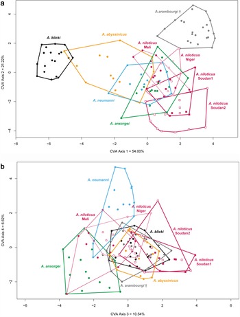
The MANOVA performed on the shape variables (first 14 axes of the PCA) also reveals significant differences between groups (p = 1.949 × 10−93) (Table 3), and the CVA allows species discrimination (Figs. 4, 5). The negative extremity of the first axis, isolating the largest species A. blicki, is characterized by a proportionally wider tooth in its anterior part and thinner in its posterior part, a significantly shorter prelobe, and a more individualized t9 located closer to the t6. Generally, A. blicki shows higher, more rounded, and individualized tubercles (t1 and t4 are generally isolated in young individuals). Arvicanthis arambourgi, located on the positive parts of the two first axes, plots apart from all the modern species (Fig. 4). On the first axis, at the opposite of A. blicki, the fossil species is characterized by a proportionally longer prelobe (but of similar size than most of the other species). On the second axis, compared to all other modern species, A. arambourgi shows a more developed t1 and a narrower posterior part (Fig. 5). Generally, the t9 is less reduced than in all the studied modern species. Arvicanthis ansorgei and A. neumanni are well discriminated against each other on the fourth axis. Arvicanthis neumanni, the smallest species, is located on the positive part of axis 4 and displays a narrower tooth with a proportionally longer prelobe, and a t6 markedly shifted posteriorly. In A. ansorgei, located in the negative parts of axes 3 and 4, the prelobe is relatively short and the t9 appears relatively well developed and slightly offset posteriorly. Arvicanthis abyssinicus and A. niloticus, generally located in the middle parts of the CVA axes, display similar molar size and morphological features. Although their molars are relatively large with a short prelobe compared to the other species, in A. abyssinicus, the tubercules appear less aligned than in A. niloticus (especially the t4, which is generally located more posteriorly).
Representation of axes 1 and 2 (a) and axes 3 and 4 (b) of the Canonical Variate Analysis (CVA) based on the first 14 axes of the PCA.

Reconstruction of the mean shapes at the extremities of the first four axes of the CVA (difference ×5 for a better visualization). Gray = negative extremity of the axes, Orange = positive extremity of the axes.

Results of the MANOVA with pairwise t-tests comparisons on the shape variables of the considered modern and fossil Arvicanthis groups. Significant p values (<0.05) are represented in bold.

Since size is an important discriminating criterion for some species, we chose to include the log of the centroid size in the CVA analysis in order to increase the efficiency of the discriminations. Applying the KNN method, both on shape and size parameters, we obtained 100% of correct classification for specimens belonging to A. neumanni and A. blicki (Table 4). The fossil species A. arambourgi is well separated from the modern groups (83.3% of correct classification). Arvicanthis abyssinicus and A. ansorgei give values of 61.5% and 66.7%, respectively. Concerning the A. niloticus group, only the population from Niger displays relatively good values of correct classification (68.8%). Most of the misclassified A. niloticus specimens fall in other A. niloticus populations, or in A. abyssinicus.
Classifications provided by the k-Nearest Neighbors (KNN) method using both size and shape variables (k = 1).

In order to explore possible population differences within A. niloticus, we performed a separate analysis on the four considered populations of A. niloticus. While the ANOVA performed on size does not show significant differences (Fig. 3; Table 2), the MANOVA performed on the shape variables reveals significant differences between these four groups (p = 7.728 × 10⁻⁹). In looking at the pairwise comparisons (Table 5), the two populations from Sudan can be distinguished from the two other populations (Mali and Niger), although these differences are not significant considering the Bonferroni-corrected p-values (Table 5).
Results of the MANOVA with pairwise t-tests comparisons on the shape variables of the four Arvicanthis niloticus populations. Significant p values (<0.05) are represented in bold.

Microwear analyses
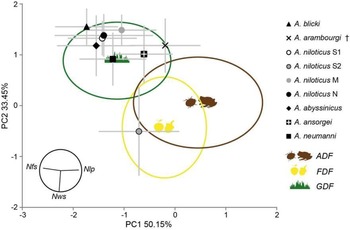
Dental microwear data quantified for each population of Arvicanthis are shown in Table 6, and results of the multivariate analyses based on the number of fine scratches (Nfs), wide scratches (Nws), and large pits (Nlp) are shown in Table 7 and Figures 6 and 7. The global results are relatively homogeneous, however, the post-hoc tests show finer differences, notably concerning the A. niloticus specimens coming from Sudan, which were split into two groups according to their origin (S1: El Sabaloga, S2: Dongola). Indeed, most of the studied specimens belonging to modern species such as A. blicki, A. niloticus (S1, Mali, Niger), A. abyssinicus, A. ansorgei, and A. neumanni fall into the GDF group (grass-dominated feeders). But the A. niloticus specimens coming from Dongola (S2) are clearly included in the FDF group (fruit-dominated feeders). Concerning the fossil specimens of A. arambourgi from Tighennif, we see that they do not fall into a precise category according to the statistical tests, even if they tend to be closer to the GDF group (grass-dominated feeders) on the PCA (Fig. 7). They also display the smallest differences with A. ansorgei according to the LSD test on the number of fine scratches.
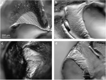
Examples of dental microwear patterns. (a) Arvicanthis blicki (MNHN-ZM-MO-1972-77), (b) A. niloticus Niger (MNHN-ZM-MO-1959-267), (c) A. niloticus Sudan (MNHN-ZM-MO-2003-511), (d) A. arambourgi (UM 7). Scale for all images shown in (a).

Visualization of the two first axes of the PCA performed on the results of the microwear analysis. ADF = animal-dominated feeders, FDF = fruit-dominated feeders, GDF = grass-dominated feeders, Nfs = number of fine scratches, Nws = number of wide scratches, Nlp = number of large pits, M = Mali, N = Niger, S1 = Sudan (El Sabaloga), S2 = Sudan (Dongola). See Gomes Rodrigues et al. (Reference Gomes Rodrigues, Merceron and Viriot2009, Reference Gomes Rodrigues, Marivaux and Vianey-Liaud2012) for the reference database (MUROIDEA).

Dental microwear data quantified for each population of Arvicanthis. Nfs = number of fine scratches, Nws = number of wide scratches, Nlp = number of large pits.

Pairwise comparison tests performed on dental microwear data. ADF = animal-dominated feeders, FDF = fruit-dominated feeders, GDF = grass-dominated feeders, Nfs = number of fine scratches, Nws = number of wide scratches, Nlp = number of large pits. Significance at α = 0.05 is indicated in bold for both LSD and HSD tests, and in normal letters for only LSD test.

DISCUSSION
Arvicanthis species discrimination, relation to diet and ecology
Considering the upper first molar size, A. blicki is the largest species, while A. neumanni is the smallest. Arvicanthis abyssinicus and A. niloticus display a similar mean size, while A. ansorgei and A. arambourgi are a bit larger. These results are in line with previous studies (Bekele et al., Reference Bekele, Capanna, Corti, Marcus and Schlitter1993; Corti and Fadda, Reference Corti and Fadda1996; Fadda and Corti, Reference Fadda and Corti2001) with, however, A. blicki displaying larger M1s relative to its skull size, thus confirming a marked macrodonty (Monadjem et al., Reference Monadjem, Taylor, Denys and Cotterill2015).
Considering both size and shape parameters, despite some overlap, we were able to efficiently discriminate the different modern and fossil Arvicanthis species. This is especially true for the three eastern African modern species A. blicki, A. neumanni, and A. abyssinicus, and the extinct northern African species A. arambourgi. The western African modern species A. ansorgei appears relatively close in size and shape to the widespread A. niloticus group.
According to some authors, differences in Arvicanthis species morphology and size may reflect adaptations to different ecological niches (Bekele et al., Reference Bekele, Capanna, Corti, Marcus and Schlitter1993; Fadda and Corti, Reference Fadda and Corti2001), especially in Ethiopia where geographical, climatic, and ecological contrasts possibly triggered the emergence of several endemic species and/or clades (Bryja et al., Reference Bryja, Colangelo, Lavrenchenko, Meheretu, Sumbera, Bryjova, Verheyen, Leirs and Castiglia2019). Fadda and Corti (Reference Fadda and Corti2001) showed that in western Africa the skull shape is mainly correlated with latitude and mean temperature, while in eastern Africa, the main factors explaining morphological variation are elevation and latitude. The close relationship with geo-climatic variables strongly suggests that morphometric variation in Arvicanthis skulls can be linked to local climatic conditions (Fadda and Corti, Reference Fadda and Corti2001). For example, the eastern African species A. abyssinicus, A. blicki, and A. neumanni, which display very different skull and molar sizes and shapes, also have different ecological requirements (Table 8). Arvicanthis abyssinicus and A. blicki are found in mid- and high-elevation grasslands of the Ethiopian High Plateau (1600–3600 m and 2500–4050 m, respectively), while A. neumanni occupies dry savanna habitats, at a maximum of 1000 m of elevation. Following the theory of a close relationship between morphology and ecology, A. ansorgei and A. niloticus, which display similar external body and molar morphologies, should thus share a similar ecology. Although they are morphologically close, they belong to two different genetic clades among the Arvicanthis genus (Bryja et al., Reference Bryja, Colangelo, Lavrenchenko, Meheretu, Sumbera, Bryjova, Verheyen, Leirs and Castiglia2019). Moreover, the literature highlights some ecological differences (Table 8): A. ansorgei occupies bush and woodland savannas and is presumably herbivorous, while A. niloticus is found across a very large geographic range (Fig. 1) and shows a broader diet. Nonetheless, the global microwear results are relatively homogeneous (most species are grass-dominated feeders), which is not very surprising, since microwear data show mostly dietary trends and are not always very discriminating within the same genus (especially among the Muridae family; Gomes Rodrigues et al., Reference Gomes Rodrigues, Merceron and Viriot2009). This was previously observed for other murids, such as Mus and Apodemus (Renaud et al., Reference Renaud, Gomes Rodrigues, Ledevin, Pisanu, Chapuis and Hardouin2015; Kerr et al., Reference Kerr, Cornette, Gomes Rodrigues, Renaud, Chevret, Tresset and Herrel2017). Indeed, the diet of the Arvicanthis species seems quite homogeneous (mainly herbivores—grass, leaves, seeds—and occasional consumption of insects) (e.g., Granjon and Duplantier, Reference Granjon and Duplantier2009; Denys et al., Reference Denys, Taylor, Aplin, Wilson, Lacher and Mittermeier2017; Hirpasa et al., Reference Hirpasa, Wondimu and Tadele2022), but because most of the data refer to A. niloticus, the precise ecology and diet of some species remain poorly known. Moreover, even if some population differences were observed (see below), no link between morphological changes and dietary changes could be established.
Ecological information (distribution, habitat, and diet) on the considered Arvicanthis species.

Therefore, the variability in M1 size and shape within Arvicanthis cannot be explained only by dietary niche and habitat. It is likely that several factors are intertwined, including joint action of environmental pressure, genetic drift, and isolation driving the morphological differences, thus confirming what had been previously stated by Corti and Fadda (Reference Corti and Fadda1996) and Fadda and Corti (Reference Fadda and Corti2001).
Geographic variations in Arvicanthis morphology and diet
Arvicanthis niloticus is an adaptable and ubiquitous species with a broader diet than the other Arvicanthis species (Table 8). This may reflect a higher ecological plasticity for this species, and/or the existence of several clades with their own ecological particularities throughout the geographical range of this group (Dobigny et al., Reference Dobigny, Tatard, Gauthier, Ba, Duplantier, Granjon and Kergoat2013; Bryja et al., Reference Bryja, Colangelo, Lavrenchenko, Meheretu, Sumbera, Bryjova, Verheyen, Leirs and Castiglia2019), the two being not exclusive. Regarding this issue, the discriminant analysis performed on the molar size and shape variables revealed some differences between the considered A. niloticus populations (Table 4, Fig. 5), separating the two Sudanese populations on one side, and the populations from Mali and Niger on the other, and highlighting morphological differences between eastern and central/western African populations. This would be in line with the existence of two different clades within this group, as evinced by Bryja et al. (Reference Bryja, Colangelo, Lavrenchenko, Meheretu, Sumbera, Bryjova, Verheyen, Leirs and Castiglia2019): A. niloticus sensu stricto in northeastern Africa, and A. niloticus clade “C2-C4” in southeastern, central, and western Africa.
Moreover, microwear analyses highlighted dietary differences between two populations of A. niloticus, but different from those highlighted by the GM analysis. Here, the populations are both coming from Sudan: specimens from El Sabaloga (S1) are grass-dominated feeders, while specimens from Dongola (S2) are fruit-dominated feeders (Fig. 7). These important differences, especially regarding the higher number of large features quantified in specimens from S2, could be explained by the fact that these specimens were found mainly in deserts, contrary to specimens from S1, which were found in less-arid semi-desertic areas (Abdel Rahman et al., Reference Abdel Rahman, Ducroz, Mitchell, Lamb, Contrafatto, Denys, Lecompte and Taylor2008). Even if both populations probably lived in grassy patches in the riparian fringe of the Nile River, living in drier environments would induce a larger consumption of hard and tough items, such as tree bark, rather than fruits (Poulet, Reference Poulet1982), but can also include consumption of insects and ingestion of grit, which might result in larger microwear features. Moreover, some large areas adjacent to the Nile in Sudan are used for agriculture. Consequently, some populations could have fed on crops, which could have resulted in the observed differences in the microwear patterns. But for the moment, we lack information on the types of crops potentially present in the regions at the time of trapping, as well as on the type of traces they could have left on the teeth. Such geographical differences in microwear within a species were also evident in other rodent taxa, such as Apodemus sylvaticus (Gomes Rodrigues et al., Reference Gomes Rodrigues, Renaud, Charles, Le Poul, Solé, Aguilar and Michaux2013; Kerr et al., Reference Kerr, Cornette, Gomes Rodrigues, Renaud, Chevret, Tresset and Herrel2017).
Previous studies have highlighted geographic differences in skull size and shape within the A. niloticus complex correlated to ecological and/or geographic parameters (e.g., Bekele et al., Reference Bekele, Capanna, Corti, Marcus and Schlitter1993; Fadda and Corti, 1998). These geographical variations and their potential link with habitat and/or species competition deserve to be explored more deeply, especially in light of the recent results on the phylogeographic structure of this group (Bryja et al., Reference Bryja, Colangelo, Lavrenchenko, Meheretu, Sumbera, Bryjova, Verheyen, Leirs and Castiglia2019).
Application to a fossil population
It is interesting to note that most divergences leading to current Arvicanthis species diversity occurred simultaneously in eastern Africa at the beginning of the Early Pleistocene (Bryja et al., Reference Bryja, Colangelo, Lavrenchenko, Meheretu, Sumbera, Bryjova, Verheyen, Leirs and Castiglia2019). This period is characterized by fundamental shifts and high variability in African climate, with alternation of warm-humid and cooler, drier, and more open conditions leading to the periodic expansion of savanna grasslands (e.g., Cerling, Reference Cerling1992; DeMenocal, Reference DeMenocal2004; Trauth et al., Reference Trauth, Larrasoana and Mudelsee2009; Levin, Reference Levin2015). Most Arvicanthis species have adapted well to savanna environments and have been sensitive to the climatic variations affecting these habitats, as have other mammalian species at various periods (e.g., Abdel Rahman et al., Reference Abdel Rahman, Ducroz, Mitchell, Lamb, Contrafatto, Denys, Lecompte and Taylor2008; Bryja et al., Reference Bryja, Granjon, Dobigny, Patzenhauerova, Konecny, Duplantier and Gauthier2010; McDonough et al., Reference McDonough, Sumbera, Mazoch, Ferguson, Phillips and Bryja2015; Kaya et al., Reference Kaya, Bibi, Žliobaitė, Eronen, Hui and Fortelius2018). The geographic range and population demographics of Arvicanthis probably followed the evolution of savannas over time. Reduction and fragmentation of savanna habitats most likely led to contraction and isolation of Arvicanthis populations. This isolation could have resulted in the disappearance of certain populations and/or speciation and/or adaptation to new habitats. Conversely, the expansion of savanna areas in other periods allowed geographic and demographic expansion of Arvicanthis. After the Early Pleistocene, repeated dispersals of Arvicanthis populations occurred to the west (Sudano-Guinean savannas) and to the south (Masai steppe) (Bryja et al., Reference Bryja, Colangelo, Lavrenchenko, Meheretu, Sumbera, Bryjova, Verheyen, Leirs and Castiglia2019), but also possibly to the north, as suggested by the first appearance of Arvicanthis remains in a northern African site dating from the Early Pleistocene (Jebel Ressas 8, Tunisia; Mein and Pickford, Reference Mein and Pickford1992). However, the route taken to reach the Maghreb is still unknown, and several hypotheses are possible: Nile valley and lybico-Egyptian route, Saharan reliefs or hydrographic network, Atlantic coast (e.g., Compton, Reference Compton2011; Garcea, Reference Garcea2012; Aulagnier et al., Reference Aulagnier, Cuzin and Thévenot2017; Scerri, Reference Scerri2017; Stoetzel, Reference Stoetzel2017).
Thus, the fossil northern African species A. arambourgi, dating to the Early–Middle Pleistocene, could have originated from an in situ speciation in the Maghreb from an ancient population that arrived from eastern Africa during the Early Pleistocene, and which did not leave any current descendant, at least in northwestern Africa (evolutionary dead-end). To date, A. arambourgi is only known in northwestern Africa and was not recorded in other regions of Africa. During subsequent wetter periods, it could have migrated southwards or eastwards in the “Green Sahara,” but it could have been in competition with other populations coming from the south and/or the east, such as A. niloticus (or its ancestor). Since the fossil record is extremely poor or nonexistent in central-western Africa, the hypothesis of the potential occurrence of A. arambourgi south of the Sahara at certain periods cannot be totally ruled out.
Paleoenvironmental data available for the Tighennif site suggest an open and arid environment of dry steppe/savanna, with persistence of some more wooded areas (Jaeger, Reference Jaeger1975; Geraads, Reference Geraads1981; Meunier et al., Reference Meunier, Stoetzel, Souttou, Sekour, Moussa, Boukhemza, Doumandji and Denys2020). Microwear analyses have shown that the Arvicanthis from Tighennif do not fall into a precise diet category; they are close to the GDF group (grass-dominated feeders), with a low number of scratches compared to extant species, but at the limit with the ADF group (animal-dominated feeders) (Fig. 7). One possibility is that Arvicanthis had a diet more oriented towards insects (or hard food in general), which is consistent with more arid environments (less available plant material, and soft food in general). This has been verified for A. ansorgei, for which aridity induces a decrease in the consumption of plants compensated by that of non-orthopteran arthropods, and the diversity of plants consumed decreases with aridity (Atteynine, Reference Atteynine2017). But for other species, such as A. niloticus, the diversity of plants consumed increases with aridity (Atteynine, Reference Atteynine2017), with a switch first to dicotyledonous plants and then almost completely to grasses during the wet and dry seasons, respectively (Taylor and Green, Reference Taylor and Green1976; Addisu and Bekele, Reference Addisu and Bekele2014). Moreover, there is an increase in insect consumption during the wet season in this species (e.g., Taylor and Green, Reference Taylor and Green1976; Rabiu and Fisher, Reference Rabiu and Fisher1989; Rabiu and Rose, Reference Rabiu and Rose1997), which makes the issue more complex, especially for fossil species.
Paleobotanical data do not show any extension of savanna environments stricto sensu as far north as in the Maghreb during the Quaternary period (e.g., Dupont and Agwu, Reference Dupont and Agwu1992; Le Houerou, Reference Le Houerou1997; DeMenocal, Reference DeMenocal2004; Hooghiemstra et al., Reference Hooghiemstra, Lézine, Leroy, Dupont and Marret2006). Therefore, the northern African populations of Arvicanthis must have adapted to other types of vegetation (probably steppes) and fed on other plant species than what they were used to in sub-Saharan savannas. The fact that A. arambourgi does not fall into a precise diet category might reflect a specific ecological niche, or an ecological plasticity that allowed it to better adapt to new habitats (climatic changes and/or geographic expansions), but this cannot be confirmed with certainty in the current state of knowledge.
CONCLUSION AND PERSPECTIVES
The GM method applied in the present study on first upper molars allowed us to efficiently discriminate various modern and fossil species belonging to the genus Arvicanthis. The Early–Middle Pleistocene fossil species A. arambourgi is distinct from the modern species, although relatively close in molar size and shape to recent specimens of the western African species A. ansorgei. However, we caution that morphological similarity does not necessarily imply phylogenetic proximity (e.g., due to homoplasy).
The microwear analysis revealed a relatively homogeneous diet for all the modern and fossil species considered (i.e., grass-dominated feeders), except the specimens from Dongola, Sudan, which appear to be mostly fruit-dominated feeders. Moreover, the studied specimens of A. arambourgi and A. ansorgei likely also included a significant component of animals (insects) in their diet.
The present work represents a promising exploratory study, and geographical differences in morphology and diet deserve to be explored more deeply. This would be especially important for species with a relatively large distribution range such as A. niloticus, but also A. neumanni (in eastern Africa: Ethiopia, Somalia, Kenya, Tanzania), A. ansorgei (in western Africa: from Senegal and Guinea to Niger and Burkina Faso), and for two species not investigated here, A. nairobae (in eastern Africa: Kenya, Tanzania) and A. rufinus (in western Africa: Ghana, Benin, Cameroon, Central African Republic).
Additional GM studies also have to be performed on recently trapped and genotyped individuals to better characterize the morphological variability within the genus Arvicanthis, and to compare the phenotype to phylogenetic and phylogeographic data (Bryja et al., Reference Bryja, Colangelo, Lavrenchenko, Meheretu, Sumbera, Bryjova, Verheyen, Leirs and Castiglia2019). Moreover, in order to maximize our chances of identifying the northern African fossil remains, which are quite rare and represented by different teeth, we should apply GM not only on M1, but on all upper and lower molars.
This work thus helps set the stage for a potential revision of the fossil remains of Arvicanthis from northern African Quaternary deposits in order to (1) clarify their specific attribution and their relationship with present-day sub-Saharan populations; (2) characterize the variations in their diet in relation to Quaternary climatic and environmental changes; and (3) deduce hypotheses on their area of origin, evolution, and dispersal routes in relation to the development of savanna-like areas that also facilitated the passage of humans across the Sahara, which will be the subject of the continuation of our project.
Acknowledgments
We acknowledge Violaine Nicolas for giving us access to the modern rodent collections of the MNHN at Paris, and Suzanne Jiquel for her welcome at the ISEM at Montpellier and for giving us access to the fossil collections of the University of Montpellier. Thanks also to Laurent Marivaux (ISEM) for kindly giving us access to his stereomicroscope, and to Maxime Cammas for his help with the distribution map of species. Finally, we thank Tyler Faith (associate editor), Ignacio A Lazagabaster, and a second anonymous reviewer for their valuable comments and corrections, which greatly improved this manuscript.
Financial Support
This study received financial support from the ATM 2019 “ArviClim” project: Arvicanthis—des climats et des hommes au Pléistocène et à l'Holocène en Afrique (Emmanuelle Stoetzel & Raphaël Cornette dir.) from the Muséum national d'Histoire naturelle (Paris, France), and benefited from scientific support from the Czech-French PHC Barrande Program (n°46665SB): Geographical distribution and diversification of small mammals in sub-Saharan Africa - integration of fossil and modern data (Violaine Nicolas & Josef Bryja dir.).
Supplementary Material
The Supplementary Material for this article can be found at https://doi.org/10.1017/qua.2023.7