The role of the immune system is to recognise and destroy pathogens while minimising damage to the host. The mammalian immune system can be broadly divided into ‘innate’ and ‘acquired/adaptive’ immunity. The innate immune system is a rapid, generic response to pathogen, involving inflammation and activation of complement and phagocytes. The acquired immune system allows the organism to remember antigen from specific pathogens and augment future responses to that antigen. T and B cells are the cells central to an adaptive response. The humoral component of this response relies on circulating antibodies. These immunoglobulins (Igs), synthesised by terminally differentiated B cells (plasma cells), have a variable region that binds antigenic epitopes in a highly specific interaction, allowing antibodies to identify and bind their unique antigen. Antibodies also have a constant (Fc) region, which interacts with some complement components or immune cells. Most cells of the immune system express receptors for the constant region of Ig. Binding of IgG-opsonised pathogens to activatory Fcγ receptors (FcγRs) on leukocytes results in pathogen clearance by release of inflammatory mediators, antibody-dependent cellular cytotoxicity and phagocytosis. Thus, FcγRs provide a link between antibody–antigen complexes and cellular effector machinery.
The immune system requires tight regulation to ensure that the response to pathogen is sufficient to protect the host, but limited so an excessive response is not damaging. The family of FcγRs plays a key role in a balanced immune response. Genetic variants of these receptors that alter expression or function have been associated with altered susceptibility to both infection and autoimmunity.
Fcγ receptors
FCGR genes
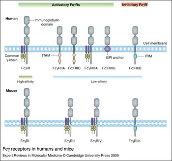
Humans and mice have orthologous FcγRs and, in both species, most of the corresponding genes are clustered in close proximity to each other in syntenic regions on chromosome 1 (Ref. Reference Nimmerjahn and Ravetch1). In mice, four IgG FcγRs have been described – FcγRI (CD64), FcγRIIb (CD32), FcγRIII (CD16) and FcγRIV – whereas in humans, six are known: FcγRI, FcγRIIA, FcγRIIB, FcγRIIC, FcγRIIIA and FcγRIIIB (Fig. 1). Based on genomic localisation and sequence similarity in the extracellular portion, mouse and human FcγRI are orthologous, as are mouse FcγRIIb and human FcγRIIB, while murine FcγRIV seems to be the orthologue of human FcγRIIIA, and mouse FcγRIII seems most closely related to human FcγRIIA. Of the human FcγRs, FcγRI is a high-affinity receptor; this review focuses on the low-affinity receptors FcγRIIA, FcγRIIB, FcγRIIC, FcγRIIIA and FcγRIIIB.
Fcγ receptors in humans and mice. In humans and mice, Fcγ receptor I (FcγRI) is the only high-affinity FcγR, while FcγRIIB/FcγRIIb is the only inhibitory receptor. Human FcγRs differ from mice in that there are four, rather than two, low-affinity activatory FcγRs. Human FcγRIIA and FcγRIIC signal directly through an immunoreceptor tyrosine-based activatory motif (ITAM), and human FcγRIIIB is linked to the membrane by a glycosylphosphatidylinositol (GPI) anchor. Most other activatory receptors (human and mouse), signal through the FcR common γ-chain; human FcγRIIIA can also signal via the ζ chain. FcγRIIB/FcγRIIb contains an immunoreceptor tyrosine-based inhibitory motif (ITIM).

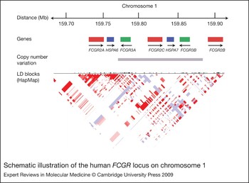
Human low-affinity FcγRs are encoded by FCGR2A, FCGR2B, FCGR2C, FCGR3A and FCGR3B (Fig. 2), and mouse low-affinity FcγRs by fcgr2b, fcgr3 and fcgr4. In both species, the genes are thought to have arisen from duplications and diversification processes, and thus have high sequence homology (Refs Reference Warmerdam2, Reference Qiu3, Reference Hughes4). In addition, there are allelic variants of several of these receptors, as well as copy number variation (CNV) of the human genes FCGR2C, FCGR3A and FCGR3B. CNV has been defined as a DNA segment that is 1 kb or larger and present at variable copy number in comparison with a reference genome (Ref. Reference Freeman5). As much as 5% of the human genome may lie within CNV regions (Refs Reference Redon6, Reference McCarroll7). It is not yet clear how such widespread CNV affects phenotypic variation or disease susceptibility. A study of expression levels of mRNA transcripts in HapMap individuals showed that CNVs contributed to 18% of the differential gene expression (Ref. Reference Stranger8). Some CNVs have been shown to be associated with common, complex diseases. For example, copy number of the CCL3LI gene has recently been associated with the autoimmune diseases systemic lupus erythematosus (SLE) (Ref. Reference Mamtani9), type 1 diabetes (Ref. Reference McKinney10) and rheumatoid arthritis (Ref. Reference McKinney10).
Schematic illustration of the human FCGR locus on chromosome 1. Sequence homology between the low-affinity FCGR genes is high, making this region difficult to study. In addition, part of this region is subject to copy number variation, as indicated by the grey bar. FCGR3B varies with FCGR2C, and independently of FCGR3A, although the exact break points have not yet been defined (Refs Reference McCarroll7, Reference Breunis94, Reference Breunis218). The linkage disequilibrium block is taken from the HapMap dataset (http://www.hapmap.org). Single-nucleotide polymorphisms (SNPs) are shown as lines across the top of the triangle. Within the triangle, red indicates that two SNPs are inherited together. This demonstrates the lack of extensive haplotype blocks and thus high degree of recombination across the FCGR locus.

In humans, CNV of FCGR3A, FCGR3B and FCGR2C combined with the high sequence homology between the two FCGR3 genes and the three FCGR2 genes has made the locus difficult to study. Few single-nucleotide polymorphisms (SNPs) in this locus have been included in genome-wide association studies, as the genotyping complexities have led to a lack of Hardy–Weinberg equilibrium in control samples, and nonmendelian inheritance in trios (Ref. Reference Estivill and Armengol11). Candidate-gene studies of polymorphisms at the FCGR locus have sometimes given conflicting results, which may be due to poor genotyping methodology, or because studies were small and therefore underpowered.
Structure and antibody binding
All FcγRs are glycoproteins with a ligand-binding α-subunit consisting of two (FcγRII and III) or three (FcγRI) C2-type extracellular Ig domains (Fig. 1). The additional Ig-like domain present in FcγRI may account for the high-affinity binding of this receptor to IgG. All FcγRs show a high degree of sequence identity in their extracellular portion (50–96%) but differ in their cytoplasmic domains (Ref. Reference Ravetch and Kinet12). FcγRIIB (in human and mouse) contains an immunoreceptor tyrosine-based inhibitory motif (ITIM) in its cytoplasmic domain, whereas human FcγRIIA and FcγRIIC contain immunoreceptor tyrosine-based activatory motifs (ITAMs) in their intracellular domain. Human FcγRIIIB does not have an intracellular domain, but is secured in the cell membrane by a glycosylphosphatidylinositol (GPI) anchor, and is thought to signal through association with complement receptor 3 (Ref. Reference Galon13), FcγRIIA (Ref. Reference Chuang, Sassaroli and Unkeless14) or lipid rafts (Ref. Reference Fernandes15). The remaining FcγRs (both mouse and human) exist as dimers consisting of the ligand-binding α-chain and the signal-transducing γ-chain molecule, known as the Fc receptor common γ-chain (Refs Reference Ravetch and Kinet12, Reference Reth16) (Fig. 1). This Fc receptor common γ-chain is distinct from the common γ-chain involved in signalling of the cytokines interleukin (IL)-2, -4, -7, -9, -13 and -15 (Ref. Reference He17).
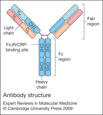
The Fc region of the IgG molecule is separated from the antigen-binding portion by a flexible hinge region, and forms two structural domains: the CH2 and CH3 domains. A short stretch of amino acid residues (234–238) of the N-terminal part of the CH2 domain, located in the lower hinge region, is directly involved in the binding to all FcγRs (Refs Reference Duncan18, Reference Lund19, Reference Canfield and Morrison20, Reference Jefferis, Lund and Pound21) (Fig. 3). Whereas FcγRI binds monomeric IgG with high affinity, the weak affinity of FcγRII, III and IV for IgG precludes binding to monomeric IgG; however, immune-complexed IgG permits more than one low-affinity receptor to bind antibody simultaneously (Refs Reference Nimmerjahn and Ravetch1, Reference Ravetch and Kinet12). Thus, despite the low affinity of each receptor for monomeric IgG, cells with multiple FcγRs have a high avidity for pathogen opsonised with a number of IgG molecules (Ref. Reference Nimmerjahn and Ravetch22).
Antibody structure. Antibodies are composed of two heavy chains (blue) and two light chains. The heavy chain determines antibody isotype (γ chain in IgG molecules). Each light chain comprises a variable (VL) region and a constant (CL) region, while each heavy chain comprises a variable (VH) region and a constant region, with three CH domains. Two CH domains of each heavy chain make up the Fc (Fragment, crystallisable) region of IgG, which mediates effector functions through binding of Fcγ receptors, C-reactive protein (CRP) and complement to the CH2 domain. The Fab (Fragment, antigen binding) region is composed of one constant and one variable domain from each heavy and light chain. The VL and VH regions are responsible for binding antigen, and are referred to as the complementarity-determining regions (CDRs).

IgG molecules are glycosylated in the CH2 domain. In humans, the majority of the Fc glycans consist of a complex biantennary structure, with a variable degree of sialylation and galactosylation (Ref. Reference Raju23). All IgG molecules contain a single conserved N-linked glycosylation site (Asn297); this helps to maintain the quaternary structure and is critical for Fc recognition (Ref. Reference Radaev and Sun24). Heterogeneity in the Fc glycans affects the binding affinity of IgG to FcγRs. For example, the presence of a core fucose reduces the affinity for FcγRIIIA by reducing the Fc protein surface available for interactions with the Asn162 glycan on the receptor (Ref. Reference Ferrara25).
FcγRs are also able to bind pentraxins, a family of innate immune mediators that includes the acute-phase proteins serum amyloid P (SAP) (Ref. Reference Bharadwaj26) and C-reactive protein (CRP) (Ref. Reference Bharadwaj27). Both SAP and CRP recognise various pathogens, including bacteria, fungi and yeast, and have similar functions to antibody in that they can both activate complement via C1q (Ref. Reference Kaplan and Volanakis28) and interact with FcγRs to induce leukocyte phagocytosis and cytokine secretion (Refs Reference Bharadwaj27, Reference Lu29). The structural mechanism for this interaction has recently been elucidated: the pentraxin-binding site overlaps with the IgG-binding site on FcγR, resulting in competition in FcγR binding, and inhibition of IgG-mediated phagocytosis by soluble pentraxins (Ref. Reference Lu29). Thus, FcγRs play a role in early, innate immune responses, as well as later, humoral responses.
Binding affinities
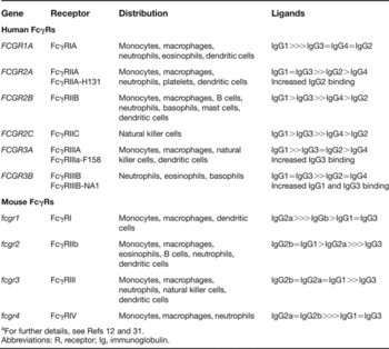
FcγRs differ in their binding affinities to IgG subtypes. Whereas FcγRI has a high affinity, the binding affinities of the other FcγRs for IgG are one to two orders of magnitude lower (Ref. Reference Ravetch and Kinet12) (Table 1). Three polymorphisms in human FCGR genes have been described that affect IgG-binding affinities (Table 1). First, a G>A point mutation in FCGR2A (rs1801274) causes an arginine to histidine amino acid substitution at position 131. In this review we refer to FcγRIIA-R131 as the allele encoding an arginine, while FcγRIIA-H131 refers to the histidine-encoding allele, and FcγRIIA-R/R131 describes homozygosity for the arginine-encoding allele. Second, a T>G SNP at nucleotide 559 (rs396991) in FCGR3A, results in either a valine or phenylalanine at amino acid position 158 (FcγRIIIA-V158 or FcγRIIIA-F158). Third, two combinations (or haplotypes) of five nucleotides (141, 147, 227, 277 and 349) within exon 3 of FCGR3B (FCGR3B*1 and FCGR3B*2) encode two isoforms termed FcγRIIIB-NA1 and FcγRIIIB-NA2 (also known as FcγRIIIb-HNA1a and FcγRIIIb-HNA1b), which differ by four amino acids. As a result, the FγRIIIB-NA1 isoform has four N-linked glycosylation sites, whereas the FcγRIIIB-NA2 isoform has six sites. An additional, rarer polymorphism, FCGR3B*3, encodes FcγRIIIB-SH (or FcγRIIIb-HNA1c), which differs from FCGR3B*2 at nucleotide 266, resulting in an alanine to aspartate substitution at amino acid 78 (Ref. Reference Bux30).
Fcγ receptors, expression and ligandsa

a For further details, see Refs Reference Ravetch and Kinet12 and Reference Ravetch, Nimmerjahn and Paul31.
Abbreviations: R, receptor; Ig, immunoglobulin.
Murine FcγRI and FcγRIV bind human IgG1 and IgG3 with affinities compatible with a potentially productive interaction (Ref. Reference Ravetch, Nimmerjahn and Paul31). Human FcγRI binds murine IgG2a and IgG3, human FcγRIIs (A, B and C) bind murine IgG1 and IgG2b, and human FcγRIII (A and B) binds murine IgG3, IgG2a and IgG2b (Ref. Reference Ravetch and Kinet12).
The ratio of activatory FcγR binding affinity to inhibitory (FcγRIIb) binding affinity has been called the A/I ratio, and is a measure of the efficacy of the IgG subclass in mediating effector responses (Ref. Reference Nimmerjahn32). In mice, these A/I ratios differ by several orders of magnitude between IgG subclasses. IgG1 has the lowest A/I ratio at 0.1, suggesting the activity of this isotype is strongly influenced by the inhibitory receptor FcγRIIb, whereas IgG2a, with an A/I of 70, is much less regulated by FcγRIIb. In models of tumour clearance and platelet depletion, IgG1 showed the most, and IgG2a the least, enhancement in biological activity in FcγRIIb-deficient mice (Ref. Reference Nimmerjahn and Ravetch33). Thus, the binding affinities of different IgG subclasses to activatory and inhibitory FcγRs impact on their ability to activate immune cells. In humans, the polymorphisms in FCGR2A, FCGR3A and FCGR3B that affect IgG binding will also impact on the A/I ratio (Table 1).
Function
Crosslinking of activatory FcγRs results in pathogen clearance by antibody-dependent cellular cytotoxicity, degranulation and phagocytosis, as well as the release of cytokines and other inflammatory mediators (Refs Reference Young, Ko and Cohn34, Reference Titus35). FcγRIIB, when crosslinked with activatory receptors (FcγR or the B cell receptor, BCR), is inhibitory, and acts to reduce cellular proliferation and calcium-mediated activation.
Activatory FcγRs on the surface of neutrophils, monocytes, macrophages and dendritic cells mediate endocytosis or phagocytosis of immune complexes, including antibody-coated microorganisms and soluble proteins. When the FcγRs are located on the surface of neutrophils, monocytes and macrophages, this process results in lysosomal degradation and cellular activation. Dendritic cells, however, also recycle phagocytosed antigen and present it to T cells via major histocompatibility complex (MHC) class I and II: in the mouse, uptake of IgG-complexed antigens via FcγRs induces dendritic cell maturation and promotes efficient MHC class I- and class II-restricted presentation of peptides from these antigens (Refs Reference Amigorena and Bonnerot36, Reference Regnault37).
A polymorphism in human FCGR2B has been described that abrogates the function of the receptor. A nonsynonymous SNP (rs1050501) in exon 5 of FCGR2B encodes a T>C transition at nucleotide 695, resulting in threonine replacing isoleucine at position 232 in the transmembrane domain. This causes exclusion of the variant receptor, FcγRIIB-T232, from lipid rafts, preventing interaction with activatory receptors and thus inhibition of cellular function (Refs Reference Floto38, Reference Kono39). Monocyte-derived macrophages from individuals homozygous for the polymorphism are more phagocytic when challenged with IgG-opsonised immune complexes, and B cells show greater activation when challenged by several stimuli (Ref. Reference Floto38).
FcγR expression: implications for immune function
FcγRs are expressed widely on immune cells, but the combination of activatory and inhibitory FcγRs expressed differs between immune cells and between species (Table 1). In myeloid cells, the inhibitory FcγR regulates the activatory FcγR, and in B cells the inhibitory FcγR regulates the BCR. The coexpression of activatory and inhibitory receptors establishes a threshold for cellular triggering by IgG antibodies. Thus, two factors may influence the degree of cellular activation in response to each IgG subclass: the binding affinity of the IgG subclass to these receptors (as discussed above), and the ratio of numbers of activatory to inhibitory receptor molecules expressed on the cell surface.
Surface expression of FcγRIIB may be altered by polymorphisms found in the promoter region of both mouse fcgr2b and human FCGR2B. A mouse promoter polymorphism that reduces expression of FcγRIIb is present in all polygenic mouse models of SLE (Refs Reference Pritchard40, Reference Jiang41). In humans, a haplotype in the FCGR2B promoter has been described that comprises two SNPs in linkage disequilibrium: the more common variant has a guanine at nucleotide position 386 and a thymine at 120 (−386G−120T), and the rarer variant has a cytosine at 386 and an adenine at 120 (−386C−120A). The effect of this haplotype is currently unclear: one group found the variant −386C−120A drove higher luciferase reporter expression (Ref. Reference Su42), but a second group investigating the −386 SNP (rs3219018) found the polymorphic form reduced luciferase activity and reduced FcγRIIB surface expression on peripheral B cells (Ref. Reference Blank43).
FcγR expression is altered by both cytokines and activation. Cell activation by the phorbol ester PMA and ditbuyryl cAMP increases FcγRII expression and phagocytosis in the human monoblastic cell line U937, with an increase in all three FcγRII RNA transcripts (Refs Reference Nambu44, Reference Cameron45). In human monocytes, interferon γ (IFN-γ), a T helper 1 (Th1) cytokine, induces the expression of FcγRI and FcγRIIA (Refs Reference Pricop46, Reference Grattage, McKenzie and Hogarth47), whereas Th2 cytokines such as IL-4 increase the expression of inhibitory FcγRIIB while downregulating all classes of activating FcγR (Ref. Reference Pricop46). In B cells, however, IL-4 decreases FcγRIIB expression (Ref. Reference Rudge48). By opposing effects on FcγR expression on different cell lineages, IL-4 promotes B cell activation but deactivates monocytes. This disparity may be due to differential activation of the FCGR genes by lineage-specific transcriptional regulators (Ref. Reference Ulgiati and Holers49).
Thus, both extrinsic factors, including cellular activation and cytokines, as well as genetic polymorphisms in FcγRs influence immune cell responses to IgG. A lower activation threshold may be beneficial in some situations, such as infection, but deleterious in others – for example, it may predispose to autoimmunity.
FcγRs in autoimmunity
FcγRs have been implicated in the pathogenesis of autoimmunity in a number of ways. First, the inhibitory receptor FcγRIIb plays an important role in maintaining self-tolerance by increasing the activatory threshold of autoreactive B cells and by facilitating the elimination of some of the autoreactive B cells generated in the bone marrow during development and in the germinal centre during somatic hypermutation. Second, FcγR ligation can profoundly affect dendritic cell maturation and mediate the internalisation of immune-complexed self-antigen, which may subsequently be presented to T cells. Third, activatory FcγRs on macrophages of the reticuloendothelial system of the spleen and liver are critical for the clearance of circulating IgG immune complexes; if these immune complexes accumulate, they may become deposited in tissues and cause inflammation, as seen in SLE. Fourth, FcγRs control the response of tissue macrophages and neutrophils to deposited immune complexes, determining the degree of local inflammation generated. Finally, FcγRs can directly mediate the pathogenic effects of autoantibodies – for example, causing red cell or platelet destruction in patients with platelet or red cell autoantibodies (Ref. Reference Cines and Blanchette50).
These effects of FcγR on susceptibility to autoimmunity do not occur in isolation but may be additive, perpetuating disease progression; for example, an excessive inflammatory response to deposited immune complexes by neutrophils and macrophages may increase tissue damage and the release of autoantigens, thus further increasing immune complex production, as well as exposing additional self-antigen in an inflammatory context, facilitating the breakdown of self-tolerance.
Mouse models of autoimmunity
Genetic studies of polygenic murine autoimmune diseases implicate FcγRs in pathogenesis. Seven independent linkage studies in murine models of autoimmune disease have identified a susceptibility locus on chromosome 1 that contains the low-affinity fcgr genes (Ref. Reference Wakeland51), although this region, syntenic to human 1q22-44, also contains other candidate genes, including complement receptor 2 and the SLAM/CD2 gene cluster, which encodes the signalling lymphocytic activation molecules, and CD2, a cell adhesion molecule found on T cells and natural killer (NK) cells (Refs Reference Boackle52, Reference Wandstrat53). Several murine autoimmune-prone inbred strains, including MRL, NZB, BXSB and NOD, have a polymorphic haplotype in the promoter region of fcgr2b (Refs Reference Pritchard40, Reference Jiang41). This polymorphism is associated with reduced FcγRIIb expression on B cells and activated macrophages (Ref. Reference Xiu54). It is clear, however, that these strains of mice have several polymorphisms predisposing to autoimmunity. Single-gene-knockout mice have helped to further elucidate the contribution of FcγRs to the development of autoimmunity.
Deficiency of the Fc receptor common γ-chain, which forms part of functional FcγRI, FcγRIII and FcγRIV, protects mice against the development of certain autoimmune diseases, including antiglomerular basement membrane antibody-mediated nephritis (Ref. Reference Suzuki55), experimental immune haemolytic anaemia and experimental immune thrombocytopaenia (Ref. Reference Clynes and Ravetch56). Collagen-induced arthritis is an experimentally induced antibody-dependent arthritis that occurs following immunisation with bovine collagen type II. H2q haplotype mice (e.g. DBA/1) are susceptible to this disease but H2b mice (e.g. C57BL/6, 129) are resistant. DBA/1 mice lacking the common γ-chain, however, are resistant to the disease despite developing similar levels of IgG anticollagen antibodies to wild-type DBA/1 mice (Ref. Reference Kleinau, Martinsson and Heyman57). These data suggest that activatory FcγRs mediate the effects of autoantibodies, but may not be involved in their generation.
FcγRIIb deficiency renders normally resistant strains of mice susceptible to several antibody-induced or immune-complex-dependent models of autoimmunity including antiglomerular basement membrane antibody-mediated nephritis (Ref. Reference Nakamura58) and collagen-induced arthritis, where deficiency in FcγRIIb renders H2b mice as susceptible as H2q mice (Ref. Reference Yuasa59). IgG1, IgG2a and IgG2b antibody responses against collagen type II are elevated in the C57BL/6 FcγRIIb-deficient animals, especially in those mice showing arthritis, suggesting the effect of FcγRIIb deficiency may be due to an effect on antibody levels. This is supported by studies in transgenic mice, where overexpression of FcγRIIb on B cells of C57BL/6 mice both reduces the IgG component of T-cell-dependent immune responses and also leads to early resolution of collagen-induced arthritis (Ref. Reference Brownlie60). These studies support a role for FcγRIIb in the control of autoantibody production via an inhibitory effect on B cells.
FcγRIIb deficiency predisposes to spontaneous as well as inducible autoimmunity: FcγRIIb-knockout mice derived on a 129/Sv/C57BL/6 background, but backcrossed for ten generations onto a C57BL/6 background, spontaneously develop hypergammaglobulinaemia, autoantibodies (to antigens such as chromatin and double-stranded DNA), and an immune-complex-mediated autoimmune disease resembling SLE, characterised by glomerulonephritis, renal failure, and a reduced life expectancy (Ref. Reference Bolland and Ravetch61). Conversely, the partial restoration of FcγRIIb levels on B cells of lupus-prone mouse strains is sufficient to restore tolerance and prevent autoimmunity (Ref. Reference McGaha, Sorrentino and Ravetch62).
FcγRIIb is also thought to contribute to the maintenance of both central and peripheral tolerance. Pre-B cells express FcγRIIb, but do not express the BCR. Aggregation of FcγRIIb on pre-B cells leads to potent inhibitory signalling and cell death, thus contributing to central tolerance by the elimination of autoreactive B cells during development (Ref. Reference Brauweiler and Cambier63). FcγRIIb also plays a role in peripheral tolerance. Antigen in the germinal centre is retained in the form of immune complexes bound to follicular dendritic cells. If B cell stimulation occurs through FcγRIIb stimulation alone (where affinity of the antigen to the BCR is low), the cell undergoes apoptosis, whereas FcγRIIb and BCR coligation results in B cell survival (if the BCR has a higher affinity for the antigen) (Ref. Reference Pearse64). Thus, FcγRIIb eliminates potentially autoreactive receptors at the price of limiting the diversity of the BCR repertoire.
FcγRIIb can also regulate plasma cell survival. Terminally differentiated plasma cells express very little BCR but high levels of FcγRIIb. In vitro, immune complexes cause plasma cell apoptosis in an FcγRIIb-dependent manner (Ref. Reference Xiang65). Plasma cells from mice prone to SLE are deficient in FcγRIIb and are therefore protected from apoptosis (Ref. Reference Xiang65). Deficiency in FcγRIIb may contribute to the survival of autoantibody-producing long-lived plasma cells.
Human FcγRs and autoimmune diseases
Systemic lupus erythematosus
SLE is characterised by the production of autoantibodies (particularly to nuclear components) and deposition of immune complexes, with inflammatory complications in multiple organs. There is a strong genetic component in the development of SLE. The concordance rate is tenfold higher in monozygotic twins (24–57%) compared with dizygotic twins (2–5%), and the sibling risk ratio (λs) is 20–29 (Refs Reference Alarcon-Segovia66, Reference Block67). The concordance rate of antinuclear antibodies is even higher in monozygotic twins, at 90% (Ref. Reference Block67). There have been several genome-wide screening studies of SLE patients attempting to identify susceptibility genes, as well as many candidate-gene association studies. Currently, 115 different genetic loci have been reported to associate with SLE, but there are conflicting reports that claim no association for 56 of these (Ref. Reference Sestak68). Nevertheless, several strong associations are consistently seen; these include genes encoding complement components, HLA region genes, and the FCGRs (reviewed in Ref. Reference Rhodes and Vyse69).
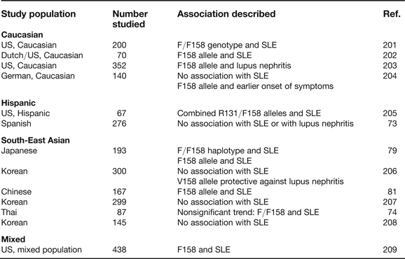
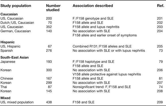
Several genetic association studies have genotyped activatory FcγR polymorphisms in case–control SLE cohorts. A meta-analysis included 12 studies of the FCGR2A SNP rs1801274 and two studies of the FCGR3A SNP rs396991. SLE was significantly associated with homozygosity for the alleles encoding lower-affinity isoforms of both FcγRIIA (FcγRIIA-R/R131) and FcγRIIIA (FcγRIIIA-F/F158) (Ref. Reference Lehrnbecher70). The effect of the FCGR2A SNP was stronger in populations of African American or South-East Asian background than in Caucasians, whereas the converse was found for the FCGR3A SNP. A second, larger meta-analysis of the FCGR2A SNP alone again suggested an association with SLE (Ref. Reference Karassa, Trikalinos and Ioannidis71). More recently, this SNP was included in a genome-wide association scan as part of an array testing 317 501 SNPs in 720 women of European ancestry with SLE and in 2337 ethnically matched controls. The FCGR2A SNP associated with SLE with a P value of 6.78 × 10⁻⁷ (Ref. Reference Harley72). Thus, there is a convincing body of evidence that the FcγRIIA-R131 allele is a risk factor for SLE. The evidence for a role of the FcγRIIIA-F158 allele is less consistent (Table 2).
FcγRIIIA polymorphisms and systemic lupus erythematosus (SLE)
Two studies of FCGR3B polymorphisms in patients with SLE have implicated the polymorphism encoding the lower-affinity isoform FcγRIIIB-NA2 in disease susceptibility (Refs Reference Gonzalez-Escribano73, Reference Siriboonrit74). These studies, however, did not take into account CNV, which has recently been described in FCGR3B (Refs Reference Redon6, Reference Aitman75, Reference Fanciulli76, Reference Hollox, Detering and Dehnugara77, Reference Breunis218). FCGR3B copy number correlates both with surface expression of FcγRIIIB and with neutrophil uptake of and adherence to immune complexes, under flow and static conditions (Ref. Reference Willcocks78). Low FCGR3B copy number may therefore result in reduced neutrophil trafficking to inflammatory lesions and an impaired ability to ingest immune complexes once there. This may explain the association of low FCGR3B copy number with SLE described in Caucasian populations (Refs Reference Fanciulli76, Reference Willcocks78).
Thus, SNPs and copy-number polymorphisms in the activatory FcγRs that decrease binding of immune complexes to leukocytes have been associated with SLE. This is consistent with the pathogenesis of SLE as a process in which there is failure to clear immune complexes.
The two polymorphisms in FCGR2B that affect expression and function of the receptor have also been associated with SLE. Two studies have shown that the FCGR2B promoter variant −386C−120A is associated with SLE in Caucasians, although it is unclear whether the polymorphism increases or decreases expression of the receptor (Refs Reference Su42, Reference Blank43). Six studies to date have examined the frequency of the function-abrogating FCGR2B transmembrane polymorphism (encoding FcγRIIB-T232) in patients with SLE (Refs Reference Siriboonrit74, Reference Kyogoku79, Reference Li80, Reference Chu81, Reference Kyogoku82, Reference Magnusson83); these studies have found an association of the FcγRIIB-T/T232 homozygous genotype with SLE in South-East Asian populations, but not in Caucasians.
ANCA-associated systemic vasculitis
ANCA-associated systemic vasculitis (AAV) comprises a group of diseases characterised by inflammation and necrosis of blood vessel walls, often associated with autoantibodies to antigens within the cytoplasm of neutrophils (antineutrophil cytoplasmic antibodies; ANCAs). ANCAs crosslink FcγRs with antigen on the surface of neutrophils to trigger superoxide production, pro-inflammatory cytokine release and degranulation (reviewed in Ref. Reference Morgan84). Recent work has suggested that autoantibodies to human lysosomal-associated membrane protein 2 (LAMP2) may be a critical factor in the pathogenesis of AAV: the study showed a direct pathological effect of these autoantibodies, likely to be mediated via FcγR (Ref. Reference Kain85).
One genetic association study found the combination of homozygosity for the low-affinity alleles – FcγRIIA-R/R131 and FcγRIIIA-F/F158 – was associated with increased disease relapse rates (Ref. Reference Dijstelbloem86). Individuals with the FcγRIIA-R/R131 genotype have a reduced ability to phagocytose Staphylococcus aureus, which provokes an IgG2 antibody response (see below) (Ref. Reference van der Pol and van de Winkel87). Chronic nasal carriage of S. aureus has been associated with higher AAV relapse rates; thus, the FcγRIIA-R/R131 genotype may predispose to AAV relapse by reducing clearance of S. aureus (Ref. Reference Stegeman88). However, a second study did not find an association between FcγRIIA genotype and AAV (Ref. Reference Tse89). The effects of CNV in FCGR3B on susceptibility to AAV are also controversial. One recent study found an association between low FCGR3B copy number and AAV (Ref. Reference Fanciulli76) but a second, larger study found the converse (Ref. Reference Willcocks78).
Thus, although in vitro data point to a role of FcγRs in mediating the actions of ANCA on neutrophil activation, genetic studies of FCGR polymorphisms are less clear.
Rheumatoid arthritis
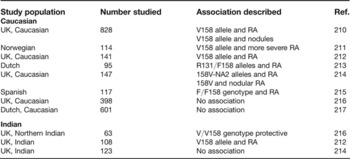
In rheumatoid arthritis, chronic inflammation in joints induces cartilage and bone destruction and leads to a progressive, deforming polyarthropathy. The serum of patients frequently contains rheumatoid factor, an IgM autoantibody directed against the Fc portion of IgG. Other IgG autoantibodies, such as anticollagen, anti-Sa and anti-cyclic citrullinated peptide (CCP) antibodies, have also been observed in rheumatoid arthritis, and it is thought these antibodies may contribute to disease pathogenesis via interactions with FcγR (Ref. Reference Menard90). Anti-CCP antibodies appear to be of particular importance: they are present in 95% of patients with rheumatoid arthritis, are detectable early in disease and are reported to predict the development of erosive rheumatoid arthritis (Ref. Reference van Venrooij, van Beers and Pruijn91). Binding of these IgG autoantibodies to FcγRIIIA may play a role in the pathogenesis of rheumatoid arthritis, as suggested by some association studies linking the higher-affinity allele FcγRIIIA-V158 with rheumatoid arthritis (Table 3).
FcγRIIIA polymorphisms and rheumatoid arthritis (RA)

Idiopathic thrombocytopaenic purpura
Idiopathic thrombocytopaenic purpura (ITP) is an autoimmune disorder of unknown aetiology, in which antiplatelet antibodies cause FcγR-mediated consumption of platelets by macrophages (Ref. Reference Cines and Blanchette50). Three studies have shown an association between the high-affinity isoform of FcγRIIIA (FcγRIIIA-V158) and ITP in children (Refs Reference Foster92, Reference Fujimoto93, Reference Breunis94). The most recent of these also found an association between ITP and a SNP in FCGR2C, which changes the common stop codon into a glutamine. Thus, in general, FCGR2C is a pseudogene, but individuals with the less common allelic form of FCGR2C express FcγRIIC on NK cells, and may also express FcγRIIC on neutrophils and monocytes, thought to be the main effector cells in ITP (Ref. Reference Breunis94).
Myasthenia gravis
In myasthenia gravis, autoantibodies against the α subunit of the nicotinic, postsynaptic acetylcholine receptor (AChR) mediate membrane lysis and accelerated receptor degradation. The resultant failure of signal transmission at the neuromuscular junction leads to muscular weakness. A study of 107 myasthenia patients found the FcγRIIA-R/R131 genotype occurred with increased frequency compared with 239 controls (Ref. Reference van der Pol95). Based on data confirming the presence of AChR-specific Th cells in the normal immune repertoire (Ref. Reference Melms96) and that AChR-containing immune complexes can enhance proliferation of these cells (Ref. Reference Melms97), the authors proposed that inefficient clearance of AChR–IgG complexes by the reticuloendothelial system may favour prolonged stimulation of autoreactive T cell clones and lead to clinical disease.
Guillain–Barré syndrome and chronic demyelinating polyneuropathy
Guillain–Barré syndrome (GBS) is an acute, inflammatory polyneuropathy, characterised histologically by demyelination or axonal damage, and clinically by a progressive, flaccid paralysis. About two-thirds of patients with GBS have serological evidence of a preceding bacterial or viral infection (Ref. Reference Hadden98). The pathogens are thought to induce specific immune responses that crossreact with peripheral nerve components, causing inflammation and subsequent neuronal damage. Most sera from GBS patients contain specific antibodies for neuronal constituents, particularly gangliosides such as GM-1 and GQ1b (Ref. Reference Ogino, Orazio and Latov99). In vitro, these anti-GM-1 antibodies have been shown to induce FcγR-dependent neutrophil degranulation, and an FcγR-mediated enhancement of phagocytosis in macrophages (Ref. Reference van Sorge100). FcγRs have been demonstrated on Schwann cells, perineural cells, and endoneurial macrophages (Ref. Reference Vedeler101). FcγRIIA polymorphisms have been studied in two GBS populations. The first study, in a Dutch population, found an increase in the FcγRIIA-H/H131 homozygous genotype in the patient group compared with the control group, and FcγRIIA-H/H131 homozygous patients had a higher risk of more severe disease than other genotypes (Ref. Reference van der Pol102). However, no such association was found in a study of Norwegian GBS patients, in whom homozygosity for the FCGR3B-NA1 allele was associated with less severe disease (Ref. Reference Vedeler103). These conflicting results may reflect small sample sizes, as the Dutch study comprised just 31 patients, and the Norwegian study 62 patients, but may also reflect a difference in the predominant preceding infectious agent in the two populations. In the Netherlands, Campylobacter jejuni is commonly associated with GBS, whereas in Norway, viral infections of the upper respiratory tract predominate (Ref. Reference Vedeler103).
Chronic inflammatory demyelinating polyneuropathy (CIDP) has similar clinical and histological features to GBS, but follows a chronic course, typically causing progressive or relapsing muscle weakness. As with GBS, the disease is thought to be mediated by autoantibodies, and often responds to treatment with intravenous immunoglobulin (IVIg). A recent study compared FcγRIIB expression on monocytes and B cells from healthy controls and patients with CIDP (Ref. Reference Tackenberg104). Untreated patients expressed lower levels of FcγRIIB, while treatment with IVIg led to increased expression. The FCGR2B promoter variant −386C−120A was present in 6 of 14 patients, compared with 1 of 26 controls, but these sample sizes are too small to draw any firm conclusions about the role of the polymorphism as a genetic risk factor in CIDP.
Multiple sclerosis
Multiple sclerosis is an inflammatory disease in which destruction of myelin and oligodendrocytes in the central nervous system leads to impaired neuronal conduction and disability in any aspect of neurological function (Ref. Reference Noseworthy105). The principal pathological features in the brain and spinal cord are focal areas of demyelinating plaques surrounded by an inflammatory infiltrate consisting of B cells, T cells, macrophages and microglia. Autoantibodies have been isolated from cerebral plaques and increased levels of circulating immune complexes have been correlated with disease severity (Ref. Reference Dasgupta106). Although FcγR polymorphisms have not been associated with susceptibility to multiple sclerosis, in a Norwegian study of 136 patients, those homozygous for the FcγRIIA-H131 allele had a significantly more benign disease course, suggesting that more effective clearance of immune complexes may be a possible mechanism for better clinical outcome in multiple sclerosis (Ref. Reference Myhr107). Homozygosity for the FCGR3B allele encoding the higher-affinity FcγRIIIB-NA1 isoform was also predictive of a more benign disease (Ref. Reference Myhr107). An Iranian study confirmed the FcγRIIA-H131 allele as a indicator of better prognosis (Ref. Reference Nikseresht108).
FcγRs in infection
The primary role of the immune system is to fight infection. Antibody plays an important role in both the early and adaptive immune response. In the early response to infection, antibody bound to pathogen may directly activate complement via the classical pathway. Alternatively, antibody-opsonised pathogen may bind to surface FcγRs on macrophages or neutrophils, facilitating phagocytosis and subsequent pathogen destruction. Antibody may also bind to pathogen-infected cells, flagging them up for destruction by NK cells, through surface FcγR engagement (so-called antibody-dependent cellular cytotoxicity). Antibodies and FcγRs also facilitate the adaptive immune response by stimulating dendritic cell maturation (thus making a more potent antigen-presenting cell) and by providing an efficient means of delivering pathogen-derived antigens to the dendritic cell for subsequent presentation. The importance of antibodies in defence against infection is highlighted by the immunodeficiency phenotype observed in patients lacking antibody (hypogammaglobulinaemia) in which there is an increased susceptibility to encapsulated bacteria, including Streptococcus and Neisseria species, as well as some protozoan infections, particularly Giardia lamblia (Ref. Reference Ballow109).
Mouse models of infection
Studies of FcγR-deficient mice in infectious models have given conflicting results. Deficiency of the Fc receptor common γ-chain did not affect susceptibility to staphylococcal septicaemia or arthritis, and bacterial load was equivalent in wild-type and γ-chain-deficient mice (Ref. Reference Gjertsson, Kleinau and Tarkowski110). However, in vitro and in vivo studies indicate that FcγRI and FcγRIII are important for phagocytosis of antibody-opsonised Bordetella pertussis, the Gram-negative bacteria causing whooping cough (Refs Reference Hellwig111, Reference Ioan-Facsinay112). Activatory FcγRs also appear to play a role in the clearance of intracellular pathogens. γ-Chain-deficient mice are more susceptible to influenza virus infection, with reduced macrophage phagocytosis and NK cell antibody-dependent cellular cytotoxicity (Ref. Reference Huber113). Following infection with Chlamydia muridarum, γ-chain-deficient mice also exhibit a delayed and reduced Chlamydia-specific Th1 cell response compared with control mice (Ref. Reference Moore114). In vitro, antichlamydial antibodies increase the rate of Th1 activation by antigen-presenting cells from FcγR-sufficient but not -deficient mice. In vivo administration of antichlamydial antibodies intranasally also greatly increased the frequency of Th1 cells in draining lymph nodes following genital chlamydial challenge.
There is also evidence to support an important role for activatory FcγRs in defence against parasitic infection. B-cell-deficient mice injected with Brugia malayi retained significant numbers of live microfilariae in their blood compared with wild-type mice (Ref. Reference Gray and Lawrence115). Treatment of these mice with hyperimmune serum within the first week of infection reduced the number of microfilariae seen. γ-Chain-deficient mice also had higher levels of microfilariae following infection, suggesting that FcγRs are mediating the protective effects of antibody, probably via antibody-dependent cellular cytotoxicity (Ref. Reference Gray and Lawrence115). In a murine malarial model, Plasmodium bergei XAT, γ-chain-deficient mice showed reduced antibody-mediated phagocytosis and increased susceptibility to infection (Ref. Reference Yoneto116).
FcγRIIb is also important in the immune response to both malarial and bacterial infection. Following infection with the rodent malaria Plasmodium chabaudi chabaudi, FcγRIIb-deficient mice develop lower parasitaemia, and less hypothermia and weight loss compared with control mice, associated with increased serum tumour necrosis factor (TNF)-α and antimalarial antibody titres (Ref. Reference Clatworthy117). FcγRIIb deficiency also increases uptake of opsonised bacteria by phagocytes, and protects against bacterial infection with both S. aureus and Streptococcus pneumoniae (Refs Reference Gjertsson, Kleinau and Tarkowski110, Reference Clatworthy and Smith118), while mice that overexpress FcγRIIb on macrophages are more susceptible to pneumococcal peritonitis and pneumonia (Ref. Reference Brownlie60). If FcγRIIb-deficient mice are immunised and challenged with high doses of S. pneumoniae, however, mortality is increased, associated with an excessive rise in pro-inflammatory cytokines (Ref. Reference Clatworthy and Smith118). This suggests a role for FcγRIIb in balancing efficient pathogen clearance against the cytokine-mediated consequences of sepsis.
Mouse models therefore suggest that FcγRs have a critical role in host defence against a range of pathogens – parasitic, viral and bacterial. As in autoimmunity, the balance of activatory FcγRs and the inhibitory FcγRIIb influences the outcome of infection. FcγRIIb deficiency increases the effects of the low-affinity FcγRs, resulting in a greater inflammatory response, but this may have a detrimental effect if it becomes excessive (Ref. Reference Clatworthy and Smith118).
Human FcγRs and infection
Encapsulated bacteria
S. pneumoniae is a major cause of human disease, particularly pneumonia, peritonitis and meningitis. The main defence against S. pneumoniae is provided by antibodies against capsular polysaccharide and cell-wall phosphocholine, which bind S. pneumoniae and mediate FcγR-dependent phagocytosis by neutrophils and macrophages (Refs Reference Briles119, Reference Mold, Rodic-Polic and Du Clos120). IgG2-deficiency predisposes to streptococcal infection, and so IgG2 is thought to play an important role in the immune response to this bacterium. Since IgG2 is a relatively inefficient activator of complement, binding to FcγRIIA is likely to be important in the immune response to streptococcal infection, particularly Fc-dependent phagocytosis. The FcγRIIA isoform FcγRIIA-H131 is the only human FcγR capable of effectively binding complexed IgG2. Genetic studies have shown an association between FcγRIIA-R/R131 homozygosity and susceptibility to invasive pneumococcal disease (Refs Reference Rodriguez121, Reference Yee122, Reference Yuan123). In vitro, neutrophils from donors homozygous for FcγRIIA-H131 have a higher phagocytic capacity for IgG-opsonised S. pneumoniae than neutrophils from FcγRIIA-R/R131 donors (Ref. Reference Rodriguez121). In vitro studies have also shown the polymorphic form of FCGR2B, FcγRIIB-T232, increases phagocytosis of antibody-opsonised S. pneumonia by monocyte-derived macrophages (Ref. Reference Floto38).
Neisseria meningitidis, a Gram-negative, encapsulated bacterium, is a commensal organism in up to 10% of healthy individuals, but in a minority of cases may cause invasive meningococcal disease, manifesting as meningitis or septicaemia (Ref. Reference Pathan, Faust and Levin124). The host response to meningococcal infection begins with the production of antibody in response to colonisation, facilitating FcγR-mediated uptake of bacteria by phagocytes (Ref. Reference Pathan, Faust and Levin124). As with S. pneumoniae, IgG2 is important in defence against N. meningitidis, and neutrophils with the FcγRIIA-R/R131 genotype phagocytose IgG2-opsonised N. meningitidis less effectively than those with FcγRIIA-H/H131 (Refs Reference Bredius125, Reference Fijen126). In a small retrospective study of 24 Dutch children who had survived fulminant meningococcal septic shock, 44% were homozygous for FcγRIIA-R131 polymorphism compared with 23% of controls (Ref. Reference Bredius127). However, in a Norwegian case–control study, no association with incidence or severity of meningococcal disease was found (Ref. Reference Smith, Vedeler and Halstensen128), while further studies found associations of FcγRIIA-R/R131 with meningococcal disease only in older individuals (Refs Reference Platonov129, Reference Domingo130, Reference Domingo131).
Thus, the FcγRIIA isoform with a low affinity for IgG2, FcγRIIA-R131, may predispose to bacterial infection, but studies have been small and larger cohorts are required to confirm this association.
Periodontitis
Periodontitis is a major cause of tooth loss in adults, resulting from both the direct effect of periodontopathic Gram-negative bacteria on the gums, and the host inflammatory response. The disease is characterised by the production of very high concentrations of serum antibodies reactive with a limited number of periodontal disease pathogens, and serum concentration of IgG2 is particularly elevated (Ref. Reference Schenkein, Barbour and Tew132). Associations between FcγR polymorphisms and susceptibility to periodontitis have been investigated in small studies in Caucasian, Asian, and African American populations (Refs Reference Loos133, Reference Chung134, Reference Kobayashi135, Reference Yasuda136, Reference Sanz137). These studies suggest that polymorphisms in the FcγR-encoding genes related to aggressive periodontitis risk are different among ethnic populations, which may reflect differences in the frequencies of mouth commensal bacteria. Larger studies, however, are needed to validate these findings.
Malaria
Malaria causes significant morbidity and mortality in many tropical countries, particularly in sub-Saharan Africa. The protective effects of antibody were shown by transfer experiments in which children with acute malaria improved after treatment with adult hyperimmune serum (Ref. Reference McGregor138). Protective antibody responses have been associated with the development of IgG1 and IgG3 subclasses, allowing opsonisation and clearance of merozoites or infected erythrocytes by macrophages and neutrophils (Refs Reference Taylor139, Reference Ndungu140). Initial studies indicated that the FcγRIIA-R/R131 genotype is associated with protection against severe malaria in children (Refs Reference Shi141, Reference Cooke142). In northwest Thailand, the FcγRIIA-131H/H genotype in combination with the FcγRIIIB-NA2 allele was associated with susceptibility to cerebral malaria (Ref. Reference Omi143). In a study of 493 Kenyan children with acute malaria, homozygotes for the FcγRIIA-R131 allele were protected against high-density parasitaemia, but no effect was seen on severe malarial anaemia (Ref. Reference Ouma144). A further study, however, found that the frequency of the FcγRIIA-R/R131 genotype was significantly higher in those with severe malaria. Levels of IgG1 and IgG3 antibodies were higher in the mild malaria compared with the severe malaria. The lowest risk of severe malaria was seen where high levels of IgG3 antibodies were combined with the H/H131 genotype (Ref. Reference Nasr145). Subsequent studies have also shown a protective effect of the FcγRIIA-H/H131 genotype against severe malaria (Refs Reference Nasr146, Reference Sinha147). These discrepancies may reflect differences in the ethnic background of the populations studied – the latter three studies included ethnically diverse populations, which are likely to have a number of different susceptibility polymorphisms.
In a study of 462 adult Thai patients with malaria, the FcγRIIIA-176F/V polymorphism was not associated with incidence of mild malaria, noncerebral severe malaria or cerebral malaria (Ref. Reference Omi148). The FCGR2B transmembrane polymorphism, rs1050501, may protect against malaria: in vitro studies have shown that macrophages with the disabled form of FcγRIIB, FcγRIIB-T232, phagocytose more opsonised Plasmodium falciparum trophozoites than those with FcγRIIB-I232 (Ref. Reference Clatworthy117). Genetic association studies have not yet been performed.
Viruses
Human immunodeficiency virus (HIV) infection, leading to acquired immune deficiency syndrome (AIDS), has become a leading cause of mortality worldwide and the main cause of death in sub-Saharan Africa. HIV-1 is a retrovirus that invades cells via CD4, a cell-surface receptor found predominantly on Th cells. Antibodies are important in host response to HIV infection, and IgG antibody subclasses differ in their ability to neutralise HIV-1 and inhibit HIV–cell fusion, with IgG1 and IgG3 being more efficient than IgG2 (Ref. Reference Scharf149). The role of FcγR in HIV infection is unclear, but in vitro studies suggest that FcγR on monocytes, when crosslinked with IgG-antibody–virus immune complexes, allow viral entry and activation of HIV gene transcription (Refs Reference Takeda, Tuazon and Ennis150, Reference Tsitsikov151). In a study of HIV-infected men in the Multicenter AIDS Cohort Study, CD4+ T cell counts fell faster in patients with the FcγRIIA-R/R131 genotype than individuals with the FcγRIIA-R/H131 or FcγRIIA-H/H131 genotypes. However, progression to AIDS was unaffected by FcγRIIA genotype, largely because FcγRIIA-H/H131 homozygotes had an increased risk of Pneumocystis jiroveci pneumonia as an AIDS-defining illness. In vitro, HIV-1 immune complexes were more efficiently internalised by monocytes from FcγRIIA-H/H131 subjects compared with FcγIIA-R/R131 subjects (Ref. Reference Forthal152). This suggests that increased uptake of HIV immune complexes by phagocytes carrying the FcγRIIA-H/H131 genotype promotes viral clearance by these cells, rather than allowing the cells to become infected. However, a Kenyan study of 448 HIV-seropositive mothers and babies found that vertical transmission of HIV was more frequent in infants with the FcγRIIA-H/H131 genotype (Ref. Reference Brouwer153), suggesting that this genotype may actually increase viral infection.
Kaposi sarcoma is the most common malignant condition associated with HIV infection. It is thought that in its early stage, Kaposi sarcoma is an angioproliferative, inflammatory lesion induced by infection with human herpes virus type 8 (HHV-8). The immunopathogenesis of Kaposi sarcoma involves the activation of lymphocytes, resulting in the production of pro-inflammatory cytokines and angiogenic factors (Ref. Reference Schalling154). NK cells are important in mediating antibody-dependent cellular cytotoxicity of both virally infected and malignant cells: their absence increases the severity of HHV-8 infections (Ref. Reference Jawahar155). FcγRIIIA is expressed on NK cells, and two studies have found the higher-affinity FcγRIIIA-V/V158 genotype is associated with the development of Kaposi sarcoma in individuals with HIV (Refs Reference Forthal152, Reference Lehrnbecher156). Thus, more avid binding of antibody to FcγRIIIA-V/V158 could lead to an increase in cytokine production, driving inflammation and angiogenesis.
Dengue viruses belong to the family Flaviviridae, genus Flavivirus, and are transmitted to humans by mosquitoes. Dengue fever is an acute viral infection characterised by fever, rash, headache, myalgia and arthralgia. Occasionally, dengue fever progresses to dengue shock syndrome (DSS), a potentially life-threatening illness associated with increased vascular permeability, haemorrhage, and shock (Ref. Reference Halstead157). Epidemiological studies show that severe complications of dengue viral infections, such as DSS, are more commonly observed during secondary infections compared with primary infections, suggesting that the presence of circulating dengue-specific antibodies constitutes a risk factor for DSS (Ref. Reference Burke158). DSS can occur in primary infections, but this is more unusual and tends to affect infants born to dengue-antibody-positive mothers (Ref. Reference Halstead157); in these infants, higher antibody levels correlate with more severe disease (Ref. Reference Kliks159). In vitro, dengue-specific IgG can enhance viral entry and infection of cells (Refs Reference Halstead157, Reference Thein160), and this effect is mediated by both FcγRI and FcγRII (Ref. Reference Littaua, Kurane and Ennis161). One study in 352 Vietnamese patients with DSS and 251 controls found a lower frequency of FcγRIIA-R131 homozygotes in the patient group, suggesting this genotype may be protected against DSS, perhaps because the weaker binding affinity reduces viral entry into cells (Ref. Reference Loke162). Effects of polymorphisms in other FcγRs have not been assessed.
Poliovirus is an RNA virus that in 90% of individuals is entirely asymptomatic. In fewer than 1% of cases, however, the virus enters the central nervous system, preferentially infecting and destroying motor neurons, leading to muscle weakness and acute flaccid paralysis (Ref. Reference Blondel163). Poliovirus enters susceptible cells through a specific poliovirus receptor, but can also be taken up by FcγRs (Ref. Reference Arita, Horie and Nomoto164), and FcγRs have been found to be expressed on both microglia and perivascular macrophages (Ref. Reference Ulvestad165). IgG3 is the main antibody subtype produced in response to poliovirus in humans (Ref. Reference Beck166). A Norwegian study of patients who contracted poliomyelitis in the 1950–1954 epidemic found the FcγRIIA-V/V158 genotype was under represented in patients with a history of acute poliomyelitis, suggesting that V/V homozygous individuals might have increased binding and clearance of antibody-opsonised poliovirus and therefore be protected against disease (Ref. Reference Rekand167).
Clinical implications/applications
Antibodies as treatment for autoimmunity, cancer and in transplantation
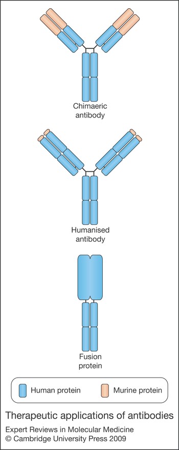
Antibody treatment, in the form of polyclonal antileukocyte antibodies, was first used in clinical practice in 1967, when Starzl showed antilymphocyte globulin could prolong the survival of a renal transplant (Ref. Reference Starzl168). Since then, antibody treatment has been refined, first by the development of monoclonal mouse anti-human antibodies, and then by chimaeric murine–human antibodies, humanised antibodies, and Fc-fusion proteins – with reducing amounts of murine protein (Fig. 4). Currently, 21 therapies have been approved by the US Food and Drug Administration (FDA), and hundreds of therapies are undergoing clinical trials. Most are concerned with immunological and oncological targets.
Therapeutic applications of antibody. Chimaeric antibodies (e.g. rituximab), are formed from human Fc (constant) regions (blue) and murine variable regions (orange). Humanised antibodies (e.g. alemtuzumab) are predominantly human, with murine complementarity-determining regions. Fusion proteins (e.g. etanercept) consist of a human Fc IgG1 fused with a human receptor.

Therapeutic antibodies may have an effect in one of four ways. First, they may block the interaction of cytokines or growth factors, such as anti-TNF treatment in rheumatoid arthritis and Crohn disease (Ref. Reference Wong169). Second, they may deliver a drug or label to a target cell, such as tositumomab (an IgG2a anti-CD20 antibody covalently bound to a radioactive isotope) in treatment of refractory follicular lymphoma (Ref. Reference Vose170). Third, therapeutic antibodies may trigger an activatory receptor, such as in the recent Phase I clinical trial of a stimulatory antibody to CD28 that had unexpected results, with healthy volunteers developing a systemic inflammatory response and multiorgan failure within 90 min of receiving the drug (Ref. Reference Suntharalingam171). Last, they may deplete cells by targeting them for antibody-dependent cellular cytotoxicity.
Commonly used depleting, or cytotoxic, antibodies include rituximab (an anti-CD20 antibody that depletes B cells and is used to treat lymphoma as well as antibody-mediated autoimmune diseases including ITP and SLE) (Refs Reference Grillo-Lopez172, Reference Looney173), trastuzumab (which targets breast cancer cells overexpressing the receptor erbB2) and alemtuzumab (which targets CD52 on the surface of mature lymphocytes and is used in the treatment of lymphoma, autoimmune disease and transplant rejection). Studies in γ-chain-deficient mice have shown that the depleting effect of these antibodies crucially depends on activating FcγRs, and that the inhibitory FcγRIIB is an important negative regulator of their activity in vivo (Refs Reference Clynes174, Reference Zhang175, Reference Uchida176). In humans, the polymorphisms in FcγRIIA and FcγRIIIA that affect IgG binding impact on the clinical efficacy of rituximab: in one study, patients homozygous for the high-affinity FcγRIIIA-158V/V genotype responded better to lymphoma treatment than those homozygous for the low-affinity allele (Ref. Reference Cartron177), and in a second study, both the FcγRIIIA-158V/V and FcγRIIA-131H/H genotypes were found to be independently associated with a better response rate and freedom from progression (Ref. Reference Weng and Levy178).
It might be possible to improve the efficacy of cytotoxic antibodies by two approaches. First, reducing the interaction of the antibody with FcγRIIB may increase its effect: in vivo and in vitro, blocking or deleting FcγRIIb on dendritic cells results in increased antitumour responses (Refs Reference Kalergis and Ravetch179, Reference Dhodapkar180). Second, the affinity of the Fc portion of the therapeutic antibody might be manipulated to increase the binding affinity to FcγRIIIA. Mutations that increase antibody binding to FcγRIIIA and lower binding to FcγRIIB result in antibodies with increased antitumour activity in vitro and in nude mice injected with human tumour cell lines (Refs Reference Shields181, Reference Lazar182). Varying antibody glycosylation can also affect binding affinity to FcγRs: removing a fucose in the sugar moiety attached to Asn297 increases the cytotoxic activity of all mouse and human IgG antibody isotypes (Ref. Reference Nimmerjahn and Ravetch33). Removing all fucose increases binding to human FcγRIIIA by 10–50-fold, but does not affect binding to FcγRIIB (Ref. Reference Ferrara25). Thus, for antibodies where FcγRIIIA is the dominant FcγR activating effector cells, removal of fucose may increase the therapeutic effect.
The therapeutic advantages of antibodies may also be utilised through Fc-fusion proteins – chimaeric proteins consisting of the effector region of a protein, such as the Fab region of an antibody or the binding region of a receptor, fused to the Fc region of an IgG molecule (Fig. 4). The Fc region mediates antibody-dependent cellular cytotoxicity, through interactions with FcγRs, and complement-dependent cytotoxicity by binding to C1q. In addition, the Fc region binds to the neonatal Fc receptor, FcRn. This receptor, which is structurally related to the MHC class I family, has no signalling capacity, and was first identified as the receptor responsible for transfer of maternal IgG across the placenta during gestation (Ref. Reference Story, Mikulska and Simister183). Subsequent studies have shown that FcRn acts as a salvage receptor, protecting IgG from lysosomal degradation and thus increasing the circulatory half-life (Ref. Reference Ghetie and Ward184). Serum half-life and effector functions of Fc-fusion proteins can therefore be modulated by engineering the Fc region to increase or reduce binding to FcRn, FcγRs and C1q. Examples of Fc-fusion proteins include etanercept, which binds to, and blocks, the TNF receptor; and abatacept, a fusion protein of CTLA4 (cytotoxic T lymphocyte antigen 4; also known as CD152), which blocks T cell costimulation, preventing T cell activation. Both these drugs have FDA approval for use in the treatment of rheumatoid arthritis (although not concurrently).
Low-affinity FcγRs and intravenous immunoglobulin
IVIg is a preparation of human polyclonal IgG obtained from pooled plasma samples taken from thousands of healthy blood donors. Although it was first utilised as replacement therapy for individuals with humoral immune deficiency (Ref. Reference Molica185), it is now increasingly used (at much higher doses) as an anti-inflammatory agent in the treatment of a variety of autoimmune conditions. It is licensed for the treatment of ITP, GBS, CIDP and Kawasaki disease, and has been used off label in myasthenia gravis, multiple sclerosis, AAV, SLE and rheumatoid arthritis.
The mechanism of action of IVIg as an immunomodulatory agent has been widely debated. Proposed mechanisms of action include the neutralisation of pathogenic autoantibodies by anti-idiotypic antibodies, the modulation of cytokine production, and the neutralisation of complement components (C3a, C5a) (Ref. Reference Basta186). These effects are mainly F(ab)2 dependent, but clinical studies in patients with ITP and in murine models of autoimmunity suggest the Fc fragment may be as potent as whole IVIg preparations (Refs Reference Debre187, Reference Bruhns188, Reference Samuelsson, Towers and Ravetch189), perhaps due to to blockade of activatory FcγRs, upregulation of FcγRIIB, or FcRn blockade.
Activatory FcγRs and IVIg
The Fc portion of IVIg may dampen an inflammatory response through the blockade of activatory FcγRs. As discussed previously, in autoimmunity, activatory FcγRs mediate many of the pathological effects of autoantibody. In a murine model of ITP, in which platelets are depleted by a monoclonal antibody, both FcγRIII deficiency and pretreatment with IVIg are protective, while FcγRI deficiency is not (Ref. Reference Samuelsson, Towers and Ravetch189). Although IVIg contains predominantly monomeric IgG (which would not be expected to significantly bind low-affinity FcγRs) it also contains small amounts of dimeric and polymeric IgG, which would bind to these receptors (Ref. Reference Teeling190). In addition, there is some evidence that monomeric IgG in IVIg may directly block FcγRII and FcγRIIIb on human neutrophils (Ref. Reference van Mirre191). Thus in ITP, treatment with IVIg may block the binding of antibody-opsonised platelets to low-affinity activatory FcγR, reducing platelet destruction by inhibiting phagocytosis.
IVIg also inhibits the pro-inflammatory response initiated by activatory FcγR crosslinking; for example, in vitro, IVIg (via soluble immune complexes) reduces IFN-γ-induced activation in both murine and human macrophages in an FcγRIII-dependent fashion (Ref. Reference Park-Min192). In this study, the mechanism of inhibition of IFN-γ-induced activation by IVIg was via the suppression of expression of the IFNGR2 subunit of the IFN receptor, and did not require the presence of FcγRIIb. In a murine model of ITP in which mice were treated with IFN-γ to exacerbate disease, the protective effects of IVIg were dependent on FcγRIII but not FcγRIIb (Ref. Reference Park-Min192). However, in a murine nephrotoxic nephritis model (without IFN-γ priming) the protective effects of IVIg are independent of FcγRIII (Ref. Reference Kaneko193). In addition to modulating the response initiated by FcγRIII binding, IVIg also appears to downregulate the expression of FcγRIV on murine splenic macrophages, thus potentially increasing the activatory threshold of these cells (Ref. Reference Kaneko193) (see next section). In patients with Kawasaki disease, a downregulation of both FcγRI and FcγRIIIA was observed on peripheral blood monocytes following IVIg treatment, which might similarly increase activation thresholds (Ref. Reference Abe194).
The inhibitory receptor FcγRIIb and IVIg
In FcγRIIb-deficient mice, IVIg is no longer effective in treating many murine models of autoimmunity including ITP, serum-induced arthritis, and nephrotoxic nephritis, suggesting an important role for the inhibitory receptor (Refs Reference Samuelsson, Towers and Ravetch189, Reference Kaneko193, Reference Crow195, Reference Kaneko, Nimmerjahn and Ravetch196). Following IVIg treatment in wild-type mice, there is an upregulation of FcγRIIb on splenic macrophages (Ref. Reference Samuelsson, Towers and Ravetch189). This increased expression of the inhibitory receptor, together with a downregulation of the activatory Fc receptor FcγRIV (Ref. Reference Kaneko193), might significantly increase the activatory threshold of an inflammatory macrophage. Modulating FcγR expression would therefore act to reduce macrophage activation and associated inflammation. More recent data confirm the relevance of these observations to humans, demonstrating an upregulation of FcγRIIb on both peripheral blood monocytes and B cells following IVIg treatment in patients with CIDP (Ref. Reference Tackenberg104). Ravetch and colleagues have suggested that IVIg indirectly increases FcγRIIb expression on effector macrophages via an action on so-called sensor macrophages, which are dependent on colony-stimulating factor (CSF)-1. IVIg is ineffective in treating autoimmunity in CSF-1-knockout mice, which are deficient in some subpopulations of monocytes and macrophages. Furthermore, CSF-1-deficient mice do not upregulate FcγRIIb on effector macrophages (Ref. Reference Bruhns188), suggesting that CSF-1-dependent sensor macrophages are required to mediate the IVIg-dependent upregulation of FcγRIIb on effector macrophages. This IVIg-binding receptor on sensor macrophages has been identified as SIGN-R1, a specific C-type lectin expressed on macrophages in the splenic marginal zone, which preferentially binds sialylated IgG (Ref. Reference Anthony197). It is estimated that around 5% of pooled serum IgG is sialylated. Deglycosylation of IVIg abrogates anti-inflammatory activity, whereas enrichment for sialylated forms significantly increases its anti-inflammatory activity such that a tenth of the dose is required to bring about the same effect (Ref. Reference Kaneko, Nimmerjahn and Ravetch196). Further studies indicate that it is a 2,6-linked sialylation at Asn297 that is required for the anti-inflammatory effect of IVIg (Ref. Reference Anthony198). In an era where the demand for IVIg significantly outstrips supply, these data suggest that an appropriately sialylated Fc fragment may be engineered to reproduce at least some of the anti-inflammatory effects of IVIg.
FcRn and IVIg
FcRn plays an important role in the regulation of the half-life of IgG, including pathogenic IgG autoantibodies. When IgG is endocytosed, it becomes bound to FcRn within acidic lysosomes. This binding prevents the degradation of IgG and leads instead to its recycling back to the surface of the cell where it is released from FcRn under neutral pH. Thus, IVIg might block the interaction between pathogenic antibody and FcRn, leading to a reduction in recycling and consequently the level of pathogenic IgG. Antibody half-life studies in animal models of ITP demonstrate that IVIg shortens the half-life of platelet-specific autoantibodies (Ref. Reference Hansen and Balthasar199). In the serum-transfer arthritis model, treatment with IVIg reduces the half-life of arthritogenic antibodies as well as joint inflammation. In FcRn-deficient mice, these protective effects of IVIg are much reduced (Ref. Reference Akilesh200). However, aglycosylated IVIg, which weakly binds classical FcγRs but has preserved affinity for FcRn, does not prevent arthritis in the serum-transfer model (Refs Reference Shields181, Reference Kaneko, Nimmerjahn and Ravetch196), arguing against a significant role for FcRn in the action of IVIg.
Conclusions
The family of FcγRs are a key part of the humoral response to infection. They also possess several properties that allow them to participate in regulation of immune activation: they differ in their binding affinities to IgG and in their expression profiles on different leukocytes under different conditions, and FcγRIIB acts to counterbalance the effects of the low-affinity activatory FcγRs. It is not surprising, therefore, that disturbances in the function or expression of these receptors should result in defective immune system function manifested as susceptibility to infection or immunity. Their role in regulation also makes the FcγRs attractive candidates for therapeutic intervention. Upregulation of FcγRIIB, for example, may reduce the deleterious inflammatory responses that occur in septic shock. In addition, the manipulation of therapeutic antibodies, already used to treat immune and oncological conditions, to improve selective FcγR binding may improve the efficacy of these treatments.
Acknowledgements and funding
Financial support was provided by the Wellcome Trust (Programme Grant Number 083650/Z/07/Z) and the NIHR Cambridge Biomedical Research Centre. The Cambridge Institute for Medical Research is in receipt of a Wellcome Trust Strategic Award (079895). M.R.C is a Wellcome Trust Intermediate Training Fellow and K.G.C.S. is a Lister Prize Fellow. We also thank the reviewers of the manuscript for their thoughtful and incisive comments.