Introduction
Vertiginous problems in children are usually misdiagnosed, and can contribute to behavioural problems, lack of co-ordination, delays in postural and motor control, and recurrent falling.Reference Nandi and Luxon1 Otitis media with effusion (OME) is one of the most common diseases in children, occurring in 40–80 per cent of this population. Approximately 90 per cent of children have OME at some time before school age.Reference Casselbrant, Furman, Mandel and Rubenstein2, Reference Alho, Oja, Koivu and Sorri3 Fifty per cent of children with serous otitis media may have balance disturbances.Reference Golz, Angel-Yeger and Parush4, Reference Grace and Pfleiderer5 Otitis media with effusion has an effect on the vestibular system due to acute labyrinthitis or negative middle-ear pressure changes. These effects occur not only during the infection but also after the infection has resolved.Reference Hamidi6
Most children with abnormal middle-ear ventilation or OME do not complain of balance disturbance or vertigo. However, their parents may describe excessive clumsiness, awkwardness and sometimes frequent falling. Moreover, pre-school children with a history of OME have significantly decreased percentile rank scores on stepping and vertical writing, which act as a measure of vestibulo-spinal function.Reference Golz, Angel-Yeger and Parush4 SzirmaiReference Szirmai7 found that OME had an effect on the bedside vestibular test results of children and adolescents, especially in the sharpened Romberg test.
Video-nystagmography abnormality has been reported in children with OME. Golz et al.Reference Golz, Westerman, Gilbert, Joachims and Netzer8 reported an overall occurrence of abnormality in 71 per cent of affected children, compared to only 4 per cent in the control group. Wang and LeeReference Wang and Lee9 reported that latencies of the air-conducted vestibular-evoked myogenic potential components P13 (the positive waveform component at 13 ms) and N23 (the negative component at 23 ms) were significantly prolonged or absent in children with OME. Bone-conducted vestibular-evoked myogenic potential testing represents an alternative technique that aims to overcome the stimulus attenuation as a result of middle-ear pathology.Reference Halmagyi and Colebatch10, Reference Brantberg, Bergenius and Tribukait11
This study aimed to assess vestibular function in children with middle-ear effusion using: the Arabic Dizziness Handicap Inventory for children (developed for use in this study), a vertigo and imbalance evaluation sheet for children (developed from several other evaluation sheets and translated into Arabic), vestibular bedside tests for children (using newly developed test material), video-nystagmography test batteries (except the caloric test), and click-evoked air- and bone-conducted vestibular-evoked myogenic potential testing.
Materials and methods
Before starting this research, consent was obtained from the family of each child. The research was approved by the Research Ethics Committee at Tanta University Hospitals (approval code 437/03/11).
This study comprised two groups of children selected from patients attending the Audiology Unit at Tanta University Hospitals. The control group consisted of 25 children aged between 5 and 12 years with bilateral normal peripheral hearing in the frequency range of 250–8000 Hz (hearing threshold level of 20 dB HL or less) and bilateral normal middle-ear function. The study group consisted of 30 children aged between 5 and 12 years with bilateral otitis media with effusion (OME), as diagnosed by tympanometry (bilateral type B tympanograms) and pure tone audiometry (bilateral conductive hearing loss). All children were normal in terms of developmental milestones, with normal speech development according to their age and normal intelligent quotients.
Exclusion criteria included: children with a non-vestibular cause of vertigo; unilateral or mixed hearing loss; and children with developmental disorders, neurological diseases or any other serious illness. Children receiving medications (for conditions other than OME) were also excluded from this study.
All children included in this study underwent the following: full audiological history-taking; otological examination; basic audiological evaluation, including pure tone audiometry using either play audiometry or voluntary thresholds according to the child's age (for both air conduction and bone conduction); the speech recognition threshold test using Arabic bisyllabic words for children (El-Mahallawi and Soliman, unpublished manuscript); the word discrimination test using an Arabic phonetically balanced word list for children (El-Mahallawi and Soliman); and immittancemetry including both tympanometry and acoustic reflex threshold measurement.
The vestibular assessments conducted are described below.
Dizziness, vertigo and imbalance evaluation
This evaluation sheet for children allows complete assessment and analysis of imbalance complaints (e.g. attack details, associated symptoms, family history and previous studies) (Appendix 1). It was adapted from several other evaluation sheets used in the assessment of children's balance (e.g. Ravid et al.Reference Ravid, Bienkovski and Eviatar12 and Niemensivu et al.Reference Niemensivu, Kentala, Wiener-Vacher and Pyykkö13).
Arabic Dizziness Handicap Inventory
This self-assessment questionnaire for children was developed for use in this study (Appendices 2 and 3). It reflects paternal observations of a child's balance performance and imbalance symptoms. It consists of 10 questions relating to the child's activities that reflect his or her balance performance. The answers to questions were scored as follows: ‘no’ = 0, ‘sometimes’ = 5 and ‘always’ = 10. The total score was then calculated as a percentage of 100.
Vestibular bedside tests
These were conducted using newly developed test materials for children (Figure 1). The tests included observation for spontaneous nystagmus, in which the child was asked to look ahead whilst he or she was engaged in a mental task.Reference Isaacson, Rubin and Goebel14 The eyes were observed by an experienced audiologist. Findings were confirmed by ocular observation for spontaneous nystagmus using video-nystagmography goggles.
Examples of materials used for the bedside evaluations of children.

An alignment test was performed by inspecting the open eyes of the child whilst they viewed a simple target. The child was instructed to follow a small target (a toy) through the full range of eye movements, including the nine cardinal directions of gaze.
In the eye tracking test, the child was asked to track a small toy that was moving slowly in horizontal and vertical directions, whilst keeping their head stationary.Reference Cass and Goebel15 Saccadic eye movements were examined by asking the child to follow a little toy that moved randomly within a card box with holes. In the gaze test, the child was asked to focus on a small toy at a central position, and then at 20 degrees to the right, left, up and down, at 20 cm distance, for about 30 seconds.Reference Driscoll, Green, Calhoun, Bailey, Newlands, Johnson and Deskin16
The head thrust test involved a passive, fast and unpredictable single head rotation, 30 degrees off the midline, whilst the child's eyes focused on a small toy. The test results were considered positive if the eyes had to make corrective saccades to re-fixate onto the target.Reference Maki-Torkko and Magnusson17 The head shake test involved either passive or active rotation of the child's head in the horizontal plane in a high frequency, low amplitude manoeuvre for 20 seconds with their closed eyes. The head was then brought to an abrupt stop and the eyes were opened and observed for nystagmus. The test results were considered abnormal if nystagmus was observed.Reference Maki-Torkko and Magnusson17
The postural control and gait tests included assessments of postural control during walking, and jumping on hard and soft ground, performed with eyes opened and closed if possible. The tandem gait test, the foam standing test, the Fukuda stepping test and the sharpened Romberg test were also conducted. In these tests, the inability of the child to perform the tests, deviation towards one side, or moving forwards or backwards were considered abnormal responses.Reference Honaker, Boismier, Shepard and Shepard18
Vestibular-evoked myogenic potentials
For both air-conducted and bone-conducted vestibular-evoked myogenic potential testing, at least two consecutive recordings were collected from each side to verify reproducibility. In each recording, the amplitude and latencies of P13 and N23 waveform components were identified.
Air-conducted vestibular-evoked myogenic potentials
Alternative click stimuli were used at 90 dB nHL with a repetition rate of 5 Hz and delivered through an insert earphone. A total of 128 sweeps were collected from each recording, with an analysis time of 0–50 ms. The responses were amplified and recorded with band pass filters of 30–3000 Hz.
The children were instructed to rotate their heads to the opposite side of the stimulated ear and flex their neck slightly downward to contract the ipsilateral sternocleidomastoid muscle. Electromyogenic activities were monitored through vestibular-evoked myogenic potentials feedback. If proper tension of the muscle was being achieved, the vestibular-evoked myogenic potential electromyogenic level feedback window showed a green level accompanied by a happy face. If the tension was not being achieved, the level dropped and a sad face was shown. Four surface electrodes were placed on middle-third of each sternocleidomastoid muscle, with a reference electrode on the ipsilateral upper sternum and a ground electrode on the forehead.
Bone-conducted vestibular-evoked myogenic potentials
The parameters for bone-conducted vestibular-evoked myogenic potentials were the same as those for air-conducted vestibular-evoked myogenic potentials, except that the stimulus was delivered through a bone vibrator that was placed over the ipsilateral mastoid process using an intensity of 58 dB nHL.
Video-nystagmography
Video-nystagmography was conducted using GN Otometrics ICS® Chartr equipment, version 5.3. A pair of infrared video goggles were placed on the patient's eye and adjusted to fit him or her firmly but comfortably. Calibration was performed by asking the child to follow a target on the light bar moving in the horizontal plane at 0.25 Hz. The tests conducted are described below.
Spontaneous nystagmus test
Eye movements were recorded for 30 seconds with visual fixation and for 30 seconds without visual fixation to confirm the findings of the prior bedside test (conducted to assess for spontaneous nystagmus).
Oculomotor tests
These included tracking, saccade and optokinetic tests. In the tracking test, children were asked to follow a target on a light bar moving in the horizontal plane at a frequency range of 0.2–0.7 Hz. At the end of the test, the eye velocity gain (the ratio of eye velocity to stimulus velocity) was calculated. A gain of less than 70 per cent was considered to be abnormal.
In the saccade test, children were asked to fixate upon a visual target on the light bar. The target jumped randomly to the right and left, with angles of eye rotation ranging from 5 to 25 degrees. Saccadic latency, accuracy and peak velocity were calculated at the end of the test. Latency of more than 280 ms, accuracy of less than 80 per cent and peak velocity of less than 300 degrees per second were considered to be abnormal.
In the optokinetic test, children were asked to follow a series of targets moving in rightwards and leftwards directions at a speed of 30 degrees per second. Optokinetic nystagmus was assessed in terms of its presence or absence. When present, it was evaluated in terms of symmetrical response, in both eyes, in rightwards and leftwards directions.
Dix–Hallpike manoeuvre
In this positioning test, the child was seated with their head 45 degrees to the right or left; their head was then pulled quickly to the head hanging position over the end of the examining table. Eye movements were observed using infrared video goggles for at least 30 seconds. The child was then returned to the sitting position with further observation of eye movement. If nystagmus was elicited in the head hanging position, the test was repeated to determine nystagmus fatigability.
Positional tests
The eye movements of the patient were recorded for 30 seconds in the supine, head right, head left, right ear down and left ear down positions to assess for the presence of nystagmus.
Neurological examination
This was performed to exclude any central or peripheral non-vestibular cause for vertigo. It included examination of: muscle tone and power, deep tendon reflexes (ankle jerk and knee jerk), and cranial nerves.
Results
Qualitative data were analysed using a chi-square test. Normally distributed quantitative data were analysed using t-tests and analyses of variance (ANOVAs) followed by a post-hoc test when the ANOVA findings were significant. For non-normally distributed data, the Mann–Whitney U test and Kruskal–Wallis test were performed, followed by a post-hoc test. A Pearson correlation was conducted to examine the relationships between two groups of quantitative, normally distributed data.
This study comprised 55 children divided into 2 groups. The control group consisted of 12 males and 13 females. Their age ranged from 4 to 12 years, with a mean age of 7.87 ± 2.3 years. All children in this group had bilateral normal peripheral hearing in the frequency range of 250–8000 Hz, with bilateral type A tympanograms. The study group consisted of 11 males and 19 females. Their age ranged from 4 to 12 years, with mean age of 7.15 ± 1.97 years. All children in this group had bilateral conductive hearing loss ranging from mild to moderate (mean loss ± standard deviation (SD) of 31.86 ± 7.3 dB), with bilateral type B tympanograms.
The ages of patients in the study and control groups were compared; this revealed no statistically significant difference between the groups (p > 0.05).
Arabic Dizziness Handicap Inventory results
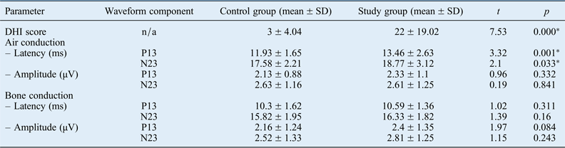
The Arabic Dizziness Handicap Inventory results for children in the control and study groups were compared using a t-test. This analysis revealed a significantly elevated score in the study group (Table I).
Dizziness handicap inventory and vemp testing results for control group and study group
*Indicates statistically significant results. VEMP = vestibular-evoked myogenic potential; SD = standard deviation; DHI = Dizziness Handicap Inventory; n/a = not applicable
Bedside test results
Analysis revealed abnormal results in the study group for the sharpened Romberg test (23.3 per cent of cases), the closed-eyes foam standing test (16.7 per cent of cases) and the tandem gait test (18.3 per cent of cases). The abnormal response was either an inability to do the test or an inability to keep the proper body position. The differences between both groups were significant (Figure 2).
Bedside test abnormalities in the study group.
The results of the study group for other bedside tests (i.e. ocular alignment, spontaneous and gaze-evoked nystagmus, saccades, and head shake and head thrust) were not significantly different from those of the control group.
Video-nystagmography results
There were no significant differences between the study group and control group in terms of the results for the oculomotor tests, the positioning test (Dix–Hallpike manoeuvre) or positional tests (p > 0.05).
Vestibular-evoked myogenic potential results
Air-conducted vestibular-evoked myogenic potentials
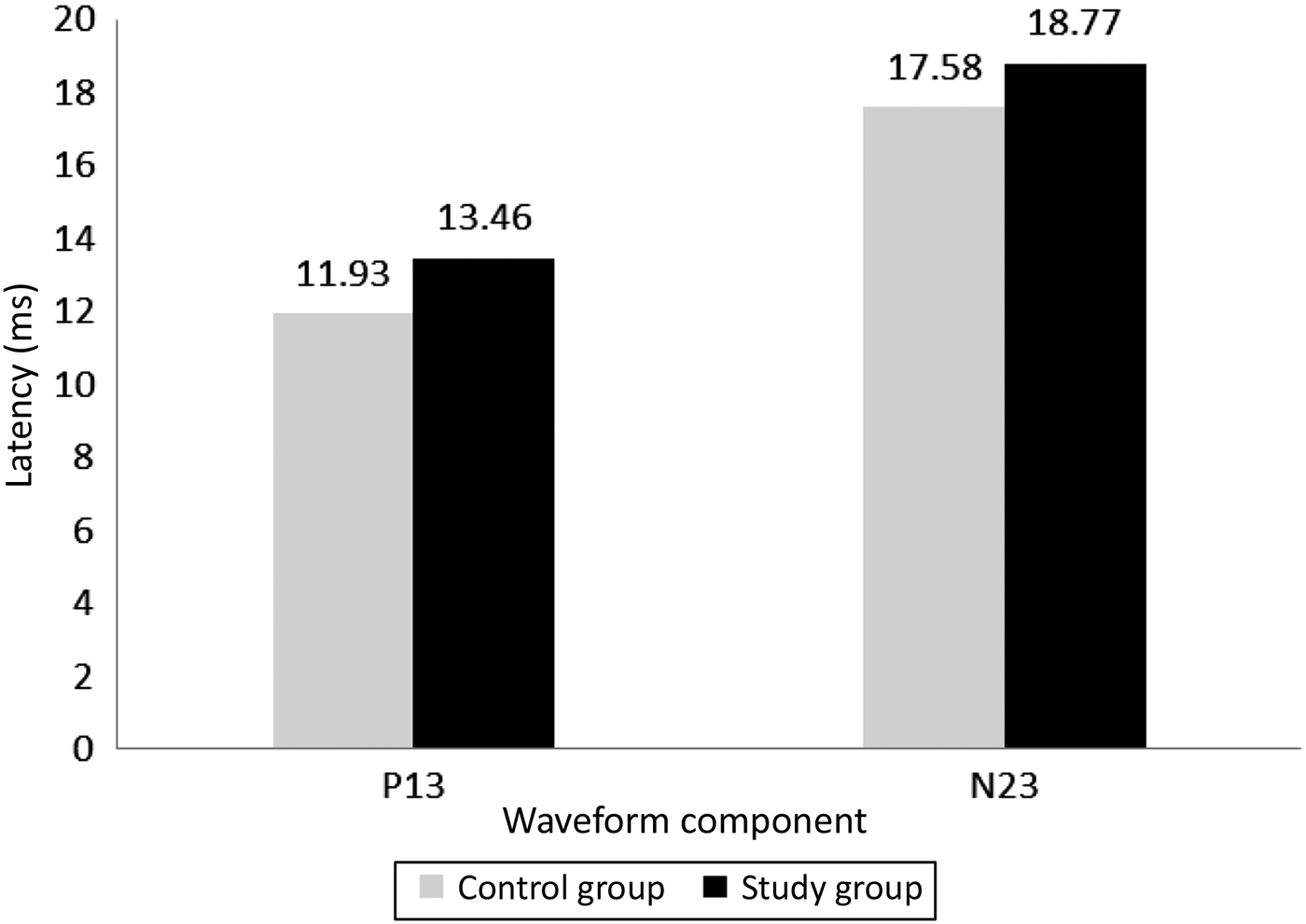
The means (± SDs) for the P13 and N23 latencies in the control group were 11.93 ± 1.65 ms and 17.58 ± 2.21 ms respectively. In the study group, air-conducted vestibular-evoked myogenic potentials were absent in 17 per cent of the patients. The means (± SDs) for P13 and N23 latencies in the remaining patients were 13.46 ± 2.63 ms and 18.77 ± 3.12 ms respectively. A comparison of both groups showed significantly delayed P13 and N23 latencies in the study group (p < 0.05) (Table II and Figure 3).
Comparison of P13 and N23 latencies for air-conducted vestibular-evoked myogenic potentials between the control group and study group.
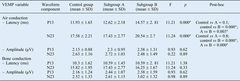
Vemp testing anova results for control group and study subgroups
Subgroup A had air–bone gaps (ABGs) of 20 dB or less; subgroup B had ABGs of more than 20 dB. *Indicates statistically significant results. VEMP = vestibular-evoked myogenic potentials; ANOVA = analysis of variance; SD = standard deviation
The means (± SDs) for P13 and N23 amplitudes were 2.13 ± 0.88 µV and 2.63 ± 1.16 µV respectively in the control group and 2.33 ± 1.10 µV and 2.61 ± 1.25 µV respectively in the study group. There was no statistically significant difference between the groups (Table I and Figure 3).
Air–bone gap effect
In this study, a 20 dB air–bone gap (ABG) was used as a cut-off level because it is the most common cut-off level used.Reference Halmagyi, Curthoys, Colebatch and Baloh19 Accordingly, the study group was subdivided into two subgroups: subgroup A with an ABG of 20 dB or less and subgroup B with an ABG of more than 20 dB.
In subgroup A, air-conducted vestibular-evoked myogenic potentials were recorded in 95 per cent of the children. The mean (± SD) P13 and N23 latencies were 12.62 ± 2.18 ms and 17.43 ± 2.77 ms respectively. The peaks had mean (± SD) amplitudes of 2.30 ± 0.95 µV and 2.72 ± 1.03 µV respectively. In subgroup B, air-conducted vestibular-evoked myogenic potentials could be recorded in 78 per cent of the children. The mean (± SD) P13 and N23 latencies were 14.57 ± 2.81 ms and 20.54 ± 2.70 ms respectively. The peaks had mean (± SD) amplitudes of 2.38 ± 1.31 µV and 2.48 ± 1.49 µV respectively (Table I and Figure 4).
Comparison of P13 and N23 latencies for air-conducted vestibular-evoked myogenic potentials between the control group and the two study subgroups. (Subgroup A had air–bone gaps (ABGs) of 20 dB or less; subgroup B had ABGs of more than 20 dB.)
Analyses of variance and post-hoc tests were conducted to compare P13 and N23 latencies and amplitudes in the control group and two study subgroups. The results showed statistically significant delayed latencies in subgroup B when compared with the control group and subgroup A. A comparison of the control group and subgroup A showed no significant difference. As regards to P13 and N23 amplitudes, there was no statistically significant difference between the control and study subgroups (p > 0.05) (Table II and Figure 4).
Bone-conducted vestibular-evoked myogenic potentials
These were recorded in 100 per cent of children in the control group and in 98 per cent of children with otitis media with effusion. Comparison of the control and study groups showed no significant differences in terms of P13 and N23 latencies or amplitudes. The size of ABG appeared to have no effect on bone-conducted vestibular-evoked myogenic potentials as there was no significant difference between the control and study subgroups (Table II).
Interaural amplitude ratio
The amplitude asymmetry ratio was calculated using the following formula: 100 × (right ear amplitude − left ear amplitude)/(right ear amplitude + left ear amplitude).Reference Rosengren, Welgampola and Colebatch20 The results showed no significant difference between the groups as regards to P13 and N23 components in either air-conducted or bone-conducted vestibular-evoked myogenic potential recordings (p > 0.05).
Correlation findings
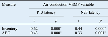
The Pearson correlation showed a significant positive correlation between air-conducted vestibular-evoked myogenic potential latencies and ABG, wherein ABG increases were associated with longer P13 and N23 latencies. In addition, there was a significant positive correlation between air-conducted vestibular-evoked myogenic potential latencies and Arabic Dizziness Handicap Inventory scores, wherein increases in inventory scores were associated with longer P13 and N23 latencies (Table III).
Pearson correlation between vemp variables, arabic dizziness handicap inventory and ABG
*Indicates statistically significant results. VEMP = vestibular-evoked myogenic potential; ABG = air–bone gap
Discussion
Paediatric vestibular disorders are serious and are often misdiagnosed. Vertiginous problems in children can contribute to behavioural problems, lack of co-ordination, delays in postural and motor control, and recurrent falling.Reference Nandi and Luxon1
It is well known that acute labyrinthitis can occur as a result of otitis media with effusion (OME). Waldron et al.Reference Waldron, Matthews and Johnson21 stated that the negative middle-ear pressure changes, transmitted through the labyrinthine windows, would lead to secondary movement of the inner-ear fluids. It has also been suggested that dizziness and vertigo in these conditions are caused by serous labyrinthitis as a result of superadded infection in a glue ear.Reference Williamson22
Arabic Dizziness Handicap Inventory
The Arabic Dizziness Handicap Inventory was developed as a measure of paternal observation of a child's balance performance; the inventory was used in this study to evaluate children with OME. The findings revealed significantly higher inventory scores for children with OME compared with healthy controls. These results are in agreement with Golz et al.,Reference Golz, Angel-Yeger and Parush4 Koyunco et al.,Reference Koyunco, Saka, Tanyeri, Seşen, Unal and Tekat23 Niemensivu et al.Reference Niemensivu, Pyykkö and Erna24 and Niemensivu et al.,Reference Niemensivu, Kentala, Wiener-Vacher and Pyykkö13 all of whom stated that the questionnaires used were sensitive tools for detecting balance disturbance in patients with OME (via the help of their parents). Golz et al.Reference Golz, Angel-Yeger and Parush4 believed that serous labyrinthitis is responsible for the vestibular disturbances in children with chronic OME, rather than pressure changes in the middle-ear cavity.
In contrast, Cohen et al.Reference Cohen, Friedman, Lai, Pellicer, Duncan and Sulek25 reported a practical difficulty in using paternal history as a proxy for a child's balance function. They stated that it was important for such questionnaires to be accompanied by other objective tests. The disparity between our results and those of Cohen et al. may be because the latter authors studied older children with both unilateral and bilateral OME.
Bedside tests
In this study, significant abnormalities were found in the results of the sharpened Romberg test, the foam standing test and the tandem gait test in the children with OME (study group) when compared to the children in the control group. These results are in agreement with those of Engel-Yeger et al.,Reference Engel-Yeger, Golz and Parush26 wherein children with OME achieved significantly lower scores in the tandem gait test and in the balance beam standing test (with closed eyes) than the control group. The authors suggested that these results were due to lower muscle strength resulting from abnormal neural interconnections between the vestibular system, the cerebellum and the spinal cord. The results of the current study (especially the sharpened Romberg test results) were also in line with those of Szirmai,Reference Szirmai7 who reported that labyrinthitis-associated OME had an effect on performance in the bedside vestibular test in children and adolescents.
Video-nystagmography
No significant abnormality was found for any of the video-nystagmography tests among the studied groups. Similarly, Engel-Yeger et al.Reference Engel-Yeger, Golz and Parush26 and Salami et al.Reference Salami, Dellepiane, Mora, Taborelli and Jankowska27 found no significant abnormalities in video-nystagmography testing. The rapid recovery from nystagmus and vertigo in children when compared to adults has been suggested to be responsible for these results.Reference Engel-Yeger, Golz and Parush26 However, some studies have reported significant abnormalities in video-nystagmography testing as regards to spontaneous and positional nystagmus.Reference Golz, Westerman, Gilbert, Joachims and Netzer8, Reference Williamson22
This disparity in the results of video-nystagmography in children may reflect the lack of normative data for young children. Moreover, children who are old enough to sit unsupported do not tolerate sitting quietly in the dark and they do not tolerate sitting still with their heads stabilised for long enough to perform the test. Therefore, even with many breaks in a test session, children cannot usually be positioned properly.24. Furthermore, Engel-Yeger et al.Reference Engel-Yeger, Golz and Parush26 reported that video-nystagmography is not a sensitive tool for the assessment of balance performance in children with OME as they are impatient and usually have numerous eye movements and blinking which interrupt the results of video-nystagmography. Salami et al.Reference Salami, Dellepiane, Mora, Taborelli and Jankowska27 also reported on the obvious difficulties encountered in trying to conduct a standard adult procedure of video-nystagmography on a paediatric patient.
In the current study, caloric testing was not undertaken, as we believed that this procedure could be traumatic for this age group, and their narrow ear canal might reduce the intensity of the stimulus. In addition, children younger than six years of age cannot co-operate well in the mental tasks essential for caloric testing. Cohen et al.,Reference Cohen, Friedman, Lai, Pellicer, Duncan and Sulek25 Golz et al.Reference Golz, Angel-Yeger and Parush4 and ValenteReference Valente28 show agreement with this point of view.
Vestibular-evoked myogenic potentials
Air-conducted vestibular-evoked myogenic potentials
These were recorded in all children in the control group (100 per cent). These results are in agreement with those of Kelsch et al.Reference Kelsch, Schaefer and Esquivel29 These authors reported normative data for vestibular-evoked myogenic potentials in young children using 90 dB nHL clicks, wherein the P13 and N23 latencies were 11.3 ± 1.3 ms and 17.6 ± 1.4 ms respectively. Air-conducted vestibular-evoked myogenic potentials were only recorded in 73 per cent of the children in the study group, however (in 44 out of a total of 60 ears). Similar findings were reported by Wang and Lee,Reference Wang and Lee9 who detected vestibular-evoked myogenic potentials responses in only 67 per cent of children with OME.
A comparison of the P13 and N23 latencies for both groups revealed significantly delayed latencies in the study group (children with OME) compared with the control group. This could be explained by the reduction of sound energy transmissions through the middle ear in the presence of OME, leading to delayed or even absent vestibular-evoked myogenic potential responses.Reference Wang and Lee9, Reference Honaker, Boismier, Shepard and Shepard18, Reference Bath, Harris, McEwan and Yardley30, Reference Yang and Young31
Several studies have been conducted to investigate the effect of ABG on air-conducted vestibular-evoked myogenic potentials. Different cut-off levels of ABG have been used.Reference Wang and Lee9, Reference Yang and Young31 In the current study, the children with OME were allocated to subgroup A if the ABG was 20 dB or less or to subgroup B if the ABG was more than 20 dB.
Air-conducted vestibular-evoked myogenic potentials were recorded in 95 per cent of the children in subgroup A and in only 78 per cent of the children in subgroup B. The P13 and N23 latencies were delayed in both subgroups compared with the control group. This delay was significant only when subgroup B (with an ABG of more than 20 dB) was compared with the control group and subgroup A (ABG of 20 dB or less). These findings are in agreement with those of Halmagyi et al.Reference Halmagyi, Curthoys, Colebatch and Baloh19 Those authors reported that air-conducted vestibular-evoked myogenic potentials were extremely affected in patients with an ABG of more than 20 dB because of the marked effect on sound transmission, with stimulus attenuation leading to significantly delayed air-conducted vestibular-evoked myogenic potential latencies. There was no significant difference in air-conducted vestibular-evoked myogenic potential latencies between the control group and subgroup A (with an ABG of 20 dB or less) and this is also in line with Halmagyi et al.Reference Halmagyi, Curthoys, Colebatch and Baloh19
However, Yang and Young,Reference Yang and Young31 and Wang and Lee,Reference Wang and Lee9 reported no significant effect of small ABGs (10–20 dB) on vestibular-evoked myogenic potential results. This could be related to the different ABG calculation used by those authors. For example, Wang and Lee calculated the ABG as the average of the difference between air conduction and bone conduction thresholds at 500 Hz.Reference Wang and Lee9 Yang and Young calculated the average of the difference between air conduction and bone conduction thresholds at 500, 1000, 2000 and 4000 Hz, and adopted 10 dB ABG as their cut-off level.Reference Yang and Young31 In our study, we considered 20 dB ABG at these four frequencies to be our cut-off, as per Halmagyi et al.Reference Halmagyi, Curthoys, Colebatch and Baloh19
As regards to the P13 and N23 amplitudes and amplitude asymmetry ratios, there were no significant differences between the control group and the two study subgroups. This could be because of amplitude dependence on the strength of muscle contraction and not the stimulus intensity.Reference Wang and Lee9
Bone-conducted vestibular-evoked myogenic potentials
These were successfully recorded in 100 per cent of the control group children. These results are consistent with those of Sheykholeslami et al.,Reference Sheykholeslami, Murofushi, Kermany and Kaga32, Reference Sheykholeslami, Kermany and Kaga33 Welgampola et al.Reference Welgampola, Rosengren, Halmagyi and Colebatch34 and Miyamoto et al.,Reference Miyamoto, Seo, Node, Hashimoto and Sakagami35 who all also reported that bone-conducted vestibular-evoked myogenic potential latencies could occur earlier than air-conducted vestibular-evoked myogenic potentials in these children.
• Fifty per cent of children with otitis media with effusion (OME) may have some balance disturbances
• Vestibular impairment in children with OME may be due to acute labyrinthitis or negative middle-ear pressure changes
• The dizziness handicap inventory and vestibular bedside tests are valuable tools for detecting vestibular impairment in children
• Air-conducted vestibular-evoked myogenic potentials are affected in OME
• Bone-conducted vestibular-evoked myogenic potentials are useful for vestibular system evaluation, especially in the presence of conductive hearing loss
Bone-conducted vestibular-evoked myogenic potentials were successfully detected in 100 per cent of children with OME. There was no significant difference in latencies when compared with the control group, indicating that OME and ABG had no significant effect on bone-conducted vestibular-evoked myogenic potential latencies. This is in agreement with the findings of Monobe and Murofushi,Reference Monobe and Murofushi36 and Seo et al.Reference Seo, Miyamoto, Saka, Shimano, Nishida and Hashimoto37 Bone-conducted vestibular-evoked myogenic potentials were normal in children with OME, indicating a normal inferior vestibular nerve. Bone-conducted vestibular-evoked myogenic potentials can be useful for vestibular system evaluation, to overcome the attenuation of stimulation due to middle-ear pathology.Reference Seo, Miyamoto, Saka, Shimano, Nishida and Hashimoto37
Correlations
A positive correlation was found between air-conducted vestibular-evoked myogenic potential latencies and ABG, wherein increased ABG was associated with increased latencies. This could be the result of decreased sound energy transfer through the middle ear.Reference Golz, Angel-Yeger and Parush4, Reference Wang and Lee9, Reference Halmagyi, Curthoys, Colebatch and Baloh19, Reference Koyunco, Saka, Tanyeri, Seşen, Unal and Tekat23, Reference Engel-Yeger, Golz and Parush26
Moreover, a positive correlation was found between ABG and Arabic Dizziness Handicap Inventory scores, wherein increased ABG was associated with increased Arabic Dizziness Handicap Inventory scores. This reflects the parents' observation of OME effects on the child's physical activity. Golz et al.Reference Golz, Angel-Yeger and Parush4 compared clinical balance questionnaire scores with electronystagmography (ENG) results and hearing level. They found a clear-cut correlation between the abnormal findings for both tests and poor questionnaire scores. In addition, Koyunco et al.Reference Koyunco, Saka, Tanyeri, Seşen, Unal and Tekat23 revealed a correlation between children's vestibular system dysfunction scores, stance and gait test results, and ENG results. The authors reported a positive correlation, reflecting the sensitivity and importance of the paternal questionnaire.
Engel-Yeger et al.Reference Engel-Yeger, Golz and Parush26 found that their questionnaire results were highly correlated with the child's performance on the Bruininks-Oseretsky Test of Motor Proficiency balance subtest, indicating that the questionnaires are sensitive tools that can be employed to screen for disturbances in children with OME. The authors found no correlation between questionnaire results and hearing level. This could be explained by the authors' dependence on the air conduction threshold whilst ignoring ABG size.
Conclusion
This study showed that the effect of otitis media with effusion on children's balance can be easily detected by simple, non-invasive measures and procedures such as the developed Arabic Dizziness Handicap Inventory and bedside vestibular tests for children; these methods can be used easily in the out-patients clinic.
It can be concluded that the Arabic Dizziness Handicap Inventory is a valuable tool for detecting vestibular impairment in children. Moreover, vestibular bedside tests represent a sensitive, valid, easy-to-use, inexpensive, non-invasive and attractive technique for evaluating vestibular function in children. Vestibular-evoked myogenic potential testing is a well-tolerated technique for assessing vestibular function in young children, with minimal test time and reproducible results. Air-conducted vestibular-evoked myogenic potentials are affected by ABG size of more than 20 dB HL. Bone-conducted vestibular-evoked myogenic potentials are useful measures of vestibular function, especially in the presence of conductive hearing loss; bone-conducted vestibular-evoked myogenic potential testing can be used as an alternative method to elicit vestibular-dependent potentials.
Acknowledgement
The authors of this work thank Dr Ola Abed Elshohady for her part in data acquisition for this research.
Appendix 1. Dizziness, vertigo and imbalance sheet for children
Appendix 2. Arabic Dizziness Handicap Inventory for children
Appendix 3. Arabic Dizziness Handicap Inventory for children (translated)