Case report
A 10-year-old boy presented with general fatigue and shortness of breath and was transferred to our hospital. Mild intraventricular conduction disturbance was noted when he was 10 years old, and he was followed up at another hospital. According to his mother, he had exhibited hyperhidrosis since the age of 7 years.
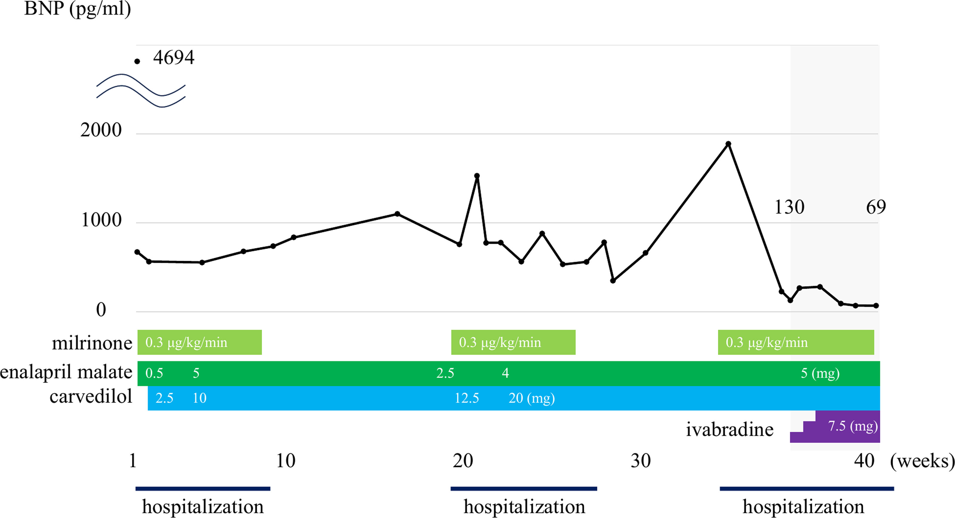
Upon admission to our hospital, the patient’s vital signs were as follows: blood pressure, 135/96 mmHg; pulse rate, 132 beats/min; respiratory rate, 51 breaths/min; body temperature, 37.9°C; and percutaneous oxygen saturation, 100% under respiration support by high-flow nasal cannula. Physical examination revealed retraction at the subcostal area, peripheral coldness, and pulsus alternans. Pulsus alternans was detected using invasive blood pressure monitoring; however, ECG monitoring showed constant QRS complexes. Chest radiography revealed a dilated heart and pulmonary congestion. The electrocardiogram showed normal sinus rhythm and mild intraventricular conduction disturbances (QRS duration 100 ms); however, electrical pulsus alternans was not observed. Transthoracic echocardiography revealed a markedly dilated left ventricle and severely impaired left ventricular contraction (ejection fraction, 19%). In addition, pulse Doppler imaging of the ascending aorta revealed alternating large and small pulses. The patient was diagnosed with dilated cardiomyopathy, and his respiratory symptoms due to congestive heart failure improved after continuous milrinone administration. He was discharged after heart failure treatment with enalapril malate (5 mg/day) and carvedilol (10 mg/day) (Figure 1). However, he had to be hospitalised again because of an acute exacerbation of heart failure. Milrinone infusion was necessary to improve his symptoms. His left ventricle dysfunction and mechanical pulsus alternans did not improve even after carvedilol titration to 20 mg/day.
Brain natriuretic peptide trend and treatment during the patient’s clinical course. The doses of milrinone, enalapril malate, carvedilol, and ivabradine are shown in boxes.

Figure 1. Long description
Panel: A line graph showing the trend of brain natriuretic peptide (BNP) levels over time with different treatments. The horizontal axis represents weeks, ranging from 1 to 40. The vertical axis represents Brain natriuretic peptide levels in picograms per milliliter, ranging from 0 to 5000. The graph includes data points connected by lines, showing fluctuations in brain natriuretic peptide levels. The treatments milrinone, enalapril malate, carvedilol, and ivabradine are shown in colored boxes below the graph, with their respective doses and durations. The graph indicates periods of hospitalization and changes in brain natriuretic peptide levels corresponding to the administration of these treatments.
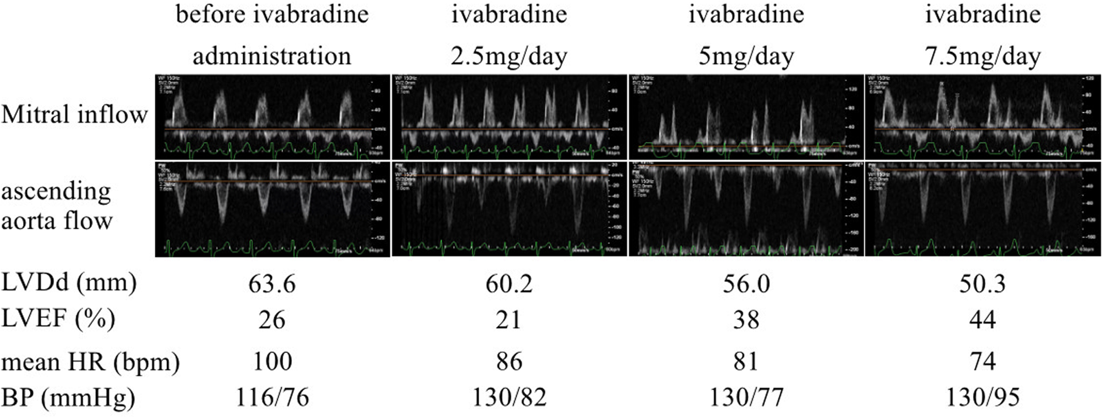
A second readmission was necessary after the acute exacerbation of heart failure induced by viral infection (Figure 1). Echocardiography showed that the early wave (E wave) and atrial wave (A wave) of mitral inflow were fused. Therefore, we assumed that his left ventricle dysfunction could be improved by lowering the heart rate with ivabradine. Given the patient’s size and age, the dose of ivabradine was started at 2.5 mg/day and gradually increased to 7.5 mg/day. Reference Bonnet, Berger, Jokinen, Kantor and Daubeney1 The patient’s mean heart rate gradually decreased, and his peripheral coldness improved. Moreover, the E and A waves of mitral inflow were clearly separated, the E/A ratio was normalised, and mechanical pulsus alternans disappeared after ivabradine was titrated to 7.5 mg/day. Left ventricle ejection fraction improved from 26% to 44% after ivabradine treatment (Figure 2). Brain natriuretic peptide levels were also reduced by ivabradine (Figure 1).
Pulse wave forms of mitral valve inflow (upper panels) and ascending aorta (lower panels), echocardiographic parameters (LV diameter at diastole [LVDd], LV ejection fraction [LVEF], and vital signs (heart rate [HR] and blood pressure [BP]) before administration and during titration of ivabradine.

We initially diagnosed the patient with dilated cardiomyopathy; however, a thorough reevaluation was performed. Blood tests revealed that his noradrenaline level was markedly elevated (22.09 pg/mL; normal range < 0.04 pg/mL). Enhanced CT revealed a mass between the inferior caval vein and the liver (Fig. S1). These results strongly suggest that the tumour produces noradrenaline and causes secondary dilated cardiomyopathy. After careful preoperative blood pressure control with cardenaline and metyrosine, the abdominal tumour was excised. The tumour was diagnosed as a paraganglioma. Genetic testing revealed a germline mutation in the SDHB gene. The final diagnoses were an abdominal noradrenaline-producing paraganglioma and catecholamine-induced dilated cardiomyopathy. The left ventricular contraction normalised after the operation, and the brain natriuretic peptide level decreased to the normal range.
Discussion
Catecholamine-induced cardiomyopathy in pheochromocytoma/paraganglioma has been reported in 8–11% of patients with pheochromocytoma and paraganglioma. Various types of cardiomyopathies, including tachycardia-induced cardiomyopathy, hypertrophic cardiomyopathy, Takotsubo cardiomyopathy, and dilated cardiomyopathy, are induced by catecholamine excess states. Reference Kumar, Pappachan and Fernandez2 Radical treatment for cardiomyopathy in pheochromocytoma/paraganglioma involves tumour excision; however, the preoperative condition, including the heart condition, should be improved as much as possible.
Four guideline-directed medical therapies, commonly referred to as the “fantastic four,” are administered to treat heart failure with reduced ejection fraction; however, ivabradine is used to treat patients with heart failure presenting with relative tachycardia. Ivabradine blocks the sodium channel HCN4 expressed specifically in the sinus node and reduces heart rate without a negative inotropic effect. Reference Bauersachs3 In addition to adult patients, ivabradine improves the left ventricular ejection fraction and clinical status in paediatric patients. Reference Bonnet, Berger, Jokinen, Kantor and Daubeney1
To the best of our knowledge, this is the first paediatric case demonstrating the effectiveness of ivabradine for cardiomyopathy in pheochromocytoma/paraganglioma. Only one report has demonstrated the effectiveness of ivabradine in treating a paraganglioma in an adult case. Reference Malaza, Brofferio, Lin and Pacak4 In this case, an inoperable metastatic paraganglioma was treated with systemic radiotherapy, which led to relative tachycardia and left ventricle dysfunction due to profound catecholamine release. Medical treatment with beta-adrenergic receptor blockers, calcium channel blockers, and catecholamine synthesis inhibitors does not relieve heart failure; however, the addition of ivabradine normalises ventricular contractions. Similar to this patient, our patient showed mild tachycardia even after treatment with an angiotensin-converting enzyme inhibitor and a beta-adrenergic receptor blocker. Because the E and A waves of the mitral inflow were fused, we considered him a good candidate for ivabradine treatment. Separation of the E and A waves of mitral inflow is crucial to obtain maximal benefit by reducing the heart rate using ivabradine. Reference Imamura and Kinugawa5 Although the mean heart rate decreases and left ventricular contraction improves after the induction of ivabradine, ivabradine should be titrated to 7.5 mg per/day to obtain a good separation of the E and A waves.
Pulsus alternans is a unique phenomenon observed in patients with left ventricle failure and can be linked to arrhythmias and sudden death. Reference Qu and Weiss6 The mechanism of pulsus alternans remains unknown; however, disturbances in cellular calcium ion signalling are considered to cause pulsus alternans. Reference Edwards and Blatter7 Kashimura and colleagues reported a case of idiopathic dilated cardiomyopathy with pulsus alternans and sinus tachycardia that was treated with ivabradine. Reference Kashimura, Ishizuka, Tanaka and Inomata8 They speculated that a heart with pulsus alternans could not keep up with tachycardia and was therefore loaded with excess calcium. The reduction in heart rate without a negative inotropic effect by ivabradine may decrease calcium overload in the heart and eliminate pulsus alternans.
In conclusion, we report a case of noradrenaline-producing paraganglioma and catecholamine-induced dilated cardiomyopathy in a paediatric patient. Ivabradine can be used as an alternative to improve left ventricular contraction and stabilise preoperative conditions.
Supplementary material
The supplementary material for this article can be found at https://doi.org/10.1017/S1047951126124184.
Acknowledgements
None.
Competing interests
Nothing to declare.
Ethical standard
This report does not involve human and/or animal experimentation.