Introduction
Staphylinidae (Coleoptera) is a taxonomically and ecologically diverse family of beetles, but phytophagy is remarkably underrepresented (Thayer Reference Thayer2005). Although there are examples in which staphylinids feed on algae (e.g., Herman Reference Herman1986) and pollen (e.g., Klimaszewski and Strum Reference Klimaszewski and Strum1991), until the discovery of Himalusa thailandensis Pace, Klimaszewski, and Center, 2010, phytophagy in the strict sense of feeding on embryophytes (i.e., terrestrial plant tissue) had only been represented by omnivorous taxa that appear to have been demonstrating atypical behaviour (Chittenden Reference Chittenden1915; Scheerpeltz Reference Scheerpeltz1927; Lipkow Reference Lipkow1966; Good and Giller Reference Good and Giller1991). In light of this, H. thailandensis is a curious example among staphylinids as an obligate phytophage.
Both adults and larvae of H. thailandensis were observed to be feeding on Paederia pilifera Hooker (Rubiaceae) in Thailand (Klimaszewski et al. Reference Klimaszewski, Pace, Center and Couture2010). Adults either chew oblong holes parallel to veins or scrape leaf surfaces. Larvae bore into leaf petioles that subsequently swell, appearing to accommodate for the growing larva. Other species of Paederia Linnaeus (P. foetida Linnaeus (skunk vine) and P. cruddasiana Prain (sewer vine)) are invasive weeds in the southern United States of America and H. thailandensis has already demonstrated promise as a valuable biological control agent (Klimaszewski et al. Reference Klimaszewski, Pace, Center and Couture2010).
In this paper we describe the larval morphology of H. thailandensis in detail. Morphology, especially mouthparts, which are used in food gathering and processing, are discussed in the context of phytophagy, and we discuss the potential adaptive significance of some of these characters. The adult morphology of the species is highly divergent, obscuring the phylogenetic relationship of Himalusa Pace, 2006 in relation to other Aleocharinae. The morphology of the larval abdominal defensive gland is discussed as a potentially useful character in placing Himalusa within the aleocharine tree of life. Habitus photographs of all life stages and line drawings of body structures are provided.
Materials and methods
Morphological study
Specimens were observed using an Olympus SZX7 stereo- and an Olympus BX51 compound microscope (Olympus, Tokyo, Japan). Illustrations were made using a camera lucida, Olympus U-DA, mounted on the compound microscope. Scale bars were drawn using an Olympus slide micrometer. Body measurements were made using a stereomicroscope ocular micrometer calibrated with an Olympus stage micrometer. Images were taken with a Visionary Digital imaging system (www.duninc.com) and stacked using CombineZM (http://combinezm.software.informer.com).
Larval specimens were prepared by digesting internal tissue in 10% KOH at ambient temperature overnight and were subsequently thoroughly washed in distilled water. If fat tissue remained, specimens were further cleared in glacial lactic acid for a variable amount of time and washed in distilled water. Some larvae were stained with chlorazol black for increased contrast. Staining was visually monitored until a desired colour contrast was obtained, then briefly washed in distilled water.
Cleared and stained specimens were placed in dilute glycerin (25% EtOH, 25% distilled H2O, 50% glycerin) and the solution was allowed to evaporate, leaving the glycerin and specimens. Once observations were complete, glycerin immersed specimens were washed in 70% EtOH and mounted permanently as described below.
For permanent mounts, cleared specimens were brought successively to an ethanol concentration of 100% and then placed into euparal on a glass slide. The lightly sclerotised abdomen collapses under the viscosity of euparal without a large enough opening to allow for the exchange of internal EtOH and surrounding mounting medium. For this reason, the heads of dehydrated larval specimens were immediately removed upon transfer to euparal. Pins were inserted in the cervix, and used to trace the membrane attached to the occipital foramen, severing the connective cervical membrane.
Larval chaetotaxy terminology follows Ashe and Watrous (Reference Ashe and Watrous1984). Tergite and sternite II are illustrated because they represent complete chaetotaxy among homologisable abdominal segments (II–VII are serially homologous).
Statistical analyses and plots were conducted in R (R Core Team 2013), operated through RStudio (2012). The three dimensional plot was generated using the Scatterplot3d package (Ligges and Mächler Reference Ligges and Mächler2003).
Specimen collection and rearing
Insects were collected 7 km north of Kui Buri, at Prachuap Kiri Kan, Thailand (12°7.940'N, 99°51.076'E) by A.D. Wright during 17–18 October 2009 and 25 October 2010. The caged beetles were exposed to four to five whole potted P. pilifera plants for 7–10 days. They readily fed and laid eggs on the leaves (Fig. 20). The plants were then removed and placed in a screened enclosure (BioQuip Bug Dorm-2®; BioQuip Products, Rancho Dominguez, California, United States of America). The eggs hatched and the larvae burrowed into the petioles. The plants were placed in individual containers after 7–10 days, or when larval emergence was observed. Containers were clear plastic boxes (Pioneer Plastics, North Dixon, Kentucky, United States of America) measuring 31.3×23.0×10.2 cm with a slot at the base. A 6.35×5.0 cm opening was cut into one side and covered with screen. A P. pilifera vine was inserted through the slot, which was plugged by tightly wrapping cotton around the stem. Each plant was monitored daily for the larval emergence. Larvae were then placed in a 148-mL (40-dram) vial containing a uniform mixture of 100 mL of Atami CocoFiber®, (Atami B.V., Huisbergenweg, The Netherlands), 5 mL of sand, and 20 mL of deionised water. A dental wick was inserted into and protruded on either side of the vial lid to absorb excess moisture. Vials were checked daily for adult emergence.
To obtain larvae of known age, a leaf with full-term eggs was placed on a fresh P. pilifera leaf. This leaf remained attached to a vine, which passed through a large plastic petri dish. The dish had been placed over a single node of a P. pilifera vine. The vine entered and exited through holes on opposite sides of the closed dish with a single node bearing two leaves within the dish. The leaf petioles were examined daily to detect larval entry points. Petioles were monitored and larvae extracted from the chambers on each day of development to determine the instar corresponding with each day of age. Head capsule width and body length of each larva was measured.
Himalusa thailandensis Pace, Klimaszewski, and Center, 2010
Larval description
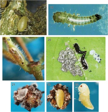
Instar I. Head and thorax relatively larger than abdomen; body (Fig. 18) uniformly tapering posteriorly. Mean body length 1.13 mm (0.87–1.24 mm, n=11), mean head width 0.25 mm, 0.23–0.26 mm, n=11).
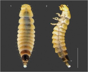
Instar III. Body rotund (Figs. 1–2), approaching eruciform, mean body length 3.16 mm (2.77–3.57 mm, n=11), overall lightly sclerotised, all appendages and urogomphi reduced in size. Sclerites pale yellow, abdominal segment IX black, segments VIII and X dark brown, membranous areas cream coloured.
Himalusa thailandensis third-instar habitus. 1. Dorsal. 2. Lateral. Scale bar=1 mm.

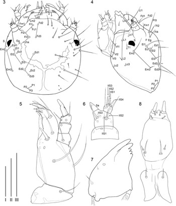
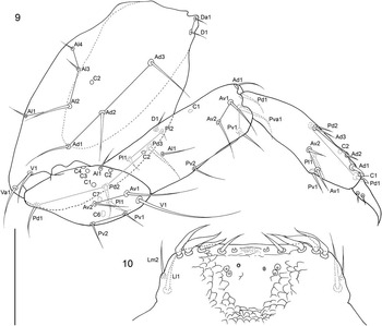
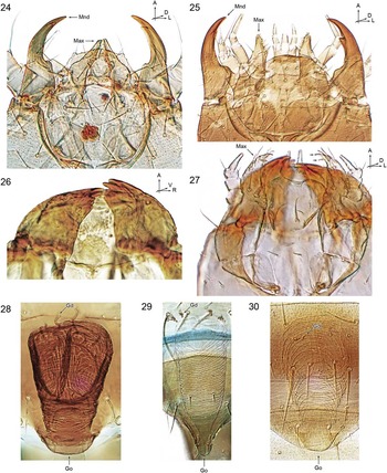
Head subcircular in dorsal view (Figs. 1, 3) (mean head width=0.38 mm, 0.36–0.41 mm, n=11), subglobose in lateral view (Figs. 2, 4), vertex (i.e., epicranial region of Ashe and Watrous (Reference Ashe and Watrous1984)) dorsally depressed and appearing as an indentation of dorsal profile in lateral view (Figs. 2, 4) due to dorsal inflation of anterior-epicranial region; antennae arising from large projections on the head that are two times longer than the basal antennomere; Fd1, Fc1, L3, P4, T2, and V1 absent; clypeal region (anterior of frontal and posterior of labral region of Ashe and Watrous (Reference Ashe and Watrous1984)) slightly dorsally inflated in lateral view (Figs. 2, 4). Stemma (ocellus of Ashe and Watrous (Reference Ashe and Watrous1984)) (Figs. 1–4) globose and anteriorly directed. Antennae (Figs. 1–4, 6) reduced in length and stout. Labrum (Figs. 1, 3) sub-rectangular; Lm1 absent. Epipharynx (Fig. 10) with a U-shaped region surrounding middle with imbricate texture oriented anteriorly; paired sensory structure apicomedially with pseudopore-like structures medially, a pair of small medial pores and two pairs of larger submedial pores present; apicolaterally with three pairs of adorally curved fusiform setae. Mandibles (Figs. 7, 26) extremely broad basally with points of muscle attachment widely divergent, broadly triangular in dorsal perspective; pre-apical tooth equal in size and occupying a plane dorsal to apical tooth, three small teeth present basal to pre-apical. Maxillae (Figs. 5, 27) with palpomere I large, subequal in length and width to II; mala with three conspicuous non-setal teeth ventrally, six setae and inconspicuous imbricate cuticular texture dorsally. Labial complex (Fig. 8) lightly sclerotised; palpomeres reduced in length, ligula short and subequal in length to palpomere I; mentum ovular in outline; submentum consisting of a pair of poorly sclerotised parallel sided sclerites.
Himalusa thailandensis third-instar body parts. 3. Head, dorsal. 4. Head, lateral. 5. Maxilla, left ventral. 6. Antenna, right dorsal. 7. Mandible, left dorsal. 8. Labial complex, ventral. Scale bars: I=0.05 mm (6–7), II=0.2 mm (3–4), III=0.1 mm (5, 8).

Himalusa thailandensis third-instar body parts. 9. Metathoracic leg, left anterior view. 10. Epipharynx. Scale bar=0.1 mm.

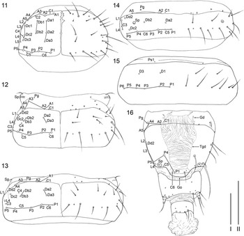
Pronotum (Fig. 11) semicircular, sclerotised area transverse with rounded anterior margin; Db3 absent. Mesonotum (Fig. 12) transverse. Mesosternal spiracle laterally produced with a seta anteroventrally; C2, Da1, Db1, Dc1, and Dc3 absent. Metanotum (Fig. 13) strongly transverse; C2, Da1, Db1, Dc1, Dc3, and A5 absent.
Himalusa thailandensis third-instar body parts. 11. Pronotum. 12. Mesonotum. 13. Metanotum. 14. Tergite II. 15. Sternite II. 16. Abdominal segments VIII–X. Scale bars: I=0.2 mm (11–15), II=0.2 mm (16).

Abdomen: Tergite II (Fig. 14) strongly transverse; C2, C4, Da1, Da3, Db1, Db3, Dc1, Dc3, Dd1, Dd3, A1, A3, A4, L1–3, and L5 absent. Sternite II (Fig. 15) strongly transverse; C1 and D2 absent. Tergite VIII (Figs. 16, 28) subquadrate with gland opening bluntly produced posteromedially, area of disk surrounding gland opening particularly darkly sclerotised (Figs. 1–2); C2–5, Da1–3, Db1–3, Dc1–3, A1, A3, L1, L4, L5, P2, and P3 absent. Abdominal defensive gland reservoir large, well defined, with ribbed texture throughout; four kinked and apically bifid gland ducts present (Figs. 16, 28). Urogomphi short, size overall extremely reduced. Pygopod without anal hooks.
Legs (Fig. 9) with femur, tibia and tarsungulus relatively shortened in length. Coxa with additional setae Da1 and Va1; trochanter with Al2 and C2 absent; femur with additional seta Pva1; tarsungulus with two basal setae on anterior surface.
Eggs and oviposition. Eggs are ovoid and approximately 0.6 mm in length (Fig. 17). Females deposit eggs in regions that have been fed on and damaged by adults, often adjacent to the midrib (Fig. 20). Eggs are not inserted into the tissue but deposited on the surface. Depending on the surface landscape of the damaged foliar area, an egg may be fully visible to obscured by surrounding tissue. Females do not demonstrate any additional signs of intentional maternal concealment, which may be found in other aleocharines that feed on macro-fungi (Ashe Reference Ashe1987). It is unknown whether females somehow anchor the eggs onto the substrate.
Images of Himalusa thailandensis during laboratory rearing. 17. Egg deposited at a node. 18. Instar I, dorsal. 19. Swollen stem housing a larva. 20. Adults mating on a leaf, adjacent to adult feeding damage. 21. External view of a pupal chamber, showing soil particulates that were incorporated during silk cocoon creation. 22. Pupa exposed by removing half of the pupal chamber. 23. Pupa, lateral.

Eclosion and larval development. First instars wander from their oviposition site towards the base of the leaf where they bore into the petiole near the node (Fig. 19). The entry hole is marked by frass and serves as a convenient sign of petiole occupation.
As the larva feeds within the confines of a petiole and develop, the petiole swells around the larva as if to accommodate the growing larva. At the end of feeding, third instars exit the swollen petiole and migrate to the ground where they pupate. Feeding sites fill with a white, latex-like substance after the larvae exit, but it is unclear if the larvae feed on this substance (also see the “Phytophagy in the genus Himalusa and tribe Himalusini” section below).
Pupae and pupation. Larvae that have migrated down to the ground burrow into the soil to pupate. Individuals in the laboratory were observed to descend around 1 cm below the surface, but this figure is believed to change depending on environmental conditions (e.g., humidity). Cocoons were spun in excavations underground, incorporating local substrate (Figs. 21–22). Pupae (Fig. 23) are exarate and typical of Aleocharinae.
Development. Developmental time, measured from oviposition to adult eclosion, lasted ~30 days. The total time for development is longer compared to the 15–17 day period of other Aleocharinae for which data are available (Ashe Reference Ashe1985; Eldredge Reference Eldredge2012; Echegaray and Cloyd Reference Echegaray and Cloyd2013).
Three instars were observed, which is typical for Staphylinidae (Thayer Reference Thayer2005). Because H. thailandensis larvae are cryptic and possess potential as a biological control agent against invasive sewer vine and skunk vine, we sought to confirm that all instars were identified. We calculated Dyar’s Ratio (Dyar Reference Dyar1890); the fraction by which instars proportionally increase in size. The ratio, or regression slope was devised as a predictive tool for identifying missing instars. Plotting the regression slope against mean head capsule width suggested that no instars were missed, confirming a total of three instars (Supplementary Figs. S1–S2).
Klimaszewski et al. (Reference Klimaszewski, Pace, Center and Couture2010) suggested that there might be size dimorphism in H. thailandensis adults. We might expect this size dimorphism to be expressed in developing larvae. Head width standard deviations were observed to increase in successively older instars (Fig. S1). We tested for no difference between instar II and III head width variance, although ideally we would test for a bimodal distribution in instar III, but a small sample size limited the study to comparing variances. We found no statistical significance between instar II and III head width variance (F=0.41, P=0.19). We suggest that a larger sample size be considered before ruling out the possibility that third instars demonstrate early signs of size dimorphism.
Host range. Larvae appear to be host specific to Paederia pilifera, because immatures did not feed on other plants offered in the laboratory. Adults, on the other hand, appear to be somewhat polyphagous, at least under laboratory conditions, feeding on various species (E.D.M., personal observation). Host partitioning between adults and larvae are known to occur in mycophagous staphylinids, presumably as a means to reduce generational conflict (Ashe Reference Ashe1984a, Reference Ashe1984b; Hanley and Goodrich Reference Hanley and Goodrich1995), but whether laboratory observations for H. thailandensis is evidence for such behaviour is unclear and warrants additional study.
Discussion
Phytophagy in the genus Himalusa and tribe Himalusini
An unidentified Himalayan Himalusa species (possibly the described Himalayan species, H. annaurensis Pace, 2006) was observed to be feeding on a species of Paederia (M. Rayamajhi, personal communication, Invasive Plant Research Laboratory, United States Department of Agriculture, Agricultural Research Service, Fort Lauderdale, Florida, United States of America). This observation lends evidence that all Himalusa may be phytophagous. Additionally, Maruyama et al. (Reference Maruyama, Yamamoto and Eldredge2014) noted similarities in mouthpart morphology among genera of Himalusini, potentially evidencing a phytophagous diet for all Himalusini. This may be indication that additional Himalusini species have potential for use in biological control, which is a promising prospect.
Potential adaptations to phytophagy
Larvae of H. thailandensis demonstrate morphologies that appear to be adaptive to its peculiar phytophagous lifestyle. Morphological divergences putatively adaptive to phytophagy are changes in overall body form and modification in mouthpart structures.
Larvae of Aleocharinae are generally active foragers, and have comparatively slender bodies with more or less long appendages, i.e., campodeiform (e.g., Ashe and Watrous Reference Ashe and Watrous1984; Thayer et al. Reference Thayer, Ashe and Hanley2004; Eldredge Reference Eldredge2012). Himalusa thailandensis, on the other hand, has an extremely rotund body form (Figs. 1–2) and short, compact appendages (Figs. 1–2, 6, 9), approaching an eruciform body form which is atypical for aleocharines. In particular, larvae of H. thailandensis have rotund, compact heads (Figs. 2–4), reduced antennae (Fig. 6), and short, compact legs (Fig. 9). This overall compact body form suggests an adaptation towards feeding within the sub-dermal refuge of a leaf petiole. Tight spaces may have selected for appendages of conservative length and a rotund body may reflect loss of an active lifestyle (similar to ectoparasitic Aleochara Gravenhorst, 1802 (e.g., White and Legner Reference White and Legner1966; Lin and Shiao Reference Lin and Shiao2013) and social insect symbiotic Lomechusina Fleming, 1821 (Wheeler Reference Wheeler1911; K.T.E., personal observation) (also see Paulian Reference Paulian1941).
Mouthparts are a primary means for food gathering and processing for staphylinids (Betz et al. Reference Betz, Thayer and Newton2003), which is derived in H. thailandensis, relative to the ancestral condition for the subfamily. Mandibular morphology among larvae of “higher” Aleocharinae sensu Ashe (Reference Ashe2005) is quite uniform (Figs. 24–25). In general, mandibles are elongate with a narrow base and adorally curved apex. A subapical tooth is usually present and the apical margin of the tooth is sometimes serrate. Finally, the ginglymus-condyle axis is usually oblique (see Tikhomirova Reference Tikhomirova1973).
Images of slide mounted Aleocharinae third instars. 24. Homalota Mannerheim species anterior portion of the head. 25. Bolitocharina anterior portion of the head. 26. Himalusa thailandensis mandibles. 27. Himalusa thailandensis mouthparts. 28. Himalusa thailandensis abdominal defensive gland, dorsal. 29. Homalota species abdominal defensive gland, dorsal. 30. Bolitocharina abdominal defensive gland, dorsal. A, anterior; D, dorsal; V, ventral; L, left; R, right; Mnd, mandible; Max, maxilla.

In contrast to ancestral mandibular forms in “higher” aleocharines (K.T.E., personal observation), H. thailandensis mandibles are extremely robust and stout (Figs. 7, 26). The base is broadened, subapical tooth large with three blunt teeth basally, and the ginglymus-condyle axis subvertical. The mandibles functionally articulate on a more or less horizontal plane, compared to a slightly oblique plane as in most aleocharines.
Aleocharinae are plesiomorphically predacious (Hansen Reference Hansen1997; Hunt et al. Reference Hunt, Bergsten, Levkanicova, Papadopoulou, St. John and Wild2007) with a varying mycophagous component to their diet (Klimaszewski et al. Reference Klimaszewski, Morency, Labrie, Séguin, Langor and Work2013; K.T.E., personal observation). A largely invariable larval diet may be contributing to the uniform mandibular morphology in larval aleocharines. The mandibles of larval H. thailandensis, on the other hand, resemble those of some adult Osoriinae and Oxytelinae (Staphylinidae), which are thought to be adaptive for saprophagy, where the mandibles are used for scraping and squeezing (Tikhomirova Reference Tikhomirova1973). Assuming that mandibular morphology is a good first approximation of diet and how the organism handles specific food items, this mandibular similarity to osoriines and oxytelines may indicate that H. thailandensis larvae are saprophages within the swollen petiolar confines of Paederia. This serves to explain why larvae are able to complete development in one location of the plant, where the larvae may be feeding on the latex-like substance that fills feeding sites after they exit for pupation (also see the “Eclosion and larval development” section above).
As a final note, the malar apex sports three teeth-like cuticular projections in H. thailandensis (Fig. 27). Such structures are not present in other aleocharines (Figs. 24–25). This malar morphology may work in concert with the specialised mandibles to deal with the derived diet in H. thailandensis.
Phylogenetic placement of Himalusa
Morphological divergence of both adults and larvae obscures the phylogenetic placement of H. thailandensis. Based on the tribal concepts of Ashe (Newton et al. Reference Newton, Thayer, Ashe and Chandler2001) and Seevers (Reference Seevers1978), which are based on adult characters, the presence of a maxillary pseudosegment in Himalusa suggests a close relationship with Hoplandriini: whereas overall similarity of the aedeagus and mouthparts, especially the labium, suggests that Himalusa is closely related to Placusini. While these adult characters provide conflicting assessments of the placement of Himalusa, consideration of a potentially important larval character may provide a clearer and more reliable placement. In certain groups of gland-possessing aleocharines (i.e., “higher” aleocharines of Ashe (Reference Ashe2005)), the larval abdominal defensive gland opening protrudes posteriorly, and the glandular reservoir is characteristically large, heavily sclerotised and ribbed, with gland sac ducts kinked and apically bifid (Figs. 28–30). Hereafter, we term this condition the “Homalota-type larval abdominal defensive gland” (Homalota-type LADG) (see Frank and Thomas (Reference Frank and Thomas1984) for a list of additional groups that may possess a Homalota-type LADG – categorised as taxa with a “prominently protuberant gland opening with a large internal reservoir”).
Presence of a Homalota-type LADG in Himalusa supports the close relationship of this genus with other similar LADG-bearing groups: Autaliini; Bolitocharina (Ashe Reference Ashe1990); Gyrophaenina (Paulian Reference Paulian1941; Ashe Reference Ashe1986); Homalotina (Fig. 29; Paulian Reference Paulian1941; Nikitsky Reference Nikitsky1980); Leptusina (Kemner Reference Kemner1918); Liparocephalini (Ahn Reference Ahn1997); Silusina (Fürst von Lieven Reference Fürst von Lieven1999). Importantly, Osswald et al. (Reference Osswald, Bachmann and Gusarov2013), in a molecular phylogenetic study found a monophyletic group consisting of Autaliini+Diglottini+Himalusini+Bolitocharina (Homalotini)+Gyrophaenina (Homalotini)+Silusina (Homalotini). All of these taxa possess a Homalota-type LADG except for Diglottini (Kemner (Reference Kemner1925) oddly did not mention the presence or form of the gland in his Diglotta Champion, 1887 larval description, despite his detailed illustration of the structure in an earlier work (Kemner Reference Kemner1918)). However, two other Homalota-type LADG-possessing taxa, including the subtribe Homalotina itself and Liparocephalini, were recovered outside of this group (Osswald et al. Reference Osswald, Bachmann and Gusarov2013). Additionally, a 28s-rDNA gene tree (K.T.E., personal observation) recovered a monophyletic Autaliini+Bolitocharina+Gyrophaenina+Himalusini+Homalotina+Silusina (Diglottini was not sampled): again, all of these groups possess a Homalota-type LADG. The results of Osswald et al. (Reference Osswald, Bachmann and Gusarov2013) and the 28 s dataset are reasonably congruent with the presence of a Homalota-type LADG, and Homalotini sensu Ashe (in Newton et al. Reference Newton, Thayer, Ashe and Chandler2001) and Seevers (Reference Seevers1978), potentially indicating that presence of a Homalota-type LADG has strong phylogenetic signal.
Larval morphology among gland-possessing aleocharines is relatively conservative (K.T.E., personal observation), with much of the variation manifested in chaetotaxy and the placement of epidermal glands (Ashe and Watrous Reference Ashe and Watrous1984). For this reason, we believe recognition of a Homalota-type LADG is an important addition to the number of phylogenetic characters available for aleocharine larvae.
Acknowledgements
The first author thanks A.E.Z. Short (University of Kansas) for financial support. The authors thank J. Parker (Columbia University) for critical review and advice on the manuscript, M. Maruyama (Kyûshû University Museum) for sharing unpublished observations on related genera and helpful discussions on characters and taxonomy, J.H. Frank (University of Florida) for constructive discussion about aleocharine larvae, and last but not least, T. Théry (Université de Montréal) for graciously providing a French translation for the abstract.
Supplementary Material
For supplementary material referred to in this article, please visit http://dx.doi.org/doi:10.4039/tce.2016.1