Introduction
Sinusitis in adults is common, but orbital complications are usually associated with paediatric rather than adult patients. There is a debate as to the difference between orbital apex syndrome and posterior orbital cellulitis. In the latter condition, a segment of the optic nerve in the superior orbital fissure is affected.
This paper reports a rare occurrence of isolated sphenoid sinusitis with associated presentation of posterior orbital cellulitis. The subtlety of eye signs seen in the condition necessitates a high index of clinical suspicion in order to avoid missing the diagnosis.
Case report
A 29-year-old woman presented as an acute medical admission with a 6-week history of progressive, severe, retro-orbital and peri-orbital right eye pain. The pain was throbbing in nature, increasing in severity, and associated with headaches, nausea and vomiting.
The patient's vision was assessed by the ophthalmologists, revealing blurring, reduced acuity and associated photophobia. There were no other signs of meningism.
Initial examination demonstrated right-sided ptosis and tenderness on palpation of the orbit. The patient had demonstrably reduced acuity but maintained normal colour vision.
Inflammatory markers were within normal limits.
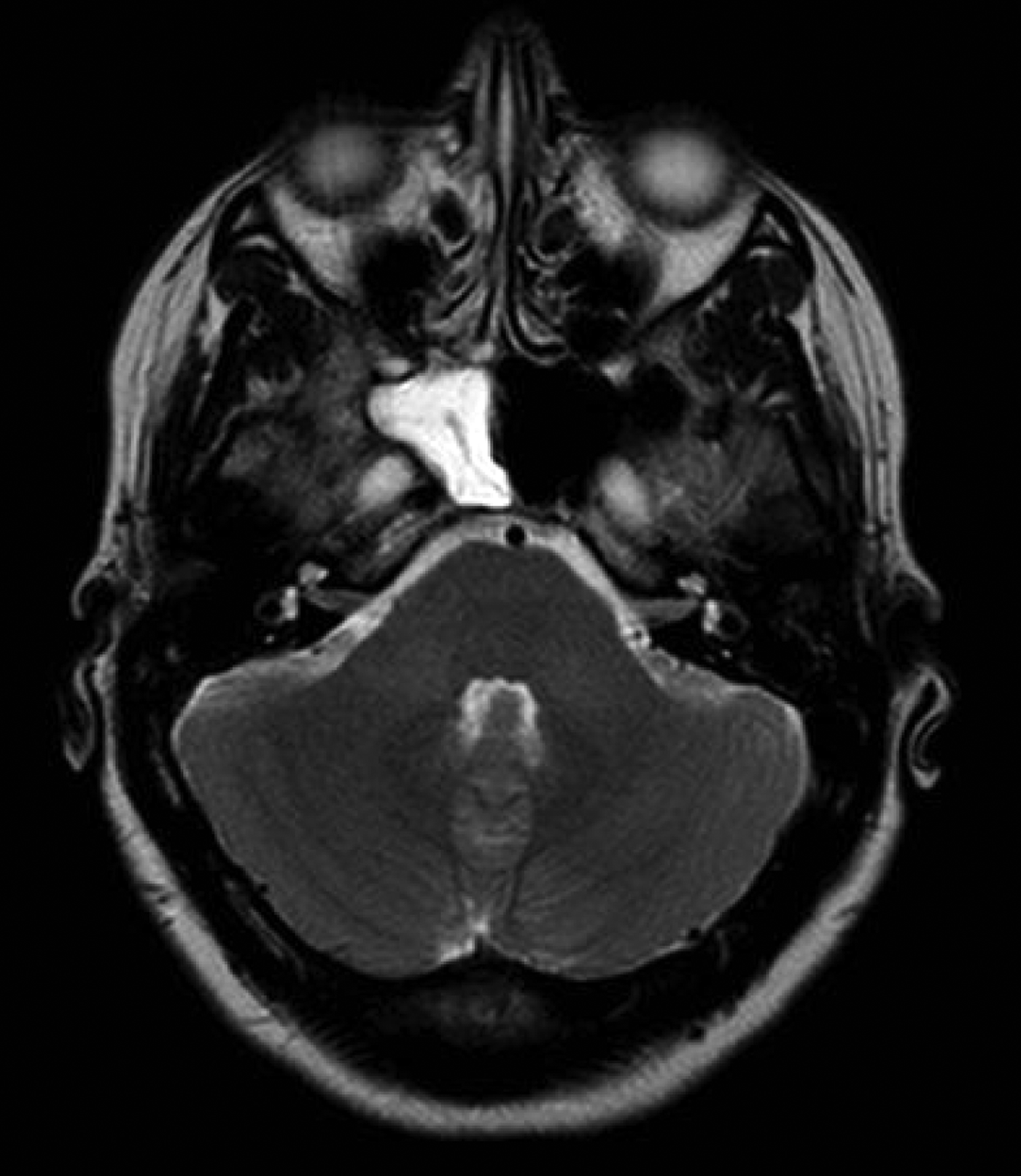
An urgent magnetic resonance imaging brain scan was performed. This showed right sphenoid sinusitis, with sphenoidal and frontal sinus mucosal thickening (Figure 1).
Axial magnetic resonance image showing right sphenoid sinusitis, with opacification and right sphenoid and frontal sinus mucosal thickening.

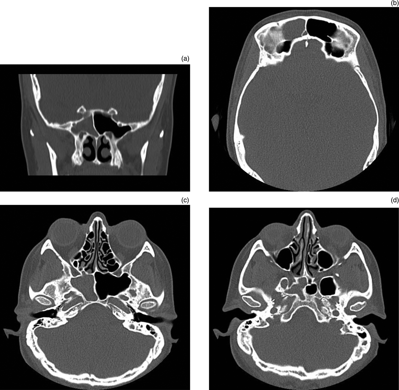
Computed tomography (CT) scanning of the sinuses was requested to further define the mucosal disease. This demonstrated right frontal and sphenoid sinus opacification only, with no evidence of maxillary, ethmoidal or contralateral disease (Figure 2).
(a) Coronal computed tomography (CT) scan showing right sphenoiditis, with clear ethmoidal sinuses. (b) Axial CT showing right frontal sinus opacification. (c) Axial CT showing sphenoid sinusitis, with clear ethmoid sinuses. (d) Axial CT showing clear maxillary sinuses.
Following the microbiologist's advice, the patient was started on intravenous Timentin (GSK, Uxbridge, UK). After 48 hours of IV antibiotics, her vision continued to deteriorate, and her ophthalmalgia and photophobia were unchanged.
Ophthalmological reassessment revealed reduced acuity: 6/36 for the right eye, compared with 6/6 for the left. Important negative findings included no visible proptosis or chemosis, no demonstrable relative afferent pupillary defect, no reduction in external ocular movements, and no diplopia. The patient's optic discs were healthy, with no swelling or venous engorgement.
The patient began to develop red desaturation. Two diagnoses were considered by ophthalmology at this point: orbital soft tissue inflammation secondary to sinus infection, and idiopathic orbital inflammatory disease (a diagnosis of exclusion).
Right-sided sphenoidotomy and frontal sinus trephination were performed urgently. Pus under pressure drained rapidly from the right sphenoid sinus and was sent for culture and sensitivity tests. The frontal sinus was full of polypoidal mucosa.
Microbial culture grew Gram-positive bacilli only; culture of the frontal sinus sample grew no organisms after 48 hours.
Post-operatively, the patient's visual acuity improved slowly. Three months after surgery, her vision had returned to normal.
Discussion
The sphenoid sinus is lined with pseudostratified epithelium, with fewer mucus-secreting cells compared with other paranasal sinuses.Reference Van Alyea1 Predisposing factors for sphenoid sinusitis include anatomical variation, radiotherapy, immunosuppression and malignancy.Reference Holt, Standefer, Brown and Gates2–Reference Pearlman, Lawson, Biller, Friedman and Potter4 In the presented case, however, no predisposing factor was found.
Headache is usually the most common initial symptom of acute sphenoiditis.Reference Van Alyea1 In our patient, there was significant right orbital pain that was predominately retro-bulbar.
Sphenoiditis is most often non-specific and poorly localised, due to its inaccessible position. Symptoms are often non-specific.Reference Holt, Standefer, Brown and Gates2 Rhinorrhoea and post-nasal drip are common symptoms, with purulent exudates frequently seen below the middle and superior turbinates.Reference Lew, Southwick, Montgomery, Weber and Baker3 Visual loss as the presenting symptom is extremely rare, with few reports in the literature. The current report highlights an unusual presentation of a rare complication of sphenoiditis.
In our patient, ophthalmalgia and visual change, including blurring and loss of vision, were the most prominent features. Interestingly, this did not appear to be due to direct involvement of the optic nerve.
Two forms of visual loss are seen as a complication of sinusitis: (1) visual loss as a complication of orbital cellulitis, with orbital soft tissue involvement characterised by proptosis, oedema, chemosis and ophthalmoplegia;Reference Chandler, Langenbrunner and Stevens5 and (2) visual loss as a component of ‘orbital apex syndrome’, characterised by involvement of vessels and nerves passing through the superior orbital fissure.Reference Holt, Standefer, Brown and Gates2, Reference Kjoer6
In the fully developed orbital apex syndrome, visual loss is accompanied by complete ophthalmoplegia (with paresis of the IIIrd, IVth and VIth cranial nerves). However, there can be visual loss alone, with no, or only minimal, orbital inflammatory signs. This is thought to be due to a segment of the optic nerve being affected, and is considered as ‘partial’ or ‘posterior’ orbital cellulitis.
Reports of sinusitis causing orbital apex syndrome are relatively rare. The term orbital apex syndrome is applied when all structures of the optic canal and superior orbital fissure are affected. When there are minimal signs of orbital inflammation, the term ‘posterior orbital cellulitis’ has been applied.
In this context, Slavin and Glaser described three cases of spheno-ethmoiditis causing irreversible visual loss with minimal inflammation. They defined this condition as a clinical syndrome in which early, severe vision loss overshadows accompanying orbital inflammatory signs.Reference Slavin and Glaser7 However, there are no reports describing fronto-sphenoiditis presenting in this way.
A study in China assessed patients presenting with orbital apex syndrome as a complication of sphenoid disease. All the patients required sphenoid surgery, and all had visual symptoms at presentation but were misdiagnosed until imaging results became available.Reference Guo, Yuan, Liu, Xu and Guan8 As in the present case, the authors of the Chinese study emphasised that the diagnosis of orbital apex syndrome can be easily missed, due to its relatively low incidence, non-specific orbital signs and lack of supporting clinical evidence.
The absence of nasal symptoms does not preclude sphenoiditis. Theoretically, any of the structures related to the sphenoid can be involved. Proetz listed 13 structures adjacent to the sphenoid sinus which may be affected: cranial nerves II, III, IV, V and VI; dura mater; pituitary gland; cavernous sinus; internal carotid artery; the sphenopalatine ganglion and artery; and the pterygoid canal and nerve.Reference Proetz9 In the presented case, the presence of visual loss in the absence of inflammatory signs would suggest a diagnosis of posterior orbital cellulitis, with involvement of a segment of the optic nerve only.
• Posterior orbital cellulitis presenting with visual loss and peri-orbital pain is reported
• Imaging showed right frontal and sphenoid sinus opacification
• Urgent right sphenoidotomy and frontal sinus trephination were performed, following poor antibiotics response and worsening symptoms
• A high index of clinical suspicion is necessary for diagnosis
• Early, aggressive treatment is imperative to avert irreversible blindness
Various dilemmas may attend the diagnosis of acute sphenoiditis. Flexible nasendoscopy may reveal purulent drainage from the sphenoid ostium of the nasopharynx, but this was not apparent in the present case. The diagnostic study of choice is high resolution CT. This investigation may reveal the presence of fluid or opacification.
Staphylococcus aureus is the most common cause of acute sphenoiditis, followed by streptococcal species. Gram-negative bacteria and anaerobes are occasionally cultured.Reference Brook10 Fungi, especially aspergillus, must be considered in all patients, particularly the immunocompromised. In the presented case, microbial cultures of samples obtained intra-operatively were relatively unremarkable.
Uncomplicated cases of acute sphenoiditis may resolve with antibiotic therapy, if diagnosed and treated early. If symptoms continue or progress, immediate surgical drainage of the sinus is indicated, as seen in the present case.
Conclusion
Acute fronto-sphenoiditis is uncommon, especially with no ethmoidal or maxillary sinus involvement, as in the present case. A high index of clinical suspicion is necessary, as it is well documented that sphenoiditis may present with subtle symptoms and elusive physical findings. Early, aggressive treatment is imperative in order to avert irreversible blindness. Surgery is the key to preventing significant morbidity when antibiotics fail to abate visual loss.