

INTRODUCTION
Parkinson’s disease (PD) is a common late life neurodegenerative disorder consisting of motor and non-motor symptoms, such as cognitive impairment. Typical cognitive changes include slowed processing, increased forgetfulness, and difficulty with multi-tasking and working memory (Levin & Katzen, Reference Levin and Katzen1994; Zgaljardic, Borod, Foldi, & Mattis, Reference Zgaljardic, Borod, Foldi and Mattis2003). From a neural systems perspective, the traditional view is that cognitive deficits are due to reduced dopaminergic input from subcortical systems that leads to disruption of the frontal neocortex (Alexander, Delong, & Strict, Reference Alexander, DeLong and Strick1986; Dubois & Pillon, Reference Dubois and Pillon1996). Additional mechanisms that play a role in cognitive symptoms include cholinergic disruption (for a review of dopaminergic and cholinergic mechanisms see Kehagia, Barker, & Robbins, Reference Kehagia, Barker and Robbins2012) and separate co-occurring pathological processes, that may present in addition to PD pathology, such as amyloid plaques (associated with Alzheimer’s disease; AD) and possibly vascular pathology (Hilker et al., Reference Hilker, Thomas, Klein, Weisenbach, Kalbe, Burghaus and Heiss2005; Jellinger, Reference Jellinger2012).
The relationship between cardiovascular risk factors and cognitive impairment has been well documented among the normal elderly (Debette et al., Reference Debette, Seshadri, Beiser, Au, Himali, Palumbo and DeCarli2011). Cardiovascular risk factors are particularly related to declines in executive functioning, processing speed and aspects of memory. A prevailing view is that long-term cardiovascular risk factors lead to structural changes in the arteries and cerebral hypoperfusion. This chronic hypoperfusion preferentially damages subcortical white matter and disrupts frontal-subcortical systems important for cognitive functioning (Aronow et al., Reference Aronow, Fleg, Pepine, Artinian, Bakris, Brown and Kostis2011; Libon, Price, Davis Garrett, & Giovannetti, Reference Libon, Price, Davis Garrett and Giovannetti2004). Indeed, studies have shown that leukoaraiosis [LA; neuroimaging marker of white matter damage seen on T2 fluid attenuated inversion recovery (FLAIR) imaging] mediates the relationship between cognition and cardiovascular risk factors among elderly individuals (Verdelho et al., Reference Verdelho, Madureira, Moleiro, Ferro, Santos, Erkinjuntti and Wallin2010).
Only a handful of studies have examined the relationship between cognition and cardiovascular risk factors in PD. On the one hand, some studies have failed to find a relationship between hypertension and dementia status in PD patients (Beyer, Aarsland, Greve, & Larsen, Reference Beyer, Aarsland, Greve and Larsen2006; Haugarvoll, Aarsland, Wentzel-Larsen, & Larsen, 2005; Lee et al., Reference Lee, Kim, Yoo, Song, Kim, Jung and Lee2010). In contrast, different findings have emerged in more nuanced cognitive studies of large samples of nondemented PD patients. Specifically, hypertension and other cardiovascular risk factors appear to compound performance on specific cognitive domains including executive functioning (set-shifting, verbal fluency, inhibition), processing speed, and delayed episodic memory recall in non-demented PD (Jones, Malaty, Price, Okun, & Bowers, Reference Jones, Malaty, Price, Okun and Bowers2012; Jones et al., Reference Jones, Jacobson, Murphy, Price, Okun and Bowers2014).
These studies generally consisted of tremor-dominant PD patients, in their mid-to-late 60s, with an average duration of motor symptoms for approximately 10 years. The latter observations are compelling and suggest that although dopa medications, and/or autonomic changes intrinsic to PD, may lower blood pressure and reduce cardiovascular risk (Scigliano, Ronchetti, Girotti, & Musicco, Reference Scigliano, Ronchetti, Girotti and Musicco2009), they do not routinely buffer against negative cognitive sequelae associated with cardiovascular risk factors.
What remains unknown is whether the negative influence of cardiovascular risk factors on cognition is similar for PD patients and normal elderly, or whether it is compounded in PD. Satz (Reference Satz1993) discussed multiple hypotheses regarding how brain reserve (individual differences in the ability to cope to increasing amounts of damage) may play a role in the expression of symptoms among progressive disorders. He proposed that having one disease pathology may lower the threshold for symptom expression of a separate pathology. Both PD and cardiovascular disease are associated with dysfunction of similar frontal-executive systems. The lack of a control group in previous studies leaves the question of increased vulnerability to cognitive impairment among individuals with comorbid PD and cardiovascular risk unanswered. This is important for understanding factors that contribute to cognitive decline in individuals with PD.
The current study had three specific aims. The first aim tested the hypothesis that PD patients with cardiovascular risk factors take a “double cognitive hit” due to co-occurring PD-related and vascular related disturbances. Because both parkinsonian and vascular disease processes can disrupt similar frontal-subcortical circuits, we predicted that cognitive status (particularly executive function and processing speed) of PD patients would be more vulnerable to cardiovascular risk factors than normal controls due to decreased “brain reserve” (Satz, Reference Satz1993).
The second aim tested the hypothesis that cardiovascular risk factors in PD patients and controls is associated with white matter abnormalities seen as LA. Cardiovascular risk factors have a role in the development of arterial stiffness, inflammation/oxidative stress, and hypoxia which may lead to LA; however, it has been proposed that aspects of PD (such as levodopa use) may reduce risk for cerebrovascular injury (including small vessel damage; Aronow et al., Reference Aronow, Fleg, Pepine, Artinian, Bakris, Brown and Kostis2011; Libon et al., Reference Libon, Price, Davis Garrett and Giovannetti2004; Scigliano et al., Reference Scigliano, Ronchetti, Girotti and Musicco2009).
The third aim tested the hypothesis that LA is associated with frontal/executive dysfunction in PD and controls. Similar to aim 1, both parkinsonian and vascular pathology (represented as LA) can disrupt similar frontal-subcortical circuits. Therefore, we predicted that cognitive functioning in PD patients would be more vulnerable to vascular pathology than normal controls.
Lastly, as an exploratory aim, we examined the possible influence of cardiovascular risk and LA on mood/motivation symptoms and motor severity. The motivation for the exploratory aim was to examine if cardiovascular risk and LA relate to other meaningful outcomes in PD.
METHODS
Design
A cross-sectional design included a convenience sample of PD patients and older controls. Participants were recruited from the community as part of National Institutes of Health (NIH) funded, prospective investigations. Parkinson participants were additionally recruited from the University of Florida Center for Movement Disorders and Neurorestoration, and included patients undergoing evaluation for deep brain surgery (DBS). The Institutional Review Board of the University of Florida approved the study and all participants provided written informed consent.
Participants
Participants included (a) those with PD (N=67) and (b) normal controls without PD (N=61). All participants were between 50 and 85 years old. Study inclusion criteria for the PD sample included: (a) clinical diagnosis of idiopathic PD made by a movement disorder neurologist based on the presence of at least two of the four cardinal motor signs of PD (bradykinesia and either muscle rigidity, resting tremor, or postural instability) and a positive response to dopaminergic therapy, per improved Unified Parkinson’s Disease Rating Scale motor subscore (UPDRS-III; Fahn & Elton, Reference Fahn and Elton1987; Hughes, Daniel, Kilford, & Lees, Reference Hughes, Daniel, Kilford and Lees1992).
We excluded individuals with evidence of severe cognitive impairment based on the Dementia Rating Scale-R (below the 5th percentile) or the Montreal Cognitive Assessment (MOCA; scores below 17; Freitas, Simoes, Alves, & Santana, Reference Freitas, Simões, Alves and Santana2013; Jurica, Mattis, & Leitten, Reference Jurica, Mattis and Leitten2001). Additional exclusion criteria for both the PD and control participants included: (a) presence of secondary/ atypical Parkinsonism and other neurologic disorders affecting the brain such as epilepsy, significant brain injury, central nervous system tumor, stroke, multiple sclerosis; (b) previous history of significant brain surgery, such as DBS, fetal cell transplant, or pallidotomy; (c) significant psychiatric disease or substance abuse; (d) history of a verbal learning disability, color blindness, or having English as the second language.
Neurocognitive and Clinical Measures
All participants completed neurocognitive testing. To reduce the number of analyses, individual test measures were grouped, a priori, into five rational domains (attention, processing speed, executive function, delayed verbal recall, and language) following previous approaches (Jones et al., Reference Jones, Jacobson, Murphy, Price, Okun and Bowers2014; Jones, Mangal, Lafo, Okun, & Bowers, Reference Jones, Mangal, Lafo, Okun and Bowers2016). The attention/working memory tests consisted of the Forward Span and Backward Span scores of the Digit Span subtest from the Wechsler Adult Intelligence Scale-III (WAIS-III). Verbal memory measures included the 20-min delay recall scores from the Hopkins Verbal Learning Test-II (HVLT-II) and the 30-min delayed recall of the Logical memory Stories-II from the Wechsler Memory Scale-III (WMS-III). The Boston Naming Test (BNT; total correctly named without cues) and an Animal Fluency test comprised the language domain. Tests of executive functioning included the Trail Making Test - Part B, the color-word interference trial of the Stroop Color-Word, and the letter fluency test of the Controlled Oral Word Association test. Lastly, the processing speed domain included the Trail Making Test - Part A, and the Word reading section of the Stroop Color-Word task. All scores were normed based on test-specific manuals or previously published norms, and then converted to Z-scores (Heaton, Miller, Taylor, & Grant, Reference Heaton, Miller, Taylor and Grant2004). Composite scores were computed for each domain by averaging the scores of the tests within a domain to create a domain-specific composite Z-score.
Clinical measures of motor severity, duration of motor symptoms, and dopaminergic medication were collected for each individual. Motor severity was assessed with the motor score of UPDRS (Fahn & Elton, Reference Fahn and Elton1987). A trained rater measured motor UPDRS scores when patients had taken their normal dopa/PD-related medications. Duration of symptoms for each patient was obtained by self-report. The levodopa equivalency dose (LED) was used to measure the amount of dopaminergic medication used by each patient (Tomlinson et al., Reference Tomlinson, Stowe, Patel, Rick, Gray and Clarke2010). Participants also completed measures of depression (Beck Depression Inventory-II; BDI-II), apathy (Apathy Scale; AS) and long standing anxiety (trait anxiety subscale of the State-Trait Anxiety Inventory; STAI-T).
Cardiovascular Risk Factors
Cardiovascular risk was assessed with the Framingham 10 year Cardiovascular risk index (D’Agostino et al., Reference D’Agostino, Vasan, Pencina, Wolf, Cobain, Massaro and Kannel2008). The 10 year Cardiovascular risk index is based on the following variables: age, gender, body mass index, diabetes status, current smoking status, systolic blood pressure, and hypertension medication status. Each variable is weighted and the total weighted score represents the risk that an individual will experience a cardiovascular event (i.e., coronary heart disease, stroke, heart failure, etc.) within 10 years.
Imaging Protocol
All MRI scans were acquired with a Siemens 3 Tesla Verio scanner with an eight-channel head coil for: (1) two T1-weighted scans [176 contiguous slices, 1 mm3 voxels, repetition time/echo time (TR/TE)=2500/3.77 ms]; (2) T2-weighted 176 contiguous slices, 1 mm3 voxels, TR/TE=3200/ 409 ms; (3) FLAIR (176 contiguous slices, 1 mm3 voxels, TR/TE=6000/395 ms).
Magnetic resonance imaging (MRI) processing involved a specific workflow using in-house software and NIH freeware with trained reliable raters (Dice similarity coefficient intra-rater range=0.84–0.93; Inter-rater range=0.80–0.83). LA was measured with in-house macros for ImageJ with voxels thresholded and created into a three-dimensional (3D) binary mask. LA was calculated as a proportion of white matter (as seen on a T1-weighted image; with the final volume controlling for variations in total intracranial volume). Total intracranial volume and white matter volume were estimated by an automated method (FreeSurfer; Fischl, Reference Fischl2012). Total intracranial volume was defined as brain plus associated cerebral spinal fluid, where the inner table of the skull was the outer boundary of the segmented image. Furthermore, due to non-normal distributions, LA volumes were log transformed.
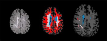
Although not a primary aim, the clinical significance of regional LA was investigated by examining regions categorized in the following manner: (1) periventricular (5 mm out from the lateral ventricle), (2) regions near the surface (5 mm underneath cortical gray matter), and (3) deep regions (remaining regions considered important for numerous frontal-subcortical connections; Price et al., Reference Price, Tanner, Schmalfuss, Brumback, Heilman and Libon2015). For each approach a “regional mask” (i.e., region defined by periventricular, deep, or infracortical) overlaid the LA mask (see Supplementary Figure 1). Regional LA was defined by the overlap of LA with the “regional mask”.
Regional white matter and leukoaraiosis. This figure depicts a T2 FLAIR slice with white matter hyperintensities (left). A slice with a “regional mask” (middle) separated into a periventricular region (blue), deep region (white), and infracortical region (red). A slice (right) depicts the regional leukoaraiosis (LA) mask (blue=periventricular LA; white=deep LA; red=infracortical LA) defined by the intersection of leukoaraiosis mask and the regional white matter mask.

Statistics
A series of multiple regression analyses were computed to inspect each aim. The first aim examined the relationship between cardiovascular risk factors and cognition. Separate regressions were conducted for each of the five cognitive domains. For each regression a single block of predictors included Group (PD or control), the Framingham 10 year Cardiovascular Risk score and an interaction term (cardiovascular risk×Group). The interaction term was computed to determine whether the association between cardiovascular risk and cognition differed among PD participants or controls.
The second aim examined the influence of cardiovascular risk factors on total LA volume. Independent variables included cardiovascular risk, group (PD vs. control) and a cardiovascular risk by group interaction term. These same analyses were repeated with regional LA volumes replacing total LA as the dependent variable.
The third aim examined the influence of total LA on cognition. Independent variables included total LA volume, group, and a LA by group interaction term. Separate regressions were conducted for each of the five cognitive domains. These same analyses were repeated with regional LA volumes replacing total LA as an independent variable.
As an exploratory aim we examined the relationship between cardiovascular risk, LA and other meaningful outcomes in PD (motor severity and mood symptoms). We conducted Pearson’s correlations to examine the relationship between cardiovascular risk, total LA, mood (BDI-II total score, AS total score, trait anxiety score from the STAI), UPDRS on motor score, duration of motor symptoms and LED, among the sub-sample of PD participants.
RESULTS
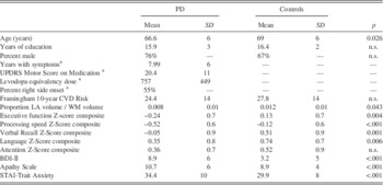
Sample characteristics are shown in Table 1. The final sample included 67 PD patients, and 61 older adult controls (total N=128). The PD and control groups did not significantly differ in terms of education or their Framingham Cardiovascular Risk score (p values >.1). Regarding cardiovascular risk, previous studies have shown the incidence rates (per 1000 people) for cardiovascular disease are 21.4 for men and 8.9 for women ages 55–64 years, and 34.6 for men and 20.0 for women ages 65–74 years (Go et al., Reference Go, Mozaffarian, Roger, Benjamin, Berry, Borden and Turner2013). As such, the degree of cardiovascular risk in the current sample was generally comparable to that of males between the ages of 55 and 75. The control group (mean age=69.0 years old) was 2 years older than the PD group (mean age=66.6 years old; t(126)=2.26; p=.026).
Sample Characteristics

a Subsample of PD patients only.
UPDRS=Unified Parkinson’s Disease Rating Scale; CVD=cardiovascular disease; LA=leukoaraiosis; WM=white matter.
Within the PD sample, 17 of the 67 participants were candidates for DBS surgery. The DBS candidates did not significantly differ from the non-DBS candidates in terms of cardiovascular risk (t(65)=0.516; p=.607) or any of the outcomes (total LA or any cognitive domain; all p values >.1).
Aim 1: Influence of Cardiovascular Risk on Cognitive Domains in PD versus Controls
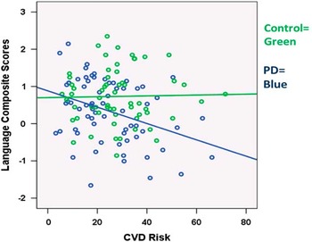
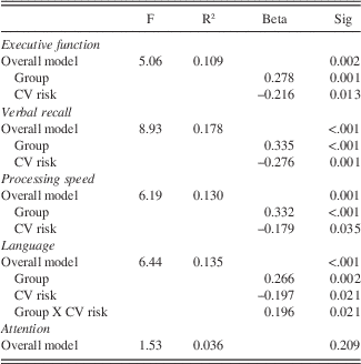
Aim 1 examined the influence of cardiovascular risk on cognitive domains among PD and control participants. As shown in Table 2, the overall regression models significantly predicted each cognitive domain except attention. Group status was a significant predictor of executive function, processing speed, verbal recall and language, meaning that PD individuals performed worse than controls for each domain. Higher cardiovascular risk scores were significantly related to worse performance on tasks of executive function, processing speed, verbal recall, and language. There was no significant Group by cardiovascular risk interaction term for executive function, verbal recall and processing speed. This meant that the detrimental influence of cardiovascular risk on cognition was largely comparable for the PD and control groups. The only exception was for the language domain (i.e., a significant Group by cardiovascular risk interaction). As shown in the Supplementary Figure 2, the interaction term can be interpreted as cardiovascular risk relating to worse language scores within the PD group only, and not the control group.
Relationship between language composite scores and cardiovascular (CV) risk among Parkinson’s patients and controls.

Results of regression analyses: Cognition and cardiovascular risk in Parkinson and control groups

Note. Only significant predictors are shown.
CV=cardiovascular.
The language composite consisted of the visual confrontation naming (BNT) and an animal fluency task. When the relationships between CVD risk and these individual tests were examined, there was a significant Group×CVD risk interaction effect on the BNT (β=-.213; p =.015) but not animal fluency (p>.05). This suggests that the interaction effect is driven by the BNT. The relationships between CVD risk and the other individual neuropsychological tests are displayed in Supplementary Table 1.
Aim 2: Influence of Cardiovascular Risk on LA in PD versus Controls
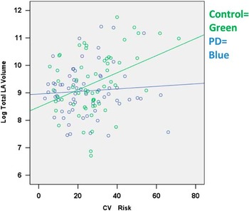
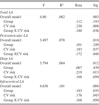
We secondarily tested the hypothesis that cardiovascular risk factors, in PD participants and controls, are associated with white matter abnormalities/LA (Table 3). The overall model (consisting of Group, CVD risk, and an interaction term) significantly predicted total LA volume. There was a significant Group by cardiovascular risk interaction (p=.036), indicating that more severe cardiovascular risk related to larger LA volume in the controls, but not PD (see Supplementary Figure 3). There was no significant main effect of Group, suggesting that PD and controls did not differ in total LA volume.
CV risk and Group interaction for total leukoaraiosis (LA). LA volume is corrected for total white matter volume and total intracranial volume. PD=Parkinson’s disease. CV=cardiovascular disease.

Leukoaraiosis and cardiovascular risk in PD and controls

CV=cardiovascular; PD=Parkinson’s disease.
Additional regression analyses examined specific regional location of LA (i.e., periventricular, deep and infracortical; Table 3). Results revealed a significant group by cardiovascular risk interaction term that related to periventricular LA, infracortical LA, and deep LA. Again, the interaction term means that more severe cardiovascular risk was related to greater LA volume (in deep, periventricular, and infracortical regions) in the controls, but not PD participants. The main effect of Group was significant in contributing to infracortical LA volume (p=.043) but no other regions, suggesting that controls had greater amounts of infracortical LA compared to PD patients.
Aim 3: Influence of LA of Cognition in PD versus Controls
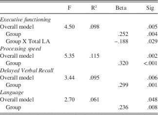
Multiple hierarchical regressions were conducted to examine the relationship between total LA and cognitive domains among PD and control participants. Independent variables included Total LA volume, group (PD or control) and the total LA volume by group interaction term. The cognitive domain was entered as the dependent variable for each regression. Results of these analyses are depicted in Table 4. The final model significantly predicted executive functioning, processing speed, language, and delayed verbal recall, but not attention (F(1,124)=.638; p=.592; R2=.015). As expected, there was a significant main effect of group, meaning that PD participants performed worse than controls across all cognitive domains, except attention. The only significant interaction (Group×Total LA) occurred with the executive functioning domain. Specifically, LA volume was related to worse executive functioning scores for controls, but not PD individuals. There was no main effect of total LA contributing to any cognitive domain. The relationship between total LA and individual cognitive domains are displayed in Supplementary Table 2.
Total leukoaraiosis and cognitive functioning

Note. Only significant predictors are shown.
LA=leukoaraiosis.
Additional exploratory hierarchical regressions were conducted with regional LA variables (periventricular, deep, or infracortical LA) replacing total LA. For all analyses, neither the main effect of regional LA, nor the regional LA by Group interaction terms were significant (all p values>.05). As such, regional measures of LA were not related to cognition in either PD patients or controls.
Post hoc analyses examined the relationship between cardiovascular risk, LA and cognitive functioning in a subsample of PD participants with and without mild cognitive impairment (MCI). MCI was classified based on Movement Disorder Society criteria (Litvan et al., Reference Litvan, Goldman, Tröster, Schmand, Weintraub, Petersen and Aarsland2012). Briefly individuals who performed 1.5 SD below the mean on at least two tests (within any domain) were classified as MCI. Pearson’s correlations revealed that neither cardiovascular risk nor LA volume (total or regional) significantly related to any cognitive domain (Supplementary Table 3).
Relationship between Cardiovascular Risk, LA, Mood, and PD Severity
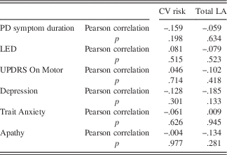
The influence of cardiovascular risk and total LA on mood (depression, apathy, anxiety), motor severity (UPDRS on medication motor score), duration of motor symptoms and LED was inspected with Pearson’s correlations (in the PD subsample only). Results showed that there were no significant relationships between LA or cardiovascular risk and any measure of mood or PD severity (Table 5).
Cardiovascular risk, PD severity, and mood symptoms

CV=cardiovascular; LA=leukoaraiosis; PD=Parkinson’s disease; UPDRS=Unified Parkinson’s Disease Rating Scale; LED=levodopa equivalency dose.
DISCUSSION
The current study examined the hypothesis that PD patients, due to decreased brain reserve, would be more vulnerable to vascular-related cognitive impairment and neuroimaging markers of small vessel disease. In general, there was limited support for interaction effects of cardiovascular risk factors and PD. Rather, the relationship between cardiovascular risk factors and cognition in PDs and controls was generally similar in terms of degree and the cognitive domains affected (except for language). Additionally, LA was associated with greater cardiovascular risk and executive dysfunction among controls, but not PD patients.
Worse cognitive functioning was associated with having a diagnosis of PD, as well as having more cardiovascular risk factors. This is an important finding because even though the effect of cardiovascular risk factors on cognition did not differ between PDs and controls (i.e., no interaction), PD patients with cardiovascular risk factors are at increased risk for cognitive impairment relative to PD patients without cardiovascular risk factors. This importance relates to the fact that certain aspects of PD (including but not limited to levodopa medication) have been suggested as being protective against cardiovascular risk factors (Nanhoe-Mahabier et al., Reference Nanhoe-Mahabier, De Laat, Visser, Zijlmans, de Leeuw and Bloem2009). Despite the fact that levodopa medication may lower blood pressure, hypertension has been observed to be present in approximately 36–38% of PD patients (Jones et al., Reference Jones, Malaty, Price, Okun and Bowers2012, Reference Jones, Jacobson, Murphy, Price, Okun and Bowers2014; Scigliano et al., Reference Scigliano, Ronchetti, Girotti and Musicco2009). Although hypertension and possibly other cardiovascular risk factors may be slightly less common among PD patients, cardiovascular risk factors are comparably detrimental to cognition among non-demented PD patients as they are among normal elderly individuals.
The current finding of a cardiovascular–cognition relationship is generally consistent with previous studies in normal elderly individuals (Aronow et al., Reference Aronow, Fleg, Pepine, Artinian, Bakris, Brown and Kostis2011; Debette et al., Reference Debette, Seshadri, Beiser, Au, Himali, Palumbo and DeCarli2011; Libon et al., Reference Libon, Price, Davis Garrett and Giovannetti2004). However, there have been some mixed findings with respect to a cardiovascular-cognition relationship in PD. Some previous studies examining the relationship between hypertension status and dementia status have failed to find a relationship in PD (Beyer et al., Reference Beyer, Aarsland, Greve and Larsen2006; Haugarvoll et al., Reference Haugarvoll, Aarsland, Wentzel‐Larsen and Larsen2005; Lee et al., Reference Lee, Kim, Yoo, Song, Kim, Jung and Lee2010). It should be noted that one cross-sectional study with PD participants did show a relationship between heart disease and increased risk for dementia (Pilotto et al., Reference Pilotto, Turrone, Liepelt-Scarfone, Bianchi, Poli, Borroni and Cosseddu2016). In contrast, previous studies have shown a relationship between cardiovascular risk, as measured by pulse pressure and individual risk factors (e.g., hypertension, diabetes), and specific cognitive domains (executive functioning, processing speed and delayed verbal recall) among non-demented PD patients (Jones et al., Reference Jones, Malaty, Price, Okun and Bowers2012, Reference Jones, Jacobson, Murphy, Price, Okun and Bowers2014; Pilotto et al., Reference Pilotto, Turrone, Liepelt-Scarfone, Bianchi, Poli, Borroni and Cosseddu2016). Discrepancies are likely due to differences in sample size as well as sensitivity of outcome measures (cognitive domains vs. dementia status).
In general, the detrimental effect of cardiovascular risk on cognition did not differ between the PD group and controls, except for the language domain. Greater cardiovascular risk was related to worse language performance among the PD group, but not the control group. In the current study, the language composite score consisted of an animal fluency task and a confrontation naming task (Boston Naming Test; BNT). When the relationships between CVD risk and the individual tests were examined, there was a significant Group by CVD risk interaction effect on the Boston Naming Task but not animal fluency. This suggests that the speeded component of the animal fluency test is unlikely to be accounting for the significant interaction effect. While, the basis for this interactive effect is unknown, it may reflect increased vulnerability of cortical regions important for language.
In general, naming impairments are associated with cortical dementias [such as Alzheimer’s disease (AD)] rather than subcortical dementias (such as vascular dementia or Parkinson’s disease dementia; Libon et al., Reference Libon, Price, Davis Garrett and Giovannetti2004). Cardiovascular risk factors are well known risk factors for AD and studies have suggested that up to 50% of patients with Parkinson’s disease dementia (PDD) meet pathological criteria for comorbid AD (Aronow et al., Reference Aronow, Fleg, Pepine, Artinian, Bakris, Brown and Kostis2011; Irwin, Lee, & Trojanowski, Reference Irwin, Lee and Trojanowski2013). Future studies are needed to clarify the mechanisms that underlie cardiovascular risk factors and language impairments in PD. Studies may additionally benefit by examining the types of errors (e.g., phonemic vs. semantic; coordinate vs. superordinate) or components/strategies (e.g., usage of clustering or switching in verbal fluency tasks; Troyer, Moscovitch, & Winocur, Reference Troyer, Moscovitch and Winocur1997) to understand if impairments represent a retrieval deficit (representing frontal-subcortical dysfunction) or a storage based naming deficit.
The relationship between cardiovascular risk factors and cognition among elderly individuals is thought to be mediated by small vessel disease and subcortical white matter damage (Aronow et al., Reference Aronow, Fleg, Pepine, Artinian, Bakris, Brown and Kostis2011; Libon et al., Reference Libon, Price, Davis Garrett and Giovannetti2004). Support for this relationship was found among the control group (LA was related to both greater cardiovascular risk and executive dysfunction), but not the PD group. A lack of a relationship between LA and cognition in the current study does not definitively exclude small vessel disease as a mechanism underlying the relationship between cognition and cardiovascular risk.
Previous studies of LA and cognition in PD samples have produced mixed results. A past review addressed this inconsistency in the literature (Bohnen & Albin, Reference Bohnen and Albin2011). The authors suggested that the relationship between LA and cognition might be more easily detected among advanced stages of PD such as PDD. To date, at least six studies have examined the relationship between LA and cognition among individuals with PD. Three of the six studies showed that LA was more common among PDD patients compared to non-demented PD patients (Beyer et al., Reference Beyer, Aarsland, Greve and Larsen2006; Lee et al., Reference Lee, Kim, Yoo, Song, Kim, Jung and Lee2010; Melzer et al., Reference Melzer, Watts, MacAskill, Pitcher, Livingston, Keenan and Anderson2013). Three other studies excluded PD patients with dementia (Dalaker et al., Reference Dalaker, Larsen, Dwyer, Aarsland, Beyer, Alves and Zivadinov2009; Ham et al., Reference Ham, Lee, Sunwoo, Hong, Sohn and Lee2016; Mak et al., Reference Mak, Dwyer, Ramasamy, Au, Tan, Zivadinov and Kandiah2015). These studies revealed inconsistent results. One study did not find a relationship between LA and MCI status or tests of executive functioning (Dalaker et al., Reference Dalaker, Larsen, Dwyer, Aarsland, Beyer, Alves and Zivadinov2009), while the other two did find a significant relationship between LA and specific cognitive tasks (semantic fluency and the Mini-Mental State Examination; Ham et al., Reference Ham, Lee, Sunwoo, Hong, Sohn and Lee2016; Mak et al., Reference Mak, Dwyer, Ramasamy, Au, Tan, Zivadinov and Kandiah2015). As such, the relationship between LA and cognition in non-demented PD patients has been variable and requires further examination.
One additional possibility for the non-significant relationship between LA and cognition in the current project is that large amounts of LA were relatively uncommon in the current sample. Threshold studies of LA have shown that cognitive impairments appear when at least 3% of the white matter contains LA (Price et al., Reference Price, Mitchell, Brumback, Tanner, Schmalfuss, Lamar and Libon2012). In the current study, only four individuals with PD had LA in at least 3% of the white matter, possibly reflecting a relatively healthy group of PD patients (in terms of cerebrovascular health) or that the PD group was approximately 2 years younger than the control group. Furthermore, past studies finding a relationship between cognition and LA have reported larger volumes of LA in their samples (up to three times larger volumes of LA compared to the current study; Dalaker et al., Reference Dalaker, Larsen, Dwyer, Aarsland, Beyer, Alves and Zivadinov2009; Melzer et al., Reference Melzer, Watts, MacAskill, Pitcher, Livingston, Keenan and Anderson2013). Therefore, one possibility for the non-significant white matter-cognition relationship in the current study is that the imaging markers lacked the sensitivity to detect the relationship.
The current study was not without limitations. First, the sample of PD patients is generally intact cognitively (within .5 SD of the normative samples). Larger studies may benefit from examining the relationship between cardiovascular risk, structural brain changes and cognition as a function of cognitive status (e.g., cognitively intact, mild cognitive impairment, or dementia). There currently is debate on whether to group/classify tests of verbal fluency (letter and animal) as tests of language or executive functioning. Movement Disorder Society consensus statements on the diagnostic criteria for PDD and PD-MCI classify tests of verbal fluency (both letter and animal fluency) under the executive function domain (Emre et al., Reference Emre, Aarsland, Brown, Burn, Duyckaerts, Mizuno and Goldman2007; Litvan et al., Reference Litvan, Goldman, Tröster, Schmand, Weintraub, Petersen and Aarsland2012). However, a recent factor analytic studies (consisting of a wide range of psychiatric and neurologic disorders) found that both verbal fluency tests load with other tests of language (Whiteside et al., Reference Whiteside, Kealey, Semla, Luu, Rice, Basso and Roper2016). To add to the debate, other studies have shown verbal fluency tests to be more related to tests of processing speed, rather than language or executive functions (McDowd et al., Reference McDowd, Hoffman, Rozek, Lyons, Pahwa, Burns and Kemper2011). The classification of verbal fluency measures in the current study is consistent with previous studies of cognitive and cardiovascular risk factors in PD (Jones et al., Reference Jones, Jacobson, Murphy, Price, Okun and Bowers2014), the diagnostic accuracy of cognitive impairment in PD (Barton et al., 2014), and previous neuropsychological models of semantic and letter fluency (see Troyer et al., Reference Troyer, Moscovitch and Winocur1997 for an overview). It is possible that differences in test assignment (e.g., grouping both verbal fluency tests into either the executive function or language domain) could influence the results.
Similarly, the attention domain was limited to one test of simple attention, and one test of working memory. Future studies may benefit by examining additional tests of working memory. Additionally, the analyses used cross-sectional data and were unable to examine longitudinal changes in cognition, white matter abnormalities or cardiovascular risk. As such, the current study was not able to clarify the directionality of the relationships. Specifically longitudinal studies would be better positioned to determine if cardiovascular factors increase the risk for cognitive impairment in PD, or if PD increases the risk for cognitive impairment related to cardiovascular disease. Longitudinal follow-up may additionally address the fact that LA was uncommon among this sample. The relationship between the progression of LA and the progression/development of cognitive impairment could also be examined in a longitudinal design. Due to the fact that large amounts of LA were not common, future studies may benefit from the use diffusion tensor imaging which may be able to detect micro-structural differences in white matter integrity not detectable by T2 FLAIR scans. Future studies may also benefit from using more refined measures of PD severity (e.g., UPDRS off-medication motor scores, dopamine transporter imaging, etc.) and examine cardiovascular risk across different stages of PD severity.
Overall, the importance of this study relates to the fact that PD and cardiovascular risk factors represent separate and independent risk factors for cognitive impairment. On average, PD patients are performing worse than their older adult counterparts, but PD patients with elevated cardiovascular risk factors may be particularly vulnerable to impairments in executive functioning, memory, processing speed, and language. Increasing our understanding of the relationship between cardiovascular risk factors and cognition may help with assessment and treatment of cognitive difficulties among individuals with PD. Cardiovascular diseases may represent a modifiable risk factor that if treated (or prevented) may reduce an individual’s risk for future cognitive decline and possibly dementia.
Acknowledgments/Study Funding
NINDS K23NS060660 (CP), R01NS082386 (CP) and in part by the Center for Movement Disorders and Neurorestoration, the National Institutes of Health/National Center for Advancing Translational Sciences (NIH/NCATS), Clinical and Translational Science Award to the University of Florida UL1TR000064, and by the American Psychological Foundation. Disclosures: Dr. Jones received research support through the American Psychological Foundation. Dr. Tanner reports no disclosures. Dr. Okun serves as a consultant for the National Parkinson Foundation, and has received research grants from NIH, NPF, the Michael J. Fox Foundation, the Parkinson Alliance, Smallwood Foundation, and the UF Foundation. Dr. Okun has previously received honoraria, but in the past 24 months has received no support from industry including travel. Dr. Okun has received royalties for publications with Demos, Manson, and Cambridge (movement disorders books). Dr. Okun has participated in CME activities on movement disorders sponsored by the USF CME office, PeerView, and by Vanderbilt University. The institution and not Dr. Okun receives grants from Medtronic and ANS/St. Jude, and the PI has no financial interest in these grants. Dr. Okun has participated as a site PI and/or co-I for several NIH, foundation, and industry sponsored trials over the years but has not received honoraria. Dr. Price receives research support through NIH and the National Science Foundation (NSF). Dr. Bowers receives research support from the NIH, Michael J. Fox Foundation, the State of Florida Ed and Ethel Moore Alzheimer’s program, and the McKnight Research Foundation.
SUPPLEMENTARY MATERIAL
To view supplementary material for this article, please visit http://dx.doi.org/10.1017/S1355617717000017