Non-technical Summary
Caryocaridids are a unique representative of the pelagic arthropod group during the Ordovician and stand out from other arthropods (ostracods, trilobites, etc.) for their remarkable pelagic abilities. Herein, we report on a species of caryocaridids, Soomicaris cedarbergensis, from the Lower Ordovician in northwestern Xinjiang, NW China, which shows the rare enrolled carapaces with the evidence of cuticular ultrastructure preserved. These caryocaridid specimens from Xinjiang provides the substantial evidence for the presence of caryocaridids in the Central Asian Orogenic Belt. This discovery suggests that S. cedarbergensis appeared as early as the Early Ordovician (late Tremadocian) and persisted until the end-Ordovician (Hirnantian) and is the longest-ranging species of known caryocaridids. The cuticle of the carapace in S. cedarbergensis is preserved in carbonate-fluorapatite, which can be divided into three mineralized lamellae (outer, middle, and inner). The outer and inner lamellae both consist of three layers, which seem to correspond to the epicuticle, exocuticle, and endocuticle of extant crustacean carapaces, respectively. The particular ultrastructure of the carapace of Ordovician caryocaridids (thin cuticle; thickened inner lamella cuticle; and large, complex oxygen supply system) probably represents an adaptation to the pelagic lifestyle during the Ordovician plankton revolution.
Introduction
Phyllocarids are an important group of Malacostraca known for their morphological diversity, prolonged fossil records, and abundant extant relatives (Rolfe Reference Rolfe, Brooks, Carpenter, Glaessner, Hahn, Hessler, Hoffman and Holthuis1969, Reference Rolfe1981; Schram and Hof Reference Schram, Hof and Edgecombe1998; Spears and Abele Reference Spears and Abele1999). However, phyllocarids have received less attention compared with other contemporaneous taxa of the phylum Arthropoda due to their relative rarity and generally poor preservation, which make it difficult to infer their habitat, size, and body structure (Sepkoski Reference Sepkoski2000). Crustacean cuticles exhibit varying levels of calcification that can differ even among different parts of the carapace of the same organism (Vega et al. Reference Vega, Dávila-Alcocer and Filkorn2005). Additionally, diagenetic processes and taphonomy have the potential to alter or even destroy evidence of the original cuticle of crustacean carapaces. Therefore, there have been few studies focused on the ultrastructure and composition of the cuticle of phyllocarids in comparison to ostracods. The known cases are mainly centered on caryocaridids and ceratiocaridids (Rolfe Reference Rolfe1962; Churkin Reference Churkin1966; Miranda Reference Miranda2002; Vannier et al. Reference Vannier, Racheboeuf, Brussa, Williams, Rushton, Servais and Siveter2003; Whittle et al. Reference Whittle, Gabbott, Aldridge and Theron2007; Liu et al. Reference Liu, Fan, Zong and Gong2023; Šilinger Reference Šilinger2023; Kovář et al. Reference Kovář, Šilinger, Fatka and Brocke2024).
Most authors have acknowledged the pelagic lifestyle of caryocaridids (e.g., Whittle et al. Reference Whittle, Gabbott, Aldridge and Theron2007; Collette and Hagadorn Reference Collette and Hagadorn2010; Liu et al. Reference Liu, Fan, Zong and Gong2022, Reference Liu, Fan, Zong and Gong2023). Ruedemann (Reference Ruedemann1934) suggested that the caryocaridids have a pelagic lifestyle based on their wide paleogeographic distribution and the relative longevity of the genus. Since then, additional scholars have attempted to demonstrate the pelagic habitat of caryocaridids based on accumulated evidence. This includes their cosmopolitan distribution, carapace morphology, abdominal morphology, and association with the shale facies (Størmer Reference Størmer1937; Chlupáč Reference Chlupáč2003; Vannier et al. Reference Vannier, Racheboeuf, Brussa, Williams, Rushton, Servais and Siveter2003). However, according to Bulman (Reference Bulman1964), the large and heavy carapace of caryocaridids prevents them from living in a pelagic environment. Additionally, caryocaridids often coexist with benthos in certain assemblages. Chlupáč (Reference Chlupáč1970) also suggested that caryocaridids are not strictly part of the plankton and likely attach to floating algal thali near the surface of water. However, Churkin (Reference Churkin1966) discovered that the carapace valves of the caryocaridids were relatively large but extremely thin. And no evidence of algal mats has been found in conjunction with caryocaridid remains (Chlupáč Reference Chlupáč1970, Reference Chlupáč2003). According to Vannier et al. (Reference Vannier, Boissy and Racheboeuf1997a), the streamlined carapace and flattened furcal rami of caryocaridids are assumed to be adaptation to their active, free-swimming lifestyle.
In extant crustaceans, the cuticular structure of the carapace appears to be closely related to lifestyle (Sohn and Kornicker Reference Sohn, Kornicker, Hanai and Ishizaki1988; Pütz and Buchholz Reference Pütz and Buchholz1991; Yamada Reference Yamada2019). Unfortunately, since Churkin (Reference Churkin1966) first studied the cuticular ultrastructure and mineralogical composition of the caryocaridid carapace, no further research has not been conducted, except for Vannier et al. (Reference Vannier, Racheboeuf, Brussa, Williams, Rushton, Servais and Siveter2003), who related the cuticular ultrastructure to the caryocaridid lifestyle. Other authors have provided brief descriptions of the cuticular ultrastructure and mineralogical composition of the carapace in caryocaridids (Miranda Reference Miranda2002; Vannier et al. Reference Vannier, Racheboeuf, Brussa, Williams, Rushton, Servais and Siveter2003; Whittle et al. Reference Whittle, Gabbott, Aldridge and Theron2007; Liu et al. Reference Liu, Fan, Zong and Gong2023; Šilinger Reference Šilinger2023; Kovář et al. Reference Kovář, Šilinger, Fatka and Brocke2024).
In recent years, we have investigated the Cambrian–Ordovician transitional strata in the Guozigou area of Xinjiang, NW China. We collected a large number of the Lower Ordovician caryocaridid specimens from the original or nearby sections reported in previous studies (Xiang and Zhang Reference Xiang and Zhang1984; Zhang Reference Zhang1987). These materials retain the rare enrolled carapace and cuticular ultrastructure of the carapace due to unique taphonomic conditions. We discuss here the carapace characteristics of caryocaridids in three preservation states. Additionally, we studied and evaluated the cuticular ultrastructure of the carapace and provide further evidence that caryocaridids are pelagic arthropods.
Materials and Methods
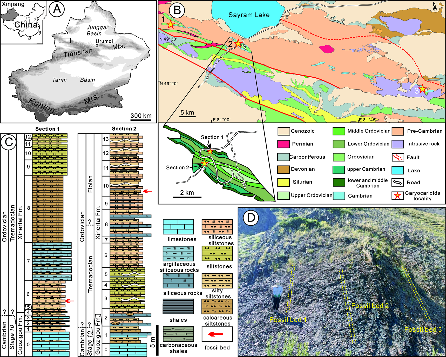
The western part of the North Tianshan Mountains in Xinjiang contains relatively continuous Ordovician strata. The most-studied area is in Guozigou (Fig. 1A,B). The Ordovician strata in the Guozigou area are divided into the Guozigou Formation (O1g), the Xinertai Formation (O1-2x), the Kekesaleixi Formation (O3kk), the Aketashi Formation (O3a), and the Hudukedaban Formation (O3h) from the bottom to the top (BGXUAR 1987c; XBGMR 1993, 1999; Li Reference Li1995; Chen et al. Reference Chen, Lin, Xu and Zhou1998). The Xinertai Formation is a collection of darkish-gray and gray siliceous, argillaceous, and carbonaceous siltstones, siliceous rocks, and sandy mudstones, with a few black shales and thin-bedded limestones (XBGMR 1999). It was most likely deposited in a shelf to slope paleoenvironment (Li Reference Li1995; XBGMR 1999). This formation yields a variety of invertebrates, including phyllocarids, graptolites, trilobites, and brachiopods (e.g., Xu and Huang Reference Xu and Huang1979; BGXUAR 1987c; Zhang Reference Zhang1987; Qiao Reference Qiao1989; XBGMR 1993, 1999).
A, Location of North Yili in North Tianshan, Xinjiang, NW China. B, Geologic map of North Yili shown in black box in A (modified from BGXUAR 1987a,b,c): 1, Jiangkunusi; 2, Guozigou; 3, Keguqinshan. C, Stratigraphic column of the Xinertai Formation in the Guozigou area (modified after Zhang Reference Zhang1987; Qiao Reference Qiao1989). D, The outcrop of three fossil beds in Bed 10 of Section 2 (geologist is 165 cm tall).

Xiang and Zhang (Reference Xiang and Zhang1984) were the first to report crustacean fossils, probably caryocaridid carapaces, in the Ordovician strata from the Guozigou area. Since then, roughly determined Caryocaris sp. has frequently been encountered in the Jiangkunusi, Guozigou, and Keguqinshan areas in later geological surveys or studies of other fossils (Zhang Reference Zhang1987; Qiao Reference Qiao1989; XBGMR 1993; Zhang Reference Zhang2010; Fig. 1B). However, no systematic description or detailed analysis of caryocaridids has been provided. The phyllocarid materials described in this paper were collected from two stratigraphic sections in the Guozigou area (Fig. 1B,C). Section 1 (44°28′47.5″N, 81°08′35.8″E) is located on the ridge between the Linkuanggou and Fenggou, where a large number of well-preserved caryocaridid fossils were found in the siliceous siltstones about 1.5 m above the bottom of Bed 6. Section 2 (44°28′11.4″N, 81°08′4.9″E) is located west of a new highway, approximately 500 m north of the Linkuanggou entrance. Three beds of caryocaridid fossils were collected from the lower part of Bed 10, and the fossils are poorly preserved and scarce (Fig. 1D). The caryocaridid-bearing levels from the Guozigou area cross three graptolite biozones, including the Adelograptus-Clonograptus (late Tremadocian, Adelograptus tenellus and Aorograptus victoriae), Tetragraptus approximatus (early Floian), and the Tshallograptus fruticosus (early Floian) Biozones, as suggested by the graptolites found in the same bed (Xu and Huang Reference Xu and Huang1979; Qiao Reference Qiao1989, Chen et al. Reference Chen, Lin, Xu and Zhou1998; XBGMR 1999; Zhang et al. Reference Zhang, Erdtmann and Feng2004, Reference Zhang, Wang, Feng, Luo and Erdtmann2005; Li et al. Reference Li, Wang and Feng2018). Our caryocaridid specimens were mainly found in the Adelograptus-Clonograptus and Tetragraptus approximatus graptolite biozones within the lower Xinertai Formation within the two sections of the Guozigou area. A total of 104 caryocaridid specimens were collected and are currently housed in the State Key Laboratory of Biogeology and Environmental Geology (BGEG) at China University of Geosciences in Wuhan.
The caryocaridid specimens were photographed using an APS-C DSLR camera and illuminated with a microscope cold-light halogen lamp. Images of enrolled specimens were taken using field emission scanning electron microscopes (Hitachi SU 8010) at the State Key Laboratory of Biogeology and Environmental Geology, China University of Geosciences, and Zeiss Axio Zoom V16 at the Nanjing Institute of Geology and Paleontology, Chinese Academy of Sciences. Microzone chemical composition analysis and wavelength dispersive element scanning of fossils were conducted using energy dispersive spectroscopy (EDS; Hitachi SU 8010). Additionally, light photomicrographs of the thin sections were taken using a Zeiss Axioscope A1 polarizing microscope at the China University of Geosciences.
Morphological Terminology Used
Lamella/lamellae, a general expression for a layered appearance of anything; lamina/laminae, the fine striations constituting a special structure in the cuticle composed of chitin–protein fibers.
Results
Description of Caryocaridids
Out of the caryocaridid specimens collected from the two sections, only 56 could be identified, including 52 incomplete carapaces, 2 telsons, and 2 furcal rami. The carapace valves of caryocaridids exhibit irregular longitudinal and oblique folds and lines due to the compression deformation. Nearly 40% of them are enrolled. The remaining specimens are highly fragmented and could not be identified.
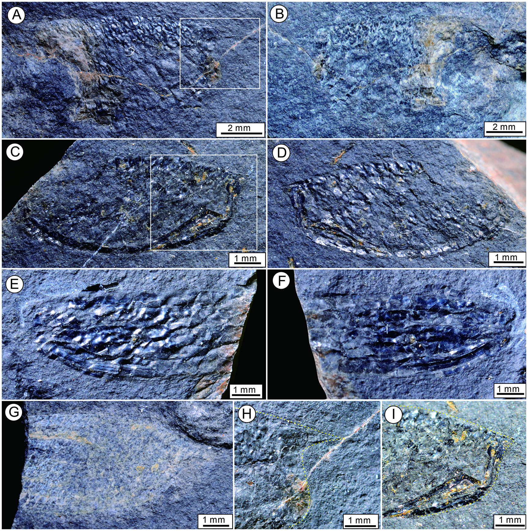
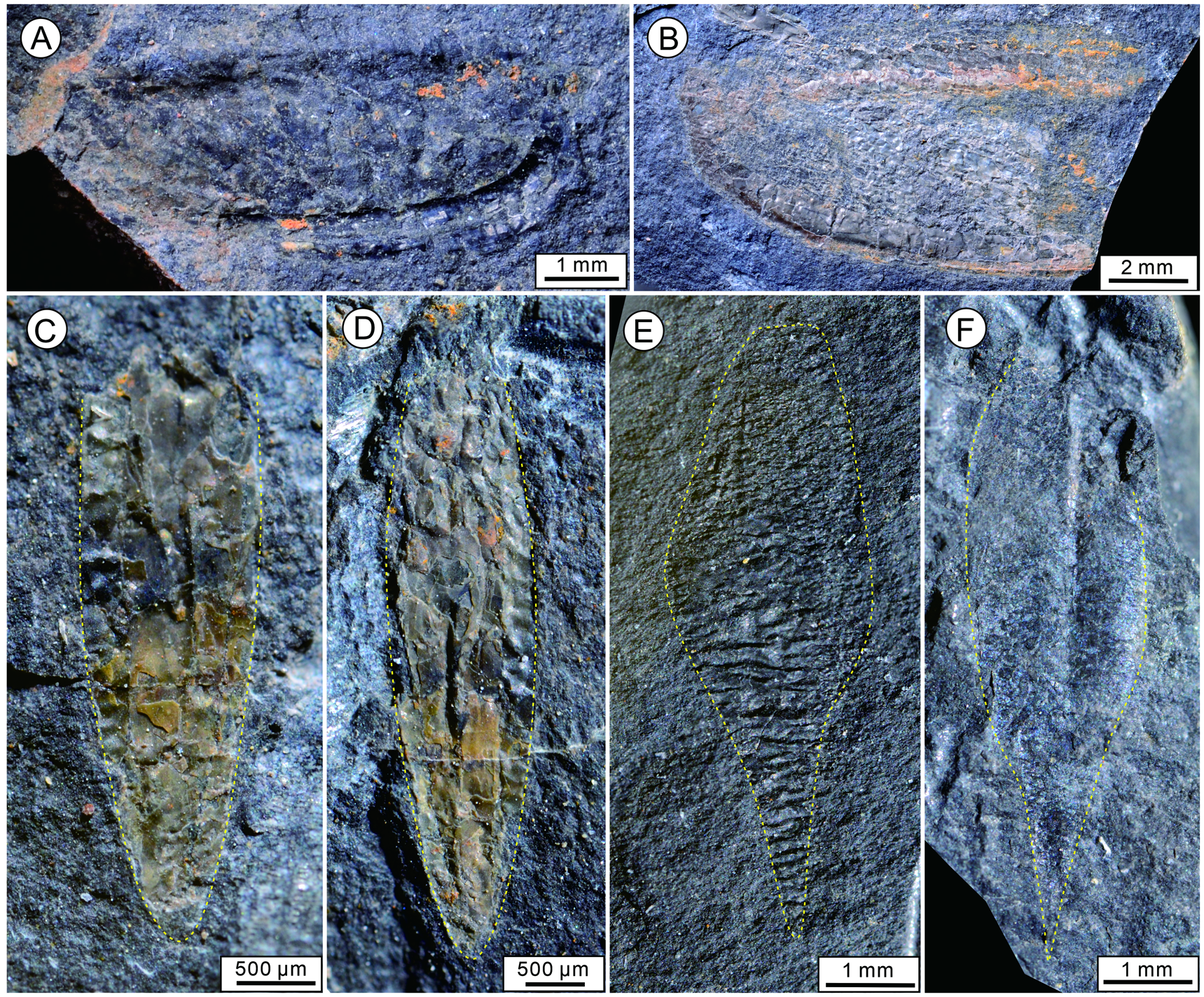
Detailed Description. The pod-shaped, bivalved carapace has a length to height ratio of 2.5. The highest point of the carapace is located in the center of the carapace. Dorsal and ventral margins are moderately convex (Figs. 2A–G, 3A,B). The anterior horn of the carapace is sharply pointed and measures 1.5–1.8 mm in length, pointing forward (Fig. 2A,B,G,H). A clear and pronounced ventral doublure with a moderate convexity and constant width of 0.4 mm runs along the ventral border (Figs. 2C–F, 3A,B). The posterior margin of carapace is curved and lacks spinules and a posteroventral spine. The posterodorsal spine is short and pointed (Figs. 2C,D,I, and 3A). The subtriangular telson is dorsoventrally flattened and has a pointed extremity (Fig. 3C,D). The maximum length of the telson is 5.0 mm, with a corresponding width of 1.2 mm near its proximal end. The furcal rami are leaf-shaped, pointed, and longer than the telson. Their outer margins lack spinelike triangular expansion and articulated spines. The maximum width of the furcal rami is located just anterior to its midlength (Fig. 3E,F). A furrow, which runs from almost the base of the spinelike expansion to the axis of the terminal posterior spine, is clearly visible (Fig. 3F). These specimens from the Guozigou area are identified as Soomicaris cedarbergensis (Whittle et al. Reference Whittle, Gabbott, Aldridge and Theron2007) based on the carapace outline, which features a long pointed anterior horn and a short pointed posterodorsal spine (Whittle et al. Reference Whittle, Gabbott, Aldridge and Theron2007; Racheboeuf and Crasquin Reference Racheboeuf and Crasquin2010).
The carapaces of Soomicaris cedarbergensis from the Lower Ordovician Xinertai Formation, Xinjiang, NW China. A, B, Incomplete right carapace, BGEG–SJ–01a,b. C, D, Incomplete left carapace, BGEG–SJ–03a,b. E, F, Incomplete right carapace, BGEG–SJ–06a,b. G, Incomplete right carapace, BGEG–SJ–02a. H, Enlargement of the white box in A, showing the anterior horn; I, Enlargement of the white box in C, showing the posterodorsal spine.

The carapaces, telsons and furcal rami of Soomicaris cedarbergensis from the Lower Ordovician Xinertai Formation, Xinjiang, NW China. A, Incomplete left carapaces, BGEG–SJ–04. B, Incomplete right carapace, BGEG–SJ–10. C, D, The well-preserved telsons, BGEG–SJ–09a,b. E, F, Incomplete furcal rami; E, BGEG–X–C6, F, BGEG–S–C9.

Geographic Distribution of Soomicaris
Caryocaridids are a well-known representative of the pelagic arthropod group during the Ordovician period. Their paleogeographic distribution became global from the Dapingian to Sandbian (Liu et al. Reference Liu, Fan, Zong and Gong2022). Before the recent description of caryocaridids from the North China Craton (Liu et al. Reference Liu, Fan, Zong and Gong2023), these arthropods were mainly found in Gondwana, Laurentia, Baltica, Avalonia, Bohemia, and South China (e.g., Chapman Reference Chapman1902, Reference Chapman1934; Woodward Reference Woodward1912; Chlupáč Reference Chlupáč1970, Reference Chlupáč2003; Jell Reference Jell1980; Shen Reference Shen1986; Racheboeuf et al. Reference Racheboeuf, Vannier and Ortega2000, Reference Racheboeuf, Crasquin and Brussa2009; Whittle et al. Reference Whittle, Gabbott, Aldridge and Theron2007; Racheboeuf and Crasquin Reference Racheboeuf and Crasquin2010). Soomicaris comprises three known species: S. cedarbergensis, S. ordosensis, and S. scanicus, which are respectively distributed across Clanwilliam in South Africa (late Hirnantian), Inner Mongolia in China (early Sandbian), and Scania in Sweden (late Tremadocian) (Möberg and Segerberg Reference Möberg and Segerberg1906; Whittle et al. Reference Whittle, Gabbott, Aldridge and Theron2007; Liu et al. Reference Liu, Fan, Zong and Gong2023). Freiberger (Reference Freiberger1947) documented the carapaces of Caryocaris curvilata in the Karatau, Kazakhstan. And then, Obut and Zuvtsov (Reference Obut, Zuvtsov and Sokolov1965) and Abdullaev and Khaletskaya (Reference Abdullaev and Khaletskaya1970) reported the Caryocaris baidamtalensis and Caryocaris sp. from the Ordovician strara of Kyrgystan and Uzbekistan respectively. Tolmacheva et al. (Reference Tolmacheva, Holmer, Popov and Gogin2004, Reference Tolmacheva, Degtyarev, Samuelsson and Holmer2008) and Nikitina et al. (Reference Nikitina, Popov, Neuman, Bassett, Holmer, Bassett and Deisler2006) also mentioned the occurrence of some carapace and telson fragments of caryocaridids or phyllocarids in the western Balkhash region and the Kol'denen River area of Kazakhstan. However, these specimens are relatively fragmented and the illustrations are poor or even missing, to the extent that further species identification is not feasible. Undoubtedly, these evidence seems to support the notion that the distribution of caryocaridids could potentially spread to the Central Asian Orogenic Belt (Tolmacheva et al. Reference Tolmacheva, Degtyarev and Ryazantsev2021). Therefore, the discovery of S. cedarbergensis in the Lower Ordovician of northwestern Xinjiang not only provides more substantial evidence for the presence of caryocaridids in the Central Asian Orogenic Belt but also further extends their distribution range within this geological area.
Soomicaris cedarbergensis, found in the Xinertai Formation in North Tianshan, is similar to the other two species of this genus co-occurring with graptolites and thin-shelled brachiopods at the same horizon (Möberg and Segerberg Reference Möberg and Segerberg1906; Liu et al. Reference Liu, Fan, Zong and Gong2023). The graptolites in the same bed suggest that S. cedarbergensis from the Guozigou area of North Tianshan, Xinjiang, is from the late Tremadocian to early Floian, occupying three graptolite biozones. Thus, S. cedarbergensis appeared as early as the late Tremadocian and persisted until the end of the Ordovician (Hirnantian), making it the longest-ranging species of known caryocaridids.
Discussion
Taphonomic Types of Carapaces
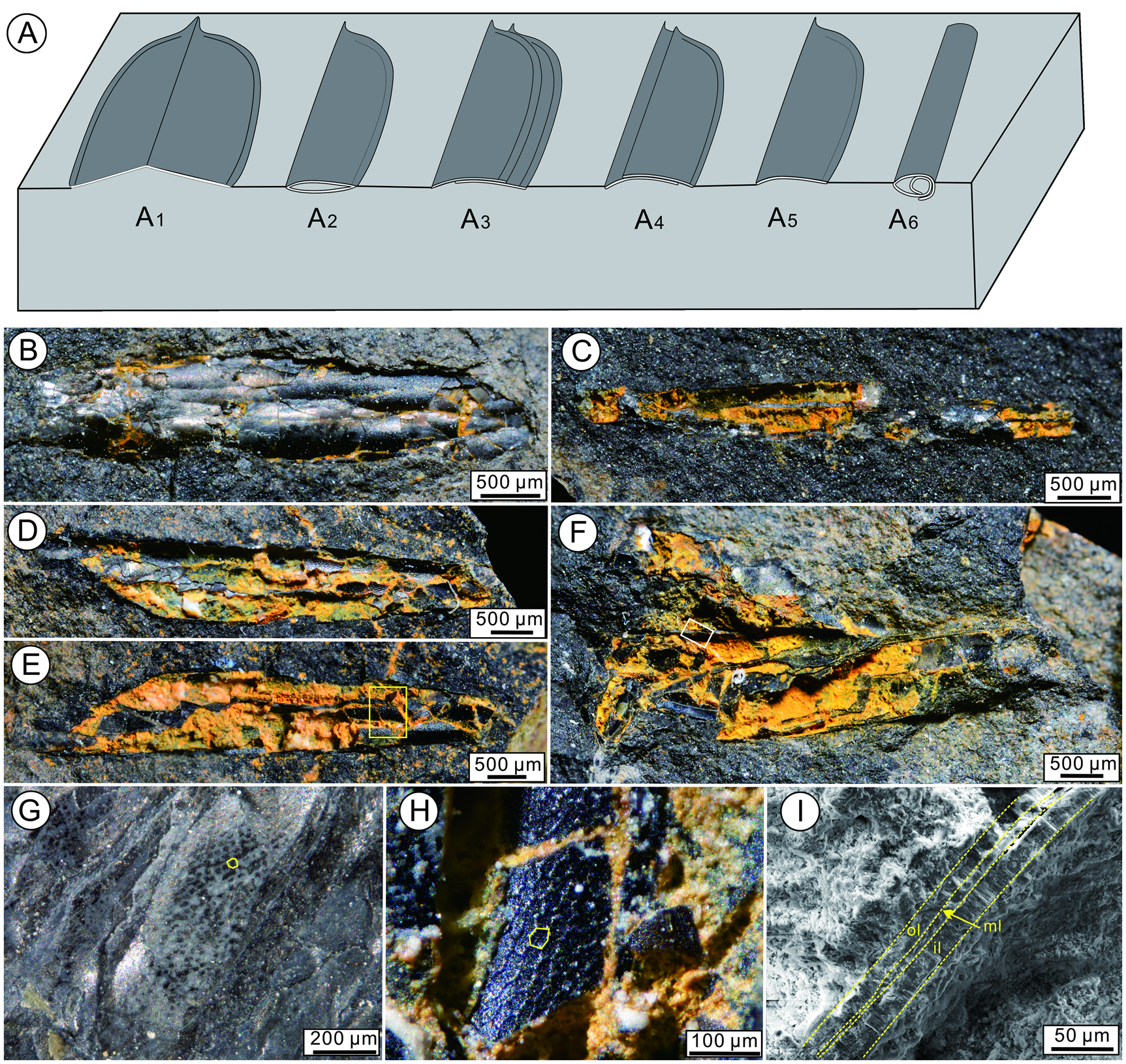
The carapaces of the reported specimens of caryocaridids exhibit varying degrees of compression deformation, which can be classified into three types: enrollment, wrinkles, and flattening (e.g., Churkin Reference Churkin1966; Chlupáč Reference Chlupáč1970; Rushton and Williams Reference Rushton and Williams1996; Vannier et al. Reference Vannier, Racheboeuf, Brussa, Williams, Rushton, Servais and Siveter2003; Fig. 4). Generally, most caryocaridid carapaces are flat in shales, siltstones, and/or siliceous rocks, but complete flattening of carapaces is not common due to their rigid strength (Chlupáč Reference Chlupáč1970). If the two carapace valves remain jointed after burial, they are eventually preserved in a butterfly shape under vertical burial (e.g., Shen Reference Shen1986: figs. 3, 4; Wang et al. Reference Wang, Hua, Wang, Gu, Chen, Zhuang, He and Li2019: fig. 3g; Liu et al. Reference Liu, Fan, Zong and Gong2023: fig. 2J; Fig. 4A1) and in the overlapping form under lateral burial (e.g., Vannier et al. Reference Vannier, Racheboeuf, Brussa, Williams, Rushton, Servais and Siveter2003: fig. 5E; Fig. 4A2). The two jointed carapace valves may be slightly separated due to bottom currents or bioturbation (e.g., Copeland Reference Copeland1967: plate 162, figs. 1, 2; Jell Reference Jell1980: fig. 5A; Liu et al. Reference Liu, Fan, Zong and Gong2023: fig. 2G,M), then compression may result in the impression of the harder border of one carapace into the valve of the opposite carapace, so that four or two parallel grooves following the ventral or dorsal margin of the carapace may be observed (e.g., Chlupáč Reference Chlupáč1970: plate I, figs. 1, 3, 5; Racheboeuf et al. Reference Racheboeuf, Vannier and Ortega2000: fig. 7C; Fig. 4A3, A4). If the two carapace valves are far apart, they are preserved in a flat, single valve form (e.g., Whittle et al. Reference Whittle, Gabbott, Aldridge and Theron2007: fig. 3A,B; Fig. 4A5). When the compression deformation is minimal, the carapace valves of caryocaridids show irregular folding and warping, resulting in irregular longitudinal and oblique folds and lines (e.g., Chapman Reference Chapman1934: plates 9, 10; Pillola et al. Reference Pillola, Piras and Serpagli2008, plate 1, fig. 4). Threse folds and lines often cause the carapace valves to break (Chlupáč Reference Chlupáč1970).
Taphonomic types of caryocaridid carapace and enrolled carapace from the Xinertai Formation, Xinjiang, NW China. A, Preservational states of caryocaridid carapace on the surface of bedding plane. A1, Carapaces preserved in the “butterfly position”; A2, jointed carapaces buried on one side; A3, A4, slightly dislocated carapaces buried on one side; A5, single carapace buried on one side; A6, enrolled carapaces. B–F, Curled incomplete carapaces, BGEG–SJ–33, 34, 35a, b, 36. G, The polygonal reticulation of a carapace, BGEG–SJ–11b; H, Enlargement of the yellow box in E, showing the polygonal reticulation. I, Natural cross-section of the carapace of the white box in F showing the three mineralized lamellae. Abbreviations: il, inner lamella; ml, middle lamella; ol, outer lamella.

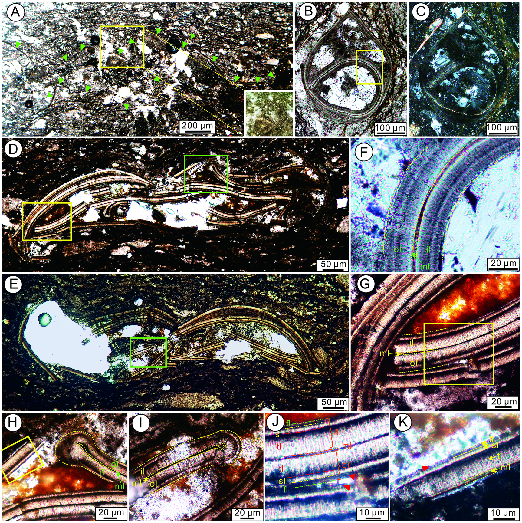
Enrollment is the rarest type, which fully demonstrates the 3D morphology of the caryocaridid carapaces. Before this paper, the only known 3D preserved specimens were Caryocaris curvilata from Alaska, North America (Churkin Reference Churkin1966), Caryocaris wrightii from Bohemia (Chlupáč Reference Chlupáč1970) and the British Isles (Rushton and Williams Reference Rushton and Williams1996; Rushton and Ingham Reference Rushton, Ingham, Fortey, Harper, Ingham, Owen, Parkes, Rushton and Woodcock2000; Vannier et al. Reference Vannier, Racheboeuf, Brussa, Williams, Rushton, Servais and Siveter2003), and some unknown caryocaridid carapaces from Texas and Nevada, North America (T. A. Hegna personal communication 2024). Approximately 40% of the total number of caryocaridid specimens found in the Guozigou area of Xinjiang are enrolled. These carapace valves are enrolled transversely and are cylindrical, tapering slightly from the middle to the ends, resembling an elongated cigar (Fig. 4A6, B–F). All enrolled specimens were partially broken. The largest caryocaridid specimen measures about 5.8 mm in length, with a height of about 0.9 mm and a length-to-height ratio of about 6.5, compared with about 2.5 in associated flattened specimens. The thin black carapace valves, with a thickness of about 40–45 μm (Figs. 5J, 6A), are supported by white calcite or loose limonite deposited between each valve (Fig. 4B–F), or they may adhere to each other to form “thick” carapace valves of two to three layers. This type of carapace is identical to the carapaces described by Churkin (Reference Churkin1966) and Chlupáč (Reference Chlupáč1970) for caryocaridids.
Light photomicrographs of the carapace of Soomicaris cedarbergensis from the Xinertai Formation, Xinjiang, NW China. A, Light photomicrograph of siliceous siltstones from Bed 6 in Section 1, BGEG–TS–01–1, green triangles indicate the carapace fragments of Soomicaris. B, C, Photomicrographs of a thin-sectioned specimen through an enrolled carapace, BGEG–TS–01–2. D, E, Random cross-section across a partially crushed specimen showing a complicated pattern of carapace valves folding, BGEG–TS–03, 04. F, G, Detailed view showing the characteristic three-layered ultrastructure of the carapace valves from the yellow boxes in B and D. H, I, Detailed view showing the closed ventral margin of carapace from the green boxes in D and E. J, K, Detailed view showing the seven-layered ultrastructure of carapace of the yellow box in G and H; red triangles indicate the laminae of the epicuticle of the carapace. Abbreviations: fl, first layer; il, inner lamella; ml, middle lamella; ol, outer lamella; sl, second layer; tl, third layer.

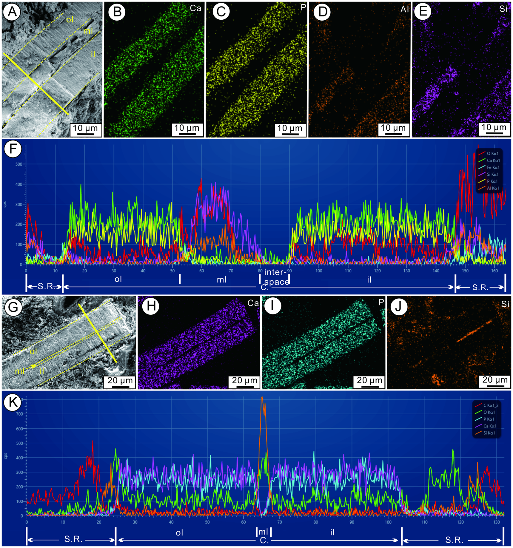
Elemental composition of the natural cross-section and thin-section carapaces by energy dispersive spectroscopy (EDS). A, The natural cross-section of a carapace showing the obvious three-layered ultrastructure. B–E, EDS maps of a natural cross-section of a carapace. F, EDS map of the carapace from the yellow line in A showing the variation in the abundance of different elements in the carapace. G, The thin section of a carapace showing the obvious three-layered ultrastructure. H–J, EDS maps of a thin section of a carapace. K, EDS map of the three-layered ultrastructure of the carapace along the yellow line in G, showing that the elemental composition of the inner and outer lamellae is obviously different from that of the middle lamella. Abbreviations: C., carapace; il, inner lamella; ml, middle lamella; ol, outer lamella; S.R., surrounding rock.

The carapace valves are typically flattened and jointed. Before sediment compaction, the carapace valves may undergo partial enrolling and folding, such as folding at the posterior ventral margin of the carapace (Fig. 2I) or complete spiral enrolling (Fig. 4B–F). Chlupáč (Reference Chlupáč1970) discovered that the carapace valves of caryocaridids can be enrolled with either the dorsal or ventral line as the axis, resulting in two types of enrollment: the ventral border is either inside the spiral with the dorsal line remaining outside, or the ventral border remains outside while the dorsal line is enrolled inside the spiral. However, these two types are challenging to observe and distinguish in actual specimens. In our study's thin-sectioned specimens, the longitudinal section of the carapace valves provides a trace of evidence that the carapace begins to roll along the dorsal hinge line from the ventral margin (Fig. 5B,C), which belongs to the first enrolled type. Racheboeuf et al. (Reference Racheboeuf, Vannier and Ortega2000) proposed that the secondary enrollment of the carapace valves in caryocaridids may be linked to the mechanical properties and architecture of the exoskeleton. Specifically, the thin and flimsy carapace with a high L:H ratio is more susceptible to enrolling. Secondary enrolling of carapace valves typically occurs after the organism's death or molting, but before compaction and fossilization (Chlupáč Reference Chlupáč1970). According to Racheboeuf et al. (Reference Racheboeuf, Vannier and Ortega2000), the exuviae or free carapaces are more susceptible to enrolling than the carapaces after natural death. This is because the soft parts of the carapaces in the latter are still attached to the exoskeleton, which act as a mechanical hindrance to the process of enrolling.
Cuticular Ultrastructure of the Carapace
The carapace valves of caryocaridids typically exhibit distinct fine lines in thin sections (Fig. 5A). Some of these lines may be bent or flexed, and in some cases, the valves may even fracture along these lines (Fig. 5D,E). Churkin (Reference Churkin1966) was the first to report on the ultrastructure of the carapace valves in caryocaridids, and he observed that the carapace was primarily composed of three distinct mineralized lamellae (Fig. 5), which resemble sandwiches. The outer (ol) and inner (il) lamellae are of similar or varying thicknesses and have a yellow-brown to orange-brown color under white polarized light (Fig. 5F–I). They also have weak fibrous structures running perpendicular to the carapace valves in some of the well-preserved specimens (Figs. 5F–I, 6A) and are separated by a black or dark-brown middle lamella (ml). The dense middle lamella can vary in thickness and lacks a fibrous structure. It has a distinct boundary with the inner and outer lamellae (Figs. 4K, 5F–K, 6A). This is why some carapaces may have a slight lamination or parting. The ventral margin of the carapace features connected inner and outer lamellae that expand into a rounded cross-section, while the middle lamella does not penetrate them (Figs. 5H,I, and 7I–L). This corresponds to the ventral doublure at the ventral margin of the carapace. In the complete enrolled carapace, the sharp end of the carapace may represent the dorsal hinged line, and two connected left and right carapace valves are enrolled along their ventral margins (Figs. 5B,C, and 7A–D). The thickness of the enrolled carapace may vary significantly in different thin sections, and the carapace valves on either side of the supposed dorsal hinge line have variable and unequal thicknesses (Churkin Reference Churkin1966: plate 65, figs. 4–6; Miranda Reference Miranda2002: fig. 6.7, 6.10; Figs. 4B,C, and 6A–D). According to Churkin (Reference Churkin1966), this phenomenon is due to the compositional division of the layered carapace valves, rather than an original difference between the two carapace valves.
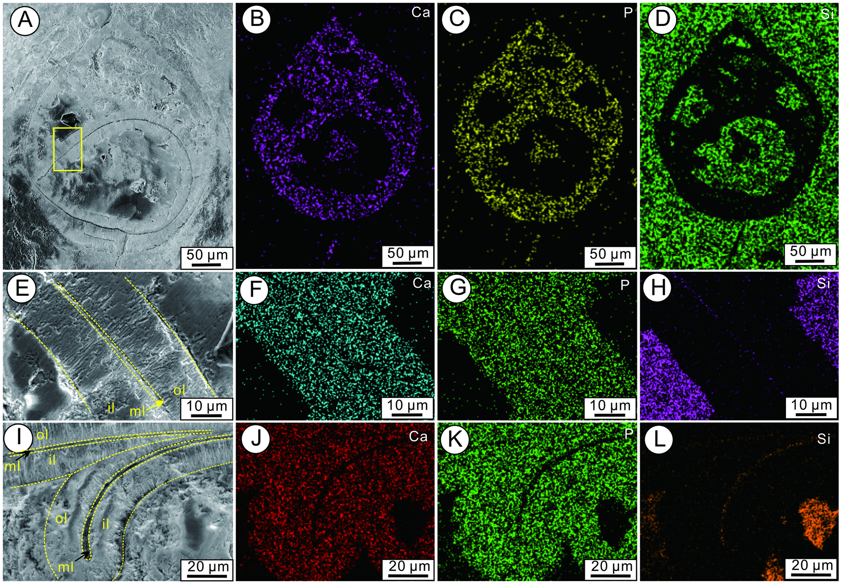
Elemental composition of the carapace by energy dispersive spectroscopy (EDS). A–D, EDS maps of a thin section through an enrolled carapace. E–H, EDS maps of the carapace showing the obvious three-layered ultrastructure of the carapace from the yellow box in A. I–L, EDS maps of the carapace showing the closed ventral margin of a carapace. Abbreviations: il, inner lamella; ml, middle lamella; ol, outer lamella.

In addition to the three mineralized lamellae observed in the carapace cuticle of the Xinjiang specimens, there is a microstratification structure consisting of three layers in the inner and outer lamellae of the carapace. The outermost first layer (fl) is yellow-brown and 2–3 μm thick, with some weak horizontal laminae (Fig. 5J,K). The second layer (sl) is yellowish and slightly thicker than the outermost layer, measuring about 4 μm (Fig. 5J,K). The thickest layer is the third layer (tl), which measures 11 to 13 μm. It has a clear boundary between the second layer and middle lamella. The third layer is dark brown to black near the second layer and pale yellow near the middle lamella (Fig. 5J,K). Both the second and third layers have weak fibrous structures that run perpendicular to the surface of the carapace valves.
The energy spectrum analysis for the natural cross-section and thin section of the carapaces in S. cedarbergensis reveals that the main elemental composition of the inner and outer lamellae is similar, as both are rich in calcium, phosphorus, and oxygen. However, the composition of the middle lamella is notably different from that of the inner and outer lamellae, with the main elements being silicon, aluminum, and oxygen (Figs. 6, 7). Therefore, the inner and outer lamellae may consist mainly of CaO and P2O5, while the middle lamella may consist mainly of SiO2 and Al2O3. This is consistent with the elemental composition of caryocaridids analyzed in previous studies (Churkin Reference Churkin1966; Miranda Reference Miranda2002; Vannier et al. Reference Vannier, Racheboeuf, Brussa, Williams, Rushton, Servais and Siveter2003), suggesting that the studied carapaces of caryocaridids may be composed of carbonate-fluorapatite (CFA). Miranda (Reference Miranda2002) suggested that the presence of a small amount of silicon and aluminum may be due to clay pollution. However, we found that these elements are consistent with the composition of the middle lamella of the carapace. Therefore, the presence of these components indicates that the cuticle of caryocaridid carapaces contains not only a large amount of phosphorus and calcium, but also a small amount of other elements.
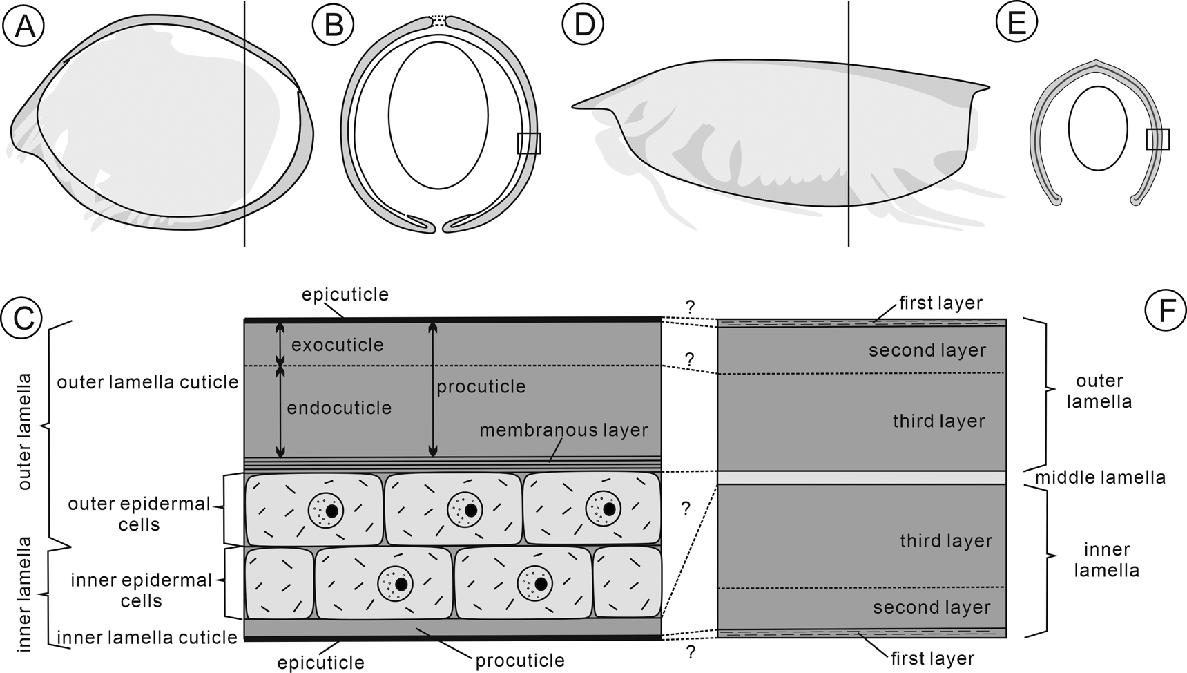
The cuticular structure of adult crustaceans typically consists of four layers: the epicuticle, exocuticle, endocuticle, and membranous layer (Dillaman et al. Reference Dillaman, Roer, Shafer, Modla, Thiel and Watling2013). The epicuticle is the outermost and thinnest layer of the cuticle (Roer and Dillaman Reference Roer and Dillaman1984), while the exocuticle, located beneath the epicuticle, is formed by stacking chitin–protein fibers (Green and Neff Reference Green and Neff1972). The endocuticle, the thickest and most calcified layer of the cuticle (Roer and Dillaman Reference Roer and Dillaman1984), is located beneath the exocuticle. Vannier et al. (Reference Vannier, Racheboeuf, Brussa, Williams, Rushton, Servais and Siveter2003) suggested that the three mineralized lamellae of the carapace valves in caryocaridids may represent the typical three-lamella cuticular structure of living crustaceans. However, the thickness and ultrastructure of the three mineralized lamellae cannot be compared with the cuticles of living and fossil crustaceans (e.g., Pütz and Buchholz Reference Pütz and Buchholz1991; Haj and Feldmann Reference Haj and Feldmann2002). In our study, the cuticular structure of the carapace valves in caryocaridids is mainly divided into three mineralized lamellae: inner, middle, and outer. The inner and outer lamellae can be further divided into three layers from the outside to the inside (Figs. 5J, 8D–E). Recent studies by Šilinger (Reference Šilinger2023) and Kovář et al. (Reference Kovář, Šilinger, Fatka and Brocke2024) have also noted the two distinguishable layers of the caryocaridid cuticle: a white and translucent layer and a mostly opaque brown-black layer. However, the figures show two translucent layers with an opaque brown-black layer stuck on top of the second translucent layer, indicating a three-layer structure (Kovář et al. Reference Kovář, Šilinger, Fatka and Brocke2024: fig. 2Cb, plate 1, figs. 9, 10).
Schematic drawing of the carapace ultrastructures of ostracods and caryocaridids. A, Internal view of right carapace valve of ostracods. B, Transverse section of the black perpendicular lines in A. C, Carapace ultrastructure of the black box in B (modified after Yamada et al. Reference Yamada, Tsukagoshi and Ikeya2004; Yamada Reference Yamada2019). D, Internal view of left carapace valve of caryocaridids. E, Transverse section of the black perpendicular lines in D. F, Carapace ultrastructure of the black box in E.

Wirkner and Richter (Reference Wirkner, Richter, Thiel and Watling2013) presented the thin section of carapace of leptostracan phyllocarids for the first time in the study of crustacean circulation and respiration. However, they did not provide a detailed explanation or discussion of these findings. Nebalia bipes has a carapace cuticular structure consisting of inner and outer lamellae cuticles. The outer lamella cuticle is much thicker than the inner lamella cuticle. Epidermal cells and hemolymph sinuses are present between the two cuticles (Wirkner and Richter Reference Wirkner, Richter, Thiel and Watling2013: fig. 14.11C). However, our current understanding of the carapace cuticle in leptostracans is still limited. For instance, we have yet to study the ultrastructure and function of the cuticle, as well as whether the inner cuticle will calcify in detail.
Extant ostracods have a complex carapace ultrastructure comprising the outer lamella (outer lamella cuticle and outer epidermal cells) and inner lamella (inner lamella cuticle and inner epidermal cells) (Bate and East Reference Bate and East1972, Reference Bate and East1975; Okada Reference Okada1982a,Reference Okadab; Vannier and Abe Reference Vannier and Abe1995; Yamada et al. Reference Yamada, Tsukagoshi and Ikeya2004; Yamada Reference Yamada2019; Fig. 8A–C). The outer lamella cuticle is significantly thicker and more calcified than the inner lamella cuticle. Calcification and thickening of the inner lamella cuticle have been observed in both living and fossil ostracod species (e.g., Olempska Reference Olempska2001, Reference Olempska2008; Yamada Reference Yamada2007; Yamada and Keyser Reference Yamada and Keyser2010). However, Becker and Adamczak (Reference Becker and Adamczak1990) argued that the marginal thickenings found in fossil species are not true calcified inner lamella cuticle, but rather infold-like structures, similar to those of myodocopans. According to Horne et al. (Reference Horne, Cohen and Martens2002), the infold structure is likely equivalent to the calcified inner lamella cuticle of ostracods. Yamada and Keyser (Reference Yamada and Keyser2010) also studied the calcification process of the inner lamella cuticle of living ostracod carapaces and found that the calcification ability of the outer epidermal cells extends to parts of the inner lamella cuticle in the adult stage, resulting in significant calcification of part of the inner lamella cuticle. Therefore, under certain conditions, the inner cuticle of some ostracod species will calcify, forming structures similar to the outer cuticle (Yamada and Keyser Reference Yamada and Keyser2010). The ultrastructure of the carapace in caryocaridids appears to be comparable to that of ostracods (Fig. 8). The outer lamella cuticle of the ostracod carapace corresponds to the outer lamella of the caryocaridid carapace. Similarly, the inner lamella cuticle of ostracods is comparable to the inner lamella of caryocaridid carapace after the inner lamella cuticle has undergone obvious calcification. The epidermal cells of ostracods appear to correspond to the middle lamella of the caryocaridid carapace, but there are significant differences in their thickness. Therefore, the cuticular ultrastructure of caryocaridid and ostracod carapaces is similar, although the inner lamella cuticle mineralization ability of caryocaridids is inferred to be much higher than that of ostracods. This high mineralization ability is necessary for caryocaridids to have their special cuticular ultrastructure (Fig. 8D,E).
Pütz and Buchholz (Reference Pütz and Buchholz1991) examined the cuticle ultrastructure of some pelagic, nektobenthic, and benthic malacostracans. They suggested that certain cuticular characteristics correlate with the lifestyle. For instance, they claimed that the degree of cuticle mineralization and the thickness of the cuticle against body volume decrease from benthic to pelagic animals. Additionally, the thickness of laminae decreases, while the number of laminae in the cuticle increases from benthic to pelagic animals. Barrande (Reference Barrande1872) and Rolfe (Reference Rolfe1962) recorded the cuticle thickness of other phyllocarid fossils. For example, the cuticle thickness of nektobenthic Ceratiocaris papilio and Ceratiocaris scharyi is 30–600 μm and 250 μm, respectively, and benthic Aristozoe regina is more than 1000 μm. The cuticle thickness of caryocaridid carapaces is typically between 15 and 35 μm (Churkin Reference Churkin1966; Miranda Reference Miranda2002; the present study; Figs. 5, 6). However, some species have carapaces as thin as 5–10 μm (Vannier et al. Reference Vannier, Racheboeuf, Brussa, Williams, Rushton, Servais and Siveter2003). Pelagic caryocaridids have a much thinner carapace cuticle compared with benthic Aristozoe and nektobenthic Ceratiocaris. This adaptation is reasonable, because caryocaridids with a light, thin carapace cuticle are better suited for a pelagic lifestyle. When the cuticle of the crustacean is deposited, the chitin–protein fibers change their direction in a helicoidal sequence, resulting in a lamellar appearance in cross-section (Bouligand Reference Bouligand1972; Roer et al. Reference Roer, Abehsera and Sagi2015). However, caryocaridid carapaces lack laminae in their natural cuticle section (Fig. 6A); instead, a weak fibrous structure exists perpendicular to the carapace valves. Vannier et al. (Reference Vannier, Wang and Coen2001: fig. 10.1) found similar patterns in the cuticle of the Devonian ostracod carapace. They claimed that the original lamellar structure of the cuticle is often destroyed and micritized, and then a similar radial ultrastructure is produced. Although caryocaridids lack a clear lamellar structure in their cuticle, similar laminae have been found in the cuticles of their closely related nektobenthic ceratiocaridids (Rolfe Reference Rolfe1962). In Paleozoic oceans, thylacocephalans, a mysterious group of pelagic, bivalved arthropods, coexisted with phyllocarids (e.g., Zatoń et al. Reference Zatoń, Filipiak, Rakociński and Krawczyński2014). Their carapaces also have a laminated ultrastructure with elongated tubular structures piercing through and slightly disturbing the cuticle laminae (Broda et al. Reference Broda, Wolny and Zatoń2015; Broda and Zatoń Reference Broda and Zatoń2017). It is inferred that the cuticle of caryocaridids develops similar laminae, but with thinner layer and a greater number of laminae than ceratiocaridids (Pütz and Buchholz Reference Pütz and Buchholz1991).
The primary function of the carapace in crustaceans is to protect the body and limbs, and this is also true for leptostracan phyllocarids (Olesen Reference Olesen, Thiel and Watling2013). According to Vannier et al. (Reference Vannier, Racheboeuf, Brussa, Williams, Rushton, Servais and Siveter2003), the carapace of caryocaridids was initially chitinous and flexible, while Benson (Reference Benson1975) noted that the marginal infold of ostracods was a means of stabilizing the carapace. Therefore, thickening the endocorneum may enhance the strength, hardness, and stability of the carapace of caryocaridids without adding too much weight, which is an obvious advantage for their pelagic lifestyle. After the carapace of leptostracans is fully developed, the inner and outer cuticles of the middle and posterior of the carapace are completely exposed to the ambient seawater (Manton Reference Manton1934; Newman and Knight Reference Newman and Knight1984; Olesen and Walossek Reference Olesen and Walossek2000). The thickened inner cuticle is preferred to counteract the erosion or damage to the carapace caused by the external environment, in addition to stabilizing the carapace.
In the carapace cuticle of caryocaridids, we observe a unique polygonal reticulate ornamentation in the inner and outer lamellae near the middle lamella, with polygonal diameters typically ranging from 10 to 20 μm (Fig. 4G,H). Such a polygonal fabric is also present in other caryocaridids (Tolmacheva et al. Reference Tolmacheva, Holmer, Popov and Gogin2004; Šilinger Reference Šilinger2023; Kovář et al. Reference Kovář, Šilinger, Fatka and Brocke2024), such as Ceratiocaris (Rolfe Reference Rolfe1962; Šilinger Reference Šilinger2023; Kovář et al. Reference Kovář, Šilinger, Fatka and Brocke2024) and Dictyocaris (Salter Reference Salter1860; Størmer Reference Størmer1935; Gensel et al. Reference Gensel, Johnson and Strother1990). Kovář et al. (Reference Kovář, Šilinger, Fatka and Brocke2024) interpreted this polygonal pattern as a common epicuticular structure of arthropods. However, the carapace surfaces of the caryocaridids in their figures are smooth (Kovář et al. Reference Kovář, Šilinger, Fatka and Brocke2024: plate 1, figs.14–18). The thylacocephalan carapaces also preserve this special polygonal ornamentation (Broda et al. Reference Broda, Wolny and Zatoń2015, Reference Broda, Rak and Hegna2020; Broda and Zatoń Reference Broda and Zatoń2017). In most cases, it is distributed on the surface of the carapace to strengthen the thin carapace (Vannier et al. Reference Vannier, Caron, Yuan, Briggs, Collins, Zhao and Zhu2007; Broda and Zatoń Reference Broda and Zatoń2017). Rolfe (Reference Rolfe1962) found a similar polygonal fabric in his study of the cuticle of Ceratiocaris, which he interpreted as being similar to the prism structure of cuticles in living crustaceans. Modern hexagonal prisms are thought to correspond to the underlying epidermal cell outlines (Dillaman et al. Reference Dillaman, Roer, Shafer, Modla, Thiel and Watling2013) and are composed of the chitin–protein complex forming the cuticle (Rolfe Reference Rolfe1962). The prismatic size of the carapace cuticle of crustaceans is typically between 4 and 15 μm, and caryocaridids typically have individual polygonal diameters of 10–20 μm. However, the individual polygon in Ceratiocaris is 52–270 μm, and as great as 800–1000 μm in Dictyocaris (Størmer Reference Størmer1935; Rolfe Reference Rolfe1962; Šilinger Reference Šilinger2023; Kovář et al. Reference Kovář, Šilinger, Fatka and Brocke2024). If such a polygonal reticulate structure corresponds to the cuticular prisms of living crustaceans, then the epidermal cells of phyllocarids will simply be too large. Vannier et al. (Reference Vannier, Williams and Siveter1997b: fig. 4C–G) reported that the carapaces of Dahlella and Nebalia have the obvious hemolymph sinuses consisting of small polygons connected to each other, forming a branching network, and the anterior and posterior margins of the carapace are not consistent. The anterior carapace of Dictyocaris has a less-developed reticular structure compared with the posterior (Salter Reference Salter1860), suggesting that the development of this structure is not uniform throughout the carapace. The hemolymph sinuses are spaces between the inner and outer cuticle that guide hemolymph flow. Each space is connected to the others and blocked by the supporting epidermis formed by the interconnecting inner and outer epidermal cells (Talbot et al. Reference Talbot, Clark and Lawrence1972; Wirkner and Richter Reference Wirkner, Richter, Thiel and Watling2013). The polygonal reticulate structure of the archaeostracans may correspond to the hemolymph sinuses of the leptostracans in shape and size (Vannier et al. Reference Vannier, Williams and Siveter1997b; Wirkner and Richter Reference Wirkner, Richter, Thiel and Watling2013). The uneven distribution of inner and outer epidermal cells is caused by the presence of hemolymph sinuses. The thickness of the middle lamella in the carapace cuticle of caryocaridids is irregular, and sometimes even disappears, which may be explained by its comparability to the epidermal cell layer (Fig. 4I). Pelagic arthropods require a higher oxygen supply to enhance and/or sustain their active metabolism for swimming, and the thin cuticle of their carapace allows for better diffusion of oxygen into the hemolymph (Perrier et al. Reference Perrier, Williams and Siveter2015). Caryocaridids have larger hemolymph sinuses in their carapace, which can accommodate more hemolymph and transport more oxygen to sustain pelagic activities.
Conclusion
This study examines the taphonomic types and ultrastructure of the caryocaridid carapace. The taphonomic types of the caryocaridid carapace can be classified into three categories (enrollment, wrinkles, and flattening) based on different compression deformations. Among these, the enrolled type is the rarest worldwide. The cuticle of the carapace in Soomicaris cedarbergensis from the Lower Ordovician at Guozigou, Xinjiang, NW China, consists of three mineralized lamellae (outer, middle, and inner). The outer and inner lamella cuticles of caryocaridids are well calcified by CFA and have weak fibrous structures running perpendicular to the carapace valves. Similar to the exoskeleton of extant crustaceans, the outer and inner lamella cuticles of caryocaridids consist of three cuticular layers: epicuticle, exocuticle, and endocuticle. This special ultrastructure of the caryocaridid carapace appears to be comparable to that of ostracods. The carapace of archaeostracans has a polygonal reticulation structure that corresponds in shape and size to the hemolymph sinuses of leptostracans. The ultrastructure of the carapace of the Ordovician caryocaridids in this study, which includes a thin cuticle, thickened inner lamella cuticle, and large hemolymph sinuses, probably represents an adaptation to a pelagic lifestyle during the Ordovician plankton revolution (Servais et al. Reference Servais, Lehnert, Li, Mullins, Munnecke, Nützel and Vecoli2008, Reference Servais, Perrier, Danelian, Klug, Martin, Munnecke and Nowak2016).
Acknowledgments
We would like to thank S. B. Liu (China University of Geosciences, Wuhan) and X. S. Zhang and W. D. Du (Fuzhou University) for their invaluable assistance during the joint fieldwork in Xinjiang. We thank S. Y. Cao, Q. Y. Xu, and L. R. Tao (China University of Geosciences, Wuhan) and H. Q. Wang (Nanjing Institute of Geology and Paleontology, Chinese Academy of Sciences) for their help in taking some of the photographs. We would like to express our gratitude to V. Kovář (Charles University) for discussing the polygonal reticulate ornamentation of the carapace with us. We also are grateful to T. A. Hegna (SUNY Fredonia) for supplying us with some literature. We greatly appreciate the critical comments and detailed suggestions provided by D. Audo and the two anonymous reviewers, as well as editor M. Hopkins, which have significantly improved the article. This research was funded by the National Natural Science Foundation of China (nos. 42072041, 41902002, 42272014, 41872034) and the Fundamental Research Founds for National University, China University of Geosciences, Wuhan (CUGDCJJ202208).
Competing Interest
The authors declare no competing interests.