




No CrossRef data available.
Article contents
Looking at comorbidity through the glasses of neuroscientific memory research: A brain-network perspective
Published online by Cambridge University Press: 29 June 2010
Abstract
As psychiatric illnesses have correlates in the brain, it is surprising that Cramer et al. make almost no reference to the brain's network character when proposing a network approach to comorbidity of psychiatric diseases. We illustrate how data from combined neuropsychological and functional and structural brain-imaging investigations could inform theoretical models about the role played by overlapping symptoms in the etiology of psychiatric comorbidity and the pathways from one disorder to another.
Information
- Type
- Open Peer Commentary
- Information
- Copyright
- Copyright © Cambridge University Press 2010
References
American Psychiatric Association (APA). (2000) Diagnostic and statistical manual of mental disorders, 4th edition, Text Revision [DSM-IV-TR]. American Psychiatric Association.Google Scholar
Beblo, T. & Herrmann, M. (2000) Neuropsychologische Defizite bei depressiven Störungen [Neuropsychological deficits in depressive disturbances]. Fortschritte der Neurologie Psychiatrie
68:1–11.CrossRefGoogle Scholar
Brand, M., Eggers, C., Reinhold, N., Fujiwara, E., Kessler, J., Heiss, W.-D. & Markowitsch, H. J. (2009) Functional brain imaging in fourteen patients with dissociative amnesia reveals right inferolateral prefrontal hypometabolism. Psychiatry Research: Neuroimaging Section
174:132–39.Google Scholar
Brotman, M. A., Rich, B. A., Guyer, A. E., Lunsford, J. E., Horsey, S. E., Reising, M. M., Thomas, L. A., Fromm, S. J. & Towbin, K. (2010) Amygdala activation during emotion processing of neutral faces in children with severe mood dysregulation versus ADHD or bipolar disorder. American Journal of Psychiatry
167:61–69.CrossRefGoogle ScholarPubMed
Casaccia-Bonnefil, P., Pandozy, G. & Mastronardi, F. (2008) Evaluating epigenetic landmarks in the brain of multiple sclerosis patients: A contribution to the current debate on disease pathogenesis. Progress in Neurobiology
86:368–78.Google Scholar
Catani, M. (2007) From hodology to function. Brain
130:602–605. [Scientific commentary]CrossRefGoogle ScholarPubMed
Diehl, B., Busch, R. M., Duncan, J. S., Piao, Z., Tkach, J. & Luders, H. O. (2008) Abnormalities in diffusion tensor imaging of the uncinate fasciculus relate to reduced memory in temporal lobe epilepsy. Epilepsia
49:1409–18.CrossRefGoogle ScholarPubMed
Ebeling, U. & von Cramon, D. (1992) Topography of the uncinate fascicle and adjacent temporal fiber tracts. Acta Neurochirurgica
115:143–48.CrossRefGoogle ScholarPubMed
Fink, G. R., Markowitsch, H. J., Reinkemeier, M., Bruckbauer, T., Kessler, J. & Heiss, W. D. (1996) Cerebral representation of one's own past: Neural networks involved in autobiographical memory. Journal of Neuroscience
16:4275–82.CrossRefGoogle ScholarPubMed
Fujie, S., Namiki, C., Nishi, H., Yamada, M., Miyata, J., Sakata, D., Sawamoto, N., Fukuyama, H., Hayashi, T. & Murai, T. (2008) The role of the uncinate fasciculus in memory and emotional recognition in amnestic mild cognitive impairment. Dementia and Geriatric Cognitive Disorders
26:432–39.CrossRefGoogle ScholarPubMed
Govindan, R. M., Behen, M. E., Helder, E., Makki, M. I. & Chugani, H. T. (2010) Altered water diffusivity in cortical association tracts in children with early deprivation identified with tract-based spatial statistics. Cerebral Cortex
20:561–69.CrossRefGoogle ScholarPubMed
Hudziak, J. J., Achenbach, T. M., Althoff, R. R. & Pine, D. S. (2007) A dimensional approach to developmental psychopathology. International Journal of Methods in Psychiatric Research
16
(Suppl. 1):S16–23.CrossRefGoogle ScholarPubMed
Lebel, C., Walker, L., Leemans, A., Phillips, L. & Beaulieu, C. (2008) Microstructural maturation of the human brain from childhood to adulthood. NeuroImage
40:1055–55.CrossRefGoogle ScholarPubMed
Lee, P. R. & Fields, R. D. (2009) Regulation of myelin genes implicated in psychiatric disorders by functional activity in axons. Frontiers in Neuroanatomy
3(1):1–8.CrossRefGoogle ScholarPubMed
Levine, B., Black, S. E., Cabeza, R., Sinden, M., McIntosh, A. R., Toth, J. P., Tulving, E. & Stuss, D. T. (1998) Episodic memory and the self in a case of isolated retrograde amnesia. Brain
121:1951–73.CrossRefGoogle Scholar
Maldonado, J. R. & Spiegel, D. (2008) Dissociative disorders. In: The American psychiatric publishing textbook of psychiatry, 5th edition, ed. Hales, R. E., Yudofsky, S. C. & Gabbard, G. O.. American Psychiatric Publishing.Google Scholar
Markowitsch, H. J., Vandekerckhove, M. M. P., Lanfermann, H. & Russ, M. O. (2003) Engagement of lateral and medial prefrontal areas in the ecphory of sad and happy autobiographical memories. Cortex
39:643–65.CrossRefGoogle ScholarPubMed
Paus, T., Keshavan, M. & Giedd, J. N. (2008) Why do many psychiatric disorders emerge during adolescence?
Nature Reviews: Neuroscience
9:947–57.CrossRefGoogle ScholarPubMed
Pessoa, L. (2008) On the relationship between emotion and cognition. Nature Reviews: Neuroscience
9:148–58.CrossRefGoogle Scholar
Phan, K. L., Orlichenko, A., Boyd, E., Angstadt, M., Coccaro, E. F., Liberzon, I. & Arfanakis, K. (2009) Preliminary evidence of white matter abnormality in the uncinate fasciculus in generalized social anxiety disorder. Biological Psychiatry
66:691–94.CrossRefGoogle ScholarPubMed
Rajkowska, G., Miguel-Hidalgo, J. J., Wei, J., Dilley, G., Pittman, S. D., Meltzer, H. Y., Overholser, J. C., Roth, B. L. & Stockmeier, C. A. (1999) Morphometric evidence for neuronal and glial prefrontal cell pathology in major depression. Biological Psychiatry
45:1085–98.CrossRefGoogle ScholarPubMed
Schacter, D. L. & Addis, D. R. (2009) On the nature of medial temporal lobe contributions to the constructive simulation of future events. Philosophical Transactions of the Royal Society of London Series B: Biological Sciences
364:1245–53.CrossRefGoogle Scholar
Sepulcre, J., Masdeu, J. C., Sastre-Garriga, J., Goni, J., Velez-de-Mendizabal, N., Duque, B., Pastor, M. A., Bejarano, B. & Villoslada, P. (2008) Mapping the brain pathways of declarative verbal memory: Evidence from white matter lesions in the living human brain. NeuroImage
42:1237–43.CrossRefGoogle ScholarPubMed
Sharot, R., Riccardi, A. M., Raio, C. M. & Phelps, E. A. (2007) Neural mechanisms mediating optimism bias. Nature
450:102–105.CrossRefGoogle ScholarPubMed
Taylor, W. D., MacFall, J. R., Gerig, G. & Krishnan, R. R. (2007) Structural integrity of the uncinate fasciculus in geriatric depression: Relationship with age of onset. Neuropsychiatric Disease and Treatment
3:669–74.Google ScholarPubMed
Tramoni, E., Aubert-Khalfa, S., Guye, M., Ranjeva, J. P., Felician, O. & Ceccaldi, M. (2009) Hypo-retrieval and hyper-suppression mechanisms in functional amnesia. Neuropsychologia
47:611–24.CrossRefGoogle ScholarPubMed
Valentino, K., Toth, S. L. & Cicchetti, D. (2009) Autobiographical memory functioning among abused, neglected, and nonmaltreated children: The overgeneral memory effect. Journal of Child Psychology and Psychiatry
50:1029–38.CrossRefGoogle ScholarPubMed
Wang, F., Kalmar, J. H., He, Y., Jackowski, M., Chepenik, L. G., Edmiston, E. E., Tie, K., Gong, G., Shah, M. P., Jones, M., Uderman, J., Constable, R. T. & Blumberg, H. P. (2009) Functional and structural connectivity between the perigenual anterior cingulate and amygdala in bipolar disorder. Biological Psychiatry
66:516–21.CrossRefGoogle ScholarPubMed
Williams, J. M. & Scott, J. (1988) Autobiographical memory in depression. Psychological Medicine
18:689–95.CrossRefGoogle ScholarPubMed
Yasmin, H., Nakata, Y., Aoki, S., Abe, O., Sato, N., Nemoto, K., Arima, K., Furuta, N., Uno, M., Hirai, S., Masutani, Y. & Ohtomo, K. (2008) Diffusion abnormalities of the uncinate fasciculus in Alzheimer's disease: Diffusion tensor tract–specific analysis using a new method to measure the core of the tract. Neuroradiology
50:293–99.CrossRefGoogle ScholarPubMed
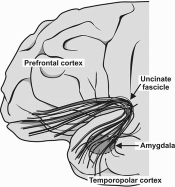
Figure 1. Lateral view of the frontal cortex showing the course of the fasciculus uncinatus.
 You have
Access
- 4
- Cited by
Comorbidity with substantial overlap of symptoms can be found in a number of disease conditions, apart from the generalized anxiety disorder (GAD) and major depressive disorder (MDD) analyzed by Cramer et al. in the target article – for example, between bipolar disorder, attention-deficit hyperactivity disorder (ADHD), and severe mood dysregulation. Furthermore, ADHD is often comorbid with bipolar disorder. “Severe mood dysregulation” is non–DSM-IV-TR terminology (Diagnostic and Statistical Manual of Mental Disorders, 4th edition, Text Revision; American Psychiatric Association 2000) that describes a condition in children which comprises a constellation of symptoms of ADHD, oppositional defiant disorder, and MDD (Hudziak et al. Reference Hudziak, Achenbach, Althoff and Pine2007).
Deficits of face-emotion processing were reported in major depressive disorder, bipolar disorder, severe mood dysregulation, and ADHD. The investigation of face-emotion processing with functional magnetic resonance imaging (fMRI) provided evidence of distinct neural correlates among youth (children and adolescents) with bipolar disorder, ADHD, and severe mood dysregulation (Brotman et al. Reference Brotman, Rich, Guyer, Lunsford, Horsey, Reising, Thomas, Fromm and Towbin2010). The neural (amygdala) activation during face-emotion processing in youths with severe mood dysregulation resembled the neural pattern reported in youths with MDD. This is of particular interest given longitudinal studies indicating that severe mood dysregulation in youths leads to subsequent depressive episodes (Brotman et al. Reference Brotman, Rich, Guyer, Lunsford, Horsey, Reising, Thomas, Fromm and Towbin2010). These results therefore provide an example of how neuropsychological investigations combined with brain imaging may scientifically test the significance of overlapping symptoms for the etiology of comorbidity and may even aid in predicting the progression from one disease to another.
Memory research data could also inform a network conceptualization of comorbidity by offering a perspective that is grounded in evidence about brain organization and development and takes into account variables such as age, gender, and developmental phase. Though not listed under the DSM-IV-TR (APA 2000) diagnostic symptom criteria, impairments of episodic-autobiographical memory are frequently described in MDD (Beblo & Herrmann Reference Beblo and Herrmann2000; Williams & Scott Reference Williams and Scott1988) or bipolar disorder. Furthermore, several psychiatric disorders (e.g., dissociative amnesia) and neuropsychiatric disorders (e.g., mild cognitive impairment [MCI], Alzheimer's dementia) characterized by alterations of episodic-autobiographical memory can be comorbid with MDD. Also, in stroke conditions, temporal lobe epilepsy, and multiple sclerosis, both MDD and episodic-autobiographical memory deficits could be present.
From a neuroscience perspective, an analysis of the nature of the co-occurrence of memory and emotional disorders includes references not only to genetic polymorphisms, but also to brain mechanisms such as extent of insult (lesion penumbra), shared vasculature, axonal innervations (branching, bifurcation) or brain metabolism (neurotransmitters, enzymatic pathways), and a desynchronization or disconnection of otherwise integrated brain networks. The latter may underlie several psychiatric symptoms, including the co-occurrence of emotional processing and episodic-autobiographical memory impairments, as illustrated further on.
It is accepted that specialization and integration characterize the human brain and that cognition and emotion are integrated through structures with a high degree of connectivity (hubs; Pessoa Reference Pessoa2008). Functional neuroimaging has provided evidence for abnormalities of functional connectivity between spatially distanced brain areas, underlying several psychiatric symptoms. Recently, the combination of functional and newer structural imaging techniques (diffusion tensor imaging) began to unearth (micro-)structural correlates for various functional connectivity dysfunctions, in particular white matter (long-range fiber tracts) abnormalities (Catani Reference Catani2007; Paus et al. Reference Paus, Keshavan and Giedd2008). Evidence for white matter changes in several psychiatric disorders comes also from genetic and anatomo-pathological research. Postmortem studies of patients with MDD revealed glial cell loss (Rajkowska et al. Reference Rajkowska, Miguel-Hidalgo, Wei, Dilley, Pittman, Meltzer, Overholser, Roth and Stockmeier1999). Dysfunctions of oligodendrocytes or genes involved in myelination have been reported both in patients with MDD and in those with bipolar disorder (Lee & Fields Reference Lee and Fields2009). Apart from genes, sex hormones are involved in a gender-differentiated modulation of white matter reorganization in adolescence (Paus et al. Reference Paus, Keshavan and Giedd2008). Environment and experience could also exert influences on white matter development, partly via epigenetic regulation of gene expression in myelinating cells (Casaccia-Bonnefil et al. Reference Casaccia-Bonnefil, Pandozy and Mastronardi2008). In children with a history of early deprivation, an overgeneral memory effect (Valentino et al. Reference Valentino, Toth and Cicchetti2009), as well as changes in fiber tracts (including the uncinate fascicle [UF]), have been described (Govindan et al. Reference Govindan, Behen, Helder, Makki and Chugani2010).
The anatomy and function of UF suggest that it may be one pathway of co-occurrence of emotional and memory disorders. UF integrates memory with emotion and links portions of the frontal and temporal lobes. Its ventromedial part connects the amygdala and uncus with the gyrus rectus and the subcallosal area (Ebeling & von Cramon Reference Ebeling and von Cramon1992) (Fig. 1). The UF partly intermingles with the anterior commissure and the inferior occipito-frontal fascicle. It matures later than other connections and may continue its development beyond 30 years (Lebel et al. Reference Lebel, Walker, Leemans, Phillips and Beaulieu2008).
Lateral view of the frontal cortex showing the course of the fasciculus uncinatus.
The ventral right UF is involved in the retrieval of episodic-autobiographical memories, in particular in ecphorizing affect-laden personal events. The UF also belongs to an emotional processing circuitry that connects the amygdala with the orbitofrontal cortex and the anterior cingulate cortex.
The role played by the right UF in the retrieval of episodic-autobiographical memories has been underlined by several studies of memory performance in patients with neurological insults as well as in normal people (Fink et al. Reference Fink, Markowitsch, Reinkemeier, Bruckbauer, Kessler and Heiss1996; Levine et al. Reference Levine, Black, Cabeza, Sinden, McIntosh, Toth, Tulving and Stuss1998). Furthermore, the relevance of the UF for declarative memory in general has received support from many researchers who described a connection between UF structural alteration and memory performance in temporal lobe epilepsy (Diehl et al. Reference Diehl, Busch, Duncan, Piao, Tkach and Luders2008), multiple sclerosis (Sepulcre et al. Reference Sepulcre, Masdeu, Sastre-Garriga, Goni, Velez-de-Mendizabal, Duque, Pastor, Bejarano and Villoslada2008), and Alzheimer's disease (Yasmin et al. Reference Yasmin, Nakata, Aoki, Abe, Sato, Nemoto, Arima, Furuta, Uno, Hirai, Masutani and Ohtomo2008). In their study of amnestic MCI, Fujie et al. (Reference Fujie, Namiki, Nishi, Yamada, Miyata, Sakata, Sawamoto, Fukuyama, Hayashi and Murai2008) reported both memory and emotional recognition impairment together with abnormalities of the UF. Consistent with the proposed role of UF in emotional processing, recent evidence suggests that disruptions in functional fronto-limbic connectivity described in MDD, bipolar disorder, and anxiety disorders may have UF microstructural changes as substrates (Phan et al. Reference Phan, Orlichenko, Boyd, Angstadt, Coccaro, Liberzon and Arfanakis2009; Taylor et al. Reference Taylor, MacFall, Gerig and Krishnan2007; Wang et al. Reference Wang, Kalmar, He, Jackowski, Chepenik, Edmiston, Tie, Gong, Shah, Jones, Uderman, Constable and Blumberg2009).
Altered connectivity between brain areas involved in memory and emotional processing may also underlie the commonly observed comorbidity between dissociative amnesia and MDD (Maldonado & Spiegel Reference Maldonado, Spiegel, Hales, Yudofsky and Gabbard2008). Evidence for functional disconnections between these areas in dissociative amnesia was provided in a study by Brand et al. (Reference Brand, Eggers, Reinhold, Fujiwara, Kessler, Heiss and Markowitsch2009). In this study functional brain imaging performed in a resting state in 14 patients with dissociative amnesia evinced malfunctions of the right temporofrontal regions with a common significant hypometabolic zone in the right inferolateral prefrontal cortex. As Tramoni et al. (Reference Tramoni, Aubert-Khalfa, Guye, Ranjeva, Felician and Ceccaldi2009) have found, these malfunctions may reflect subtle structural right-hemispheric prefrontal white matter abnormalities.
Regarding the directionality of the links between emotional disorders and episodic-autobiographical memory impairments, it seems perhaps intuitive that one pathway flows from emotional disorders to episodic-autobiographical memory impairments via a defect in emotional processing. Memory research, however, provides grounds for an additional, less intuitive pathway, which may run from episodic-autobiographical memory impairments to the emotional disorders. In this regard, recent data emphasize that episodic-autobiographical memory has a significant proscopic function. Neuroimaging studies have revealed that similar networks, which serve episodic-autobiographical memory, are engaged in self-projection and construction of future events (Schacter & Addis Reference Schacter and Addis2009). A main characteristic of patients with MDD is the inability to imagine an optimistic future (Sharot et al. Reference Sharot, Riccardi, Raio and Phelps2007), which may lead them to attempt suicide. It is plausible that this inability may reflect a disruption of the balance between the neural networks that subserve the encoding and retrieval of positive versus negative episodic-autobiographical memories (Markowitsch et al. Reference Markowitsch, Vandekerckhove, Lanfermann and Russ2003).
In conclusion, we agree that shortcomings characterize current psychometric conceptualizations of psychiatric diseases, warranting a more in-depth psychometric thinking and the advent of new conceptualizations. We suggest, however, that the development of a theoretical network approach to psychiatric diseases should rely on an iterative relationship between psychometric analysis and neuroscience, which would enable refinements of the network approach.