Introduction
Foreign accent syndrome (FAS) is a rare medical condition involving speech production abnormalities (Reference Fridriksson, Ryalls, Rorden, Morgan, George and Baylis1). FAS is characterised by the pronouncing of a patient's native language with a foreign or dialectal accent and it is usually generated by a severe brain injury, most often because of a stroke or head trauma at the language-dominant hemisphere. Other uncommon causes include multiple sclerosis, migraine and psychiatric disorders (Reference Fridriksson, Ryalls, Rorden, Morgan, George and Baylis1,Reference Munson and Heilman2). The clinical symptomatology is usually attributed to distorted articulatory planning and coordination processes (Reference Munson and Heilman2). We present the first reported case of a Greek patient with FAS secondary to a left temporal–parietal ischaemic stroke and briefly discuss possible explanations for this phenomenon.
Case report
A 76 year-old right-handed, Greek in origin, male was referred to our Emergency Unit because he had suddenly manifested changes in speech expression. The reports of other patient's relatives were also supportive of this speech disorder. The patient was Greek in origin but he was a permanent Australian resident, living in Australia for the last 50 years. As such, he had completely lost the accent of his native language, i.e. Greek, and acquired an English-Australian accent. Prior to this incident he had a typical English-Australian accent.
The neurological examination revealed that his speech resembled a mixture of Greek and English-Britain accent consistent with FAS, although he had visited only once Greece the last 15 years and never had been to the United Kingdom. Prosody was affected with evidence of mild speech rhythm changes characterised by slowed but regular rate after pauses between words and abnormalities in vowel production. There was no evidence of dysarthria. Writing and reading were normal. There was no evidence of abnormalities with comprehension, finding or repeating words and phrases. Visuo-spatial skills were also normal. Rest of the neurological examination was normal. The patient had no medical history of neurological or psychiatric diseases. He only suffered from mild well-controlled hypertension and hyperlipidaemia.
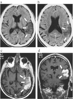
Blood counts and general biochemistry were normal. The initial brain computer tomography (CT) scan, performed at referral, was normal and the patient was admitted as inpatient with a tentative diagnosis of ischaemic stroke. Two days later, a second brain CT scan revealed a hypodense area within the left temporal–parietal region, affecting the white and grey matter, in the vascular territory branch of the left middle cerebral artery (Fig. 1a and b), suggesting a vascular insult. A brain magnetic resonance imaging (MRI) scan confirmed the presence and nature of the aforementioned ischaemic temporal lesion in the language-dominant left hemisphere, affecting the left posterior superior and middle temporal gyri, as well as the ipsilateral inferior supramarginal angular gyrus and posterior insula. The affected area impinged the adjacent left postcentral gyrus (Fig. 1c and d). Electroencephalography, on the awake state, revealed mild non-specific disturbances scattered over the left temporal–parietal area.
(a,b) CT scan. Non-contrast axial brain computer tomography image at the level of thalami (a) and at the level of bodies of the lateral ventricles (b) shows a hypodense area within the left superior temporal lobe, extending to the ipsilateral inferior supramarginal gyrus and angular gyrus (surrounded by the white arrows). (c,d) MRI scan. Axial (c) and coronal (d) post-GD T1W MRI scans, revealing the presence of an ischaemic lesion (surrounded by the white arrows) in the left temporal–parietal lobe, involving cortical and subcortical superior and middle temporal lobe, as well as the ipsilateral supramarginal and angular gyrus and posterior insula. Gadolinium enhancement of the affected areas is indicative of the subacute phase of the infarct.

Discussion
The first case of FAS was reported in 1919 by Pick, whereas to date another 60 cases have also been published (Reference Edwards, Patel and Pople3). However, the exact pathogenesis of FAS still remains vaguely defined. Some authors suggest that FAS results from dysfunction of segmental and prosodic elements of speech, while others have proposed that it is manifested secondary to disrupted motor planning of speech (Reference Edwards, Patel and Pople3–Reference Naidoo, Warriner, Oczkowski, Sévigny and Humphreys5).
Although there are several cases of FAS describing lesions in the frontal and parietal lobes, literature contains only few reports with lesions isolated in a single lobe. Typically, the lesions evoking FAS involve additional sites. Takayama et al. (Reference Takayama, Sugishita, Kido, Ogawa and Akiguchi6) described a case with FAS because of an isolated ischaemic lesion in the left precentral gyrus. We herein describe FAS caused by a stroke affecting cortical and subcortical structures of the left temporal–parietal lobe. The ischaemic lesion involved the left superior and middle temporal gyri, the ipsilateral inferior supramarginal angular gurus and posterior insula, whereas it also came in touch and impinged the left postcentral gyrus.
Concerning the pathogenetic mechanism of this phenomenon in our patient, we suggest that FAS was induced because of interrupted cortical–subcortical feedback pathways (Reference Edwards, Patel and Pople3). In such a case, the phenomenon of subcortical–cortical diaschisis may also be involved. Diaschisis, mainly described in cerebrovascular pathology, is a physiological phenomenon that is characterised by a reversible depression of functions anatomically or functionally associated to the damaged area (Reference Reggia7). Alternatively, we might assume that deficits in mirror neuron system of the primary somatosensory cortex because of affectation of the postcentral gyrus might have contributed to the clinical manifestation of FAS (Reference Mottonen, Jarvelainen, Sams and Hari8). In any case, further functional neuroimaging studies are warranted to elucidate the pathogenesis underlying FAS.