

Introduction
Non-syndromic craniosynostosis is defined as a premature fusion of one or more cranial sutures, causing cranial deformity and growth restriction (Governale, Reference Governale2015), while syndromic craniosynostosis is part of a systemic disorder and can involve several sutures, and is associated with other extra cranial anomalies, such as syndactyly, malformations of the heart, trachea and the nervous system (Vraka, Georgis & Nikas, Reference Vraka, Gerogis and Nikas2018). In this work, we specifically focus on non-syndromic craniosinostosis, which has an incidence of 4.3–5.06 cases per 10,000 births (Boulet, Rasmussen & Honein, Reference Boulet, Rasmussen and Honein2008; Singer, Bower, Southall & Goldblatt, Reference Singer, Bower, Southall and Goldblatt1999). There are several types that are defined based on the sutures involved in premature fusion, as well as by the consequent cranial form. The cranial sutures that most frequently undergo premature fusion are the sagittal, coronal, metopic and lambdoid (Fearon, Reference Fearon2014).
Imaging studies have revealed several effects of non-syndromic craniosynostosis on the brain. One study found that 50% of children with non-syndromic craniosynostosis had brain abnormalities, with the most frequent being the presence of extra-axial cerebrospinal fluid and ventriculomegaly (Magge et al., Reference Magge, Magge, Keating, Myseros, Boyajian, Sauerhammer and Oh2014). Another study showed brain abnormalities in 15% of these children, the most common being Chiari malformation (Hukki, Koljonen, Karppinen, Valanne, & Leikola, Reference Hukki, Koljonen, Karppinen, Valanne and Leikola2012). Specifically, children with right unicoronal synostosis have a different cortical and subcortical morphology compared to that of healthy children (Aldridge et al., Reference Aldridge, Kane, Marsh, Panchal, Boyadjiev, Yan and Richtsmeier2005), while anomalies in the connectivity of the posterior frontomedian cortex, anterior cingulate, parietal lobule, precuneus, angular, supramarginal and fusiform gyri have been found in children with sagittal synostosis (Beckett et al., Reference Beckett, Crooks, Lacadie, Wyk, Jou, Steinbacher and Persing2014).
Several studies have found that about 43%–50% of children with non-syndromic craniosynostosis at different ages (ranging from 2 months to 18 years old) have cognitive deficits (Kapp-Simon, Leuroux, Cunningham & Speltz, Reference Kapp-Simon, Leroux, Cunningham and Speltz2005; Magge, Westerveld, Pruzinsky & Persing, Reference Magge, Westerveld, Pruzinsky and Persing2002; Warschausky et al., Reference Warschausky, Angobaldo, Kewman, Buchman, Muraszko and Azengart2005), mainly in language function (Becker et al., Reference Becker, Petersen, Kane, Cradock, Pilgram and Marsh2005; Korpilahti, Saarinen & Hukki, Reference Korpilahti, Saarinen and Hukki2012; Shipster et al., Reference Shipster, Hearst, Somerville, Stackhouse, Hayward and Wade2003; Starr et al., Reference Starr, Collett, Gaither, Kapp-Simon, Cradock, Cunningham and Speltz2012) and academic learning (Chieffo et al., Reference Chieffo, Tamburrini, Massimi, Di Giovanni, Giansanti, Caldarelli and Di Rocco2010; Speltz et al., Reference Speltz, Collett, Wallace, Starr, Cradock, Buono and Kapp-Simon2015). Moreover, several studies have compared the cognitive and motor outcomes between children with non-syndromic craniosynostosis and healthy children. Starr et al. (Reference Starr, Kapp-Simon, Cloonan, Collett, C-radock, Buono and Speltz2007) found that children with non-syndromic craniosynostosis performed significantly worse than controls on pre- and postsurgical neurodevelopment assessments. In presurgical assessment, differences were found in mental and psychomotor development and receptive language; and in postsurgical assessment in psychomotor development, receptive language and language total scores. Starr et al. (Reference Starr, Collett, Gaither, Kapp-Simon, Cradock, Cunningham and Speltz2012) reported that in a group of 3-year olds with non-syndromic synostosis, mean scores were significantly lower in motor, mental and language developmental assessment than in controls, and the odds of children with non-syndromic craniosynostosis being in the delayed range was 1.5 to 2 times higher compared to controls. At school age, Kapp-Simon et al. (Reference Kapp-Simon, Wallace, Collett, Cradock, Crerand and Speltz2016) found statistically significant differences between controls and non-syndromic craniosynostosis children, specifically non-syndromic craniosynostosis children obtained lower scores in language (Wechsler Intelligence Scale for Children, Fourth Edition Verbal Comprehension Index) and memory ability, particularly in immediate and delayed recall, and recall of stories (Kapp-Simon et al., Reference Kapp-Simon, Wallace, Collett, Cradock, Crerand and Speltz2016). Statistically significant lower scores than controls in intelligent quotient (IQ) and academic functioning have also been reported, especially in math computation (Speltz et al., Reference Speltz, Collett, Wallace, Starr, Cradock, Buono and Kapp-Simon2015).
Studies aimed at determining the type of cognitive difference that accompanies each synostosis have yielded conflicting results. In particular, some investigations have found that children with sagittal synostosis demonstrate better cognitive performance than those with other synostosis types (Becker et al., Reference Becker, Petersen, Kane, Cradock, Pilgram and Marsh2005; Cohen et al., Reference Cohen, Cho, Nichols, Simms, Cross and Burstein2004; Korpilahti et al., Reference Korpilahti, Saarinen and Hukki2012; Starr et al., Reference Starr, Kapp-Simon, Cloonan, Collett, C-radock, Buono and Speltz2007, Reference Starr, Collett, Gaither, Kapp-Simon, Cradock, Cunningham and Speltz2012). One study involving 3-year olds found significantly better performance in children with sagittal synostosis than those with other synostosis types (unicoronal, metopic and lambdoid) in articulation and phonological skills and found that children with unicoronal, metopic or lambdoid synostosis had six times higher risk of impaired articulation and phonological skills in comparison with children with sagittal synostosis (Korpilahti et al., Reference Korpilahti, Saarinen and Hukki2012). Additionally, Starr et al. (Reference Starr, Collett, Gaither, Kapp-Simon, Cradock, Cunningham and Speltz2012) ‘found statistically significant differences in cognitive and language development between the different synostosis types, with the sagittal synostosis group outperforming lambdoid, metopic and coronal synostosis groups. In studies involving younger children below 3 years, before the corrective surgery, Cohen et al. (Reference Cohen, Cho, Nichols, Simms, Cross and Burstein2004) also found that those with sagittal synostosis obtained significantly higher scores in the Bayley Mental Development Index than those with unilateral coronal synostosis, suggesting better overall cognitive development, but found no significant differences in the Psychomotor Development Index. In contrast, Starr et al. (Reference Starr, Kapp-Simon, Cloonan, Collett, C-radock, Buono and Speltz2007) found no significant differences between children with sagittal synostosis and those with other synostosis types (metopic, unicoronal and lambdoid) in development inventories that assess cognitive and motor domains.
On the contrary, other reports have mentioned that children with sagittal and right unicoronal synostosis score significantly lower than those with other types of craniosynostosis in the Mental Development Index of Bayley Scales (Kapp-Simon et al., Reference Kapp-Simon, Leroux, Cunningham and Speltz2005), while those with lambdoid synostosis have significantly better cognitive and expressive language development (Speltz et al., Reference Speltz, Kapp-Simon, Collett, Keich, Gaither, Cradock and Cunningham2007). Finally, some studies have found that motor, language and cognitive development does not differ significantly among children with various affected sutures, for example, two studies found that children with sagittal and metopic synostosis did not differ in mental and psychomotor indexes (Da Costa et al., Reference Da Costa, Anderson, Savarirayan, Wrennall, Chong, Holmes and Meara2012, Reference Da Costa, Anderson, Holmes, Lo, Wray, Chong and Meara2013). With regard to children with multisutural craniosynostosis, Da Costa et al. (Reference Da Costa, Anderson, Holmes, Lo, Wray, Chong and Meara2013) reported scores within average range on the Psychomotor and Mental Development indexes of the Bayley Scales in pre- and postsurgical assessments. Additionally, Bellew and Chumas (Reference Bellew and Chumas2015) assessed children aged 8 to 11 years and found that the multisutural craniosynostosis group had a global mean of Verbal and Performance IQ and Full Scale IQ within the normal range, but when examined individually 33% of children had scores within low average and extremely low range in the Full Scale IQ.
There are several factors associated with the adverse cognitive outcomes found in children with non-syndromic craniosynostosis, including structural and functional brain abnormalities (Aldridge et al., Reference Aldridge, Kane, Marsh, Panchal, Boyadjiev, Yan and Richtsmeier2005; Beckett et al., Reference Beckett, Crooks, Lacadie, Wyk, Jou, Steinbacher and Persing2014; Hukki et al., Reference Hukki, Koljonen, Karppinen, Valanne and Leikola2012), type of craniosynostosis (Becker et al. Reference Becker, Petersen, Kane, Cradock, Pilgram and Marsh2005; Korpilahti et al., Reference Korpilahti, Saarinen and Hukki2012), and age at surgery (Arnaud et al., Reference Arnaud, Meneses, Lajeunie, Thorne, Marchac and Renier2002; Patel et al., Reference Patel, Yang, Hashim, Travieso, Terner, Mayes and Persing2014; Shipster et al., Reference Shipster, Hearst, Somerville, Stackhouse, Hayward and Wade2003).
In summary, preschool children with non-syndromic craniosynostosis are at risk of neuropsychological abnormalities, although there is no consensus on which type of synostosis has worse cognitive and psychomotor outcomes. If health professionals have a clear picture of the cognitive and psychomotor profile of each non-syndromic craniosynostosis type, they can provide specialized care to reduce cognitive deficiencies at critical stages of school learning. Thus, the main objective of our research was to analyze whether there are any differences in cognitive and motor performance between different non-syndromic craniosynostosis types, defined in terms of which suture is fused (sagittal, unilateral coronal, lambdoid, metopic or multisuture). The following processes were assessed: language, visuomotor skills, reasoning, processing speed, memory, arithmetic skills, overall IQ and motor skills. As suggested by previous literature, our hypothesis was that children with sagittal synostosis would perform better compared to other types of craniosynostosis.
Methods
Participants
Twenty-seven children diagnosed with non-syndromic craniosynostosis were assessed. The age of the children was between 3 and 5 years and 11 months; all had undergone surgical treatment between the age of 5 to 18 months. They were recruited from the Pediatric Neurosurgery Service at La Raza National Medical Center and were assessed after written informed consent was obtained from their parents. The present study was approved by the Ethics Committee at Iztacala Faculty of Higher Studies, Universidad Nacional Autónoma de México (Approval No.: CE/FESI/102016/1091), and was conducted in concordance with the World Medical Association Declaration of Helsinki.
Inclusion and exclusion criteria
Children with non-syndromic craniosynostosis were included, with the involvement of the following sutures: sagittal, unilateral coronal, lambdoid, metopic or multisuture (bilateral coronal, sagittal and coronal, sagittal and lambdoid). Diagnosis was made by a pediatric neurosurgeon and confirmed using computed tomography. All participants had undergone previous corrective surgery before 18 months of age. Children were excluded from the study if they had additional neurological conditions, developmental disorders, had undergone more than one neurosurgical procedure, or did not complete the assessment.
Assessment instruments
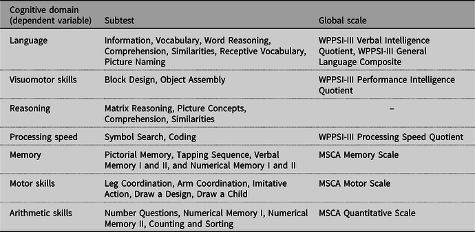
Two neuropsychological batteries were used to measure cognitive (language, visuomotor ability, reasoning, processing speed, memory, arithmetic skills and IQ) and motor variables.
Wechsler Preschool and Primary Scale of Intelligence-Third Edition
The Wechsler Preschool and Primary Scale of Intelligence-Third Edition (WPPSI-III) (Wechsler, Reference Wechsler2011) provides subtest and composite scores that integrate the Verbal, Performance and Processing Speed IQs, as well as the General Language Composite and Full Scale IQ. The test provides Mexican-appropriate norms between the age of 2 years and 6 months to 7 years and 3 months. We used all 14 subtests for children over 4 years and only 5 subtests for children under 4 years (Block Design, Information, Receptive Vocabulary Object Assembly and Picture Naming), according to the test Administration and Scoring Manual. The WPPSI-III subtests have a reliability range from .83 to .95, and the instrument possesses high correlation coefficients with previous versions of the same instrument, such as the WPPSI-R (.70–.86), as well as with the Bayley Scales of Infant Development-II (.80) (Wechsler, Reference Wechsler2011).
McCarthy Scales of Children’s Abilities
Designed for use in children between 2 years and 6 months to 8 years 6 months of age, the McCarthy Scales of Children’s Abilities (MSCA) has been adapted for the Spanish-speaking population and has appropriate normative data (McCarthy, Reference McCarthy1996). It has a reliability coefficient of 0.93. Subtests of the MSCA that integrate the Motor, Memory and Quantitative scales were used to complement the assessment provided by the WPPSI-III. MSCA scores show a low correlation with the overall IQ score of the WPPSI (McCarthy, Reference McCarthy1996), suggesting that functions assessed by both scales are complementary.
Table 1 specifies the subtests and global scales that were used as indicators of each cognitive and motor variable.
Assessment Instruments

.
Procedure
Participants were recruited from the outpatient Pediatric Neurosurgery Clinic. All parents received information regarding the rationale for the study and were asked to complete informed consent forms. Subsequently, they were given an appointment for assessment of the child. First, a structured interview was conducted with the child’s parents to collect clinical, academic, neurodevelopment and socio-economic information; this was complemented and corroborated using the patient’s clinical file. The various tests were applied in another session.
Statistical analyses
For data analysis, children were divided into five groups based on which suture was involved in each case: sagittal, unilateral coronal, metopic, lambdoid and multisuture.
To determine whether there were any relevant clinical or demographic differences between the five groups, the children’s age, sex, age at surgery (months) and parents’ education (low education level: primary or middle school education; high education level; high school or above) were compared. Fisher’s exact test was used to analyze the variables measured using a nominal scale, while all other variables were compared using the Kruskal–Wallis test.
Kruskal–Wallis tests were also used to determine whether cognitive (language, visuomotor skills, reasoning, processing speed, memory, arithmetic skills and IQ) and motor skills differed significantly between the five craniosynostosis groups. Standardized scores were used for all comparisons; for WPPSI Index Scores and IQ, mean = 100 and SD = 15, and for McCarthy Global Scales, mean = 50 and SD = 10. MSCA subtests were not used in the analysis because the instrument does not provide standardized scores for subtests, only for the global scales. When there were significant differences, post hoc analysis was performed using Dunn’s procedure with Bonferroni correction to determine which specific groups differed. Effect size was calculated using epsilon squared.
IBM SPSS Statistics 24, Chicago was used to carry out all data analyses.
Results
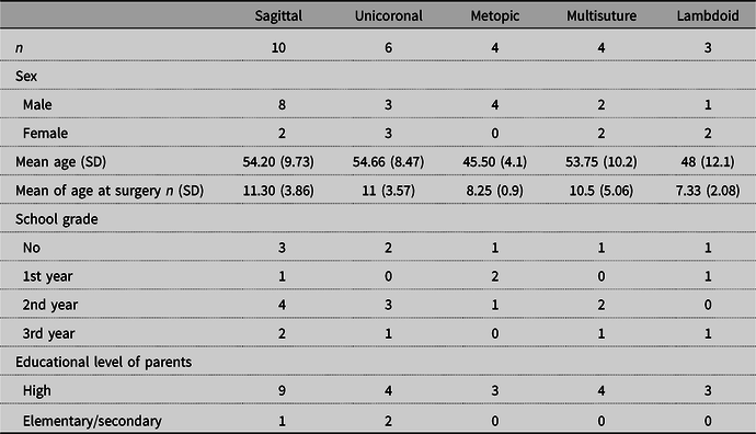
Boys comprised 67% of the sample (18/27), and the most common diagnosis was sagittal synostosis (n = 10), followed by unilateral coronal synostosis (n = 6), metopic (n = 4), lambdoid (n = 3) and multisuture (n = 4: one child with bilateral coronal synostosis, one with sagittal plus coronal and two with sagittal plus lambdoid). Sixty-two percent of the sample had scores within the normal range in all the cognitive domains assessed, according to age-appropriate normative data for the WPPSI and MSCA. The type of synostosis that had higher percentage of children with normal cognitive and motor development was sagittal synostosis.
No significant differences were found among the five synostosis groups in terms of the following sociodemographic variables: age (H(4) = 4.303, p = 0.367), age at surgery (H(4) = 4.028, p = 0.402), years of formal education (H(4) = 1.115, p = 0.892), sex (F = 4.666, p = 0.303) or parents’ education level (F = 3.180, p = 0.504) (Table 2).
Sociodemographic Characteristics of the Craniosynostosis Sample

SD, Standard deviation.
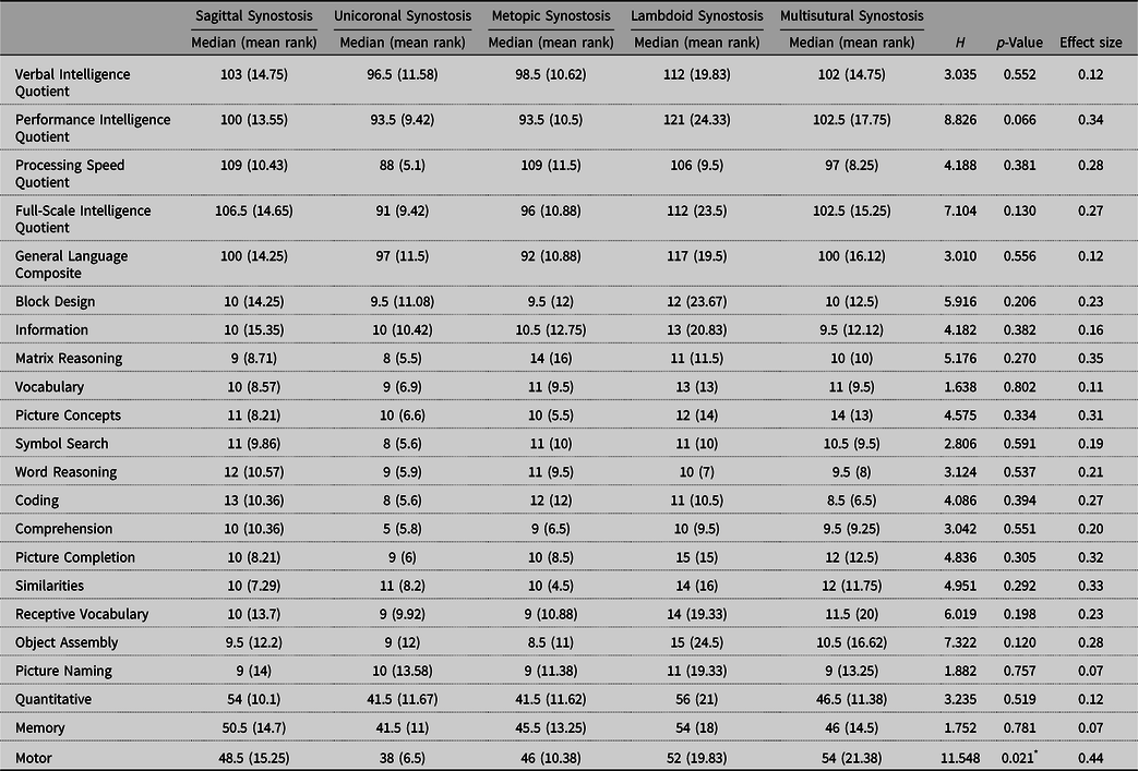
The Kruskal–Wallis test was used to determine whether the five synostosis groups differed in their cognitive or motor performance. Specifically, we compared the standardized scores of WPPSI-III indexes and subtests, as well as Motor, Memory and Quantitative scales of the MSCA. Differences were found between the groups only in the MSCA Motor Scale (Table 3). A post hoc comparison using Dunn’s method with Bonferroni corrections showed significant differences between the unicoronal and multisuture synostosis groups (p = 0.036), with the unicoronal patients presenting lower scores. The effect size was moderate.
Comparison of WPPSI-III and MSCA Subtests and Global Scales Between Craniosynostosis Types

* p < 0.05.
Discussion
The main objective of the present work was to compare the cognitive and motor performance of preschool children with different types of non-syndromic craniosynostosis. Our hypothesis was that children with sagittal synostosis would perform better in comparison with other types of craniosynostosis in cognitive and motor skills.
Despite our hypothesis based on the previous literature, we found that preschool children with sagittal synostosis had no advantage in cognitive or motor abilities compared to the other types of synostosis. The five synostosis groups scored similarly in most domains assessed except the Motor Scale, where we found a significant difference. The post hoc analysis showed specifically that the unilateral coronal synostosis group scored significantly lower than the multisuture synostosis group. Since MSCA does not provide standardized scores for single subtests, but only for global scales, we were not able to analyze and compare whether the differences correspond to fine or gross domains. Several investigations have reported that patients with unicoronal synostosis score worse than those with other types in cognitive and developmental assessments, but not in motor skills (Chieffo et al., Reference Chieffo, Tamburrini, Massimi, Di Giovanni, Giansanti, Caldarelli and Di Rocco2010; Kapp-Simon et al., Reference Kapp-Simon, Leroux, Cunningham and Speltz2005; Speltz et al., Reference Speltz, Kapp-Simon, Collett, Keich, Gaither, Cradock and Cunningham2007). Regarding the psychomotor skills, only one study (Da Costa et al., Reference Da Costa, Anderson, Holmes, Lo, Wray, Chong and Meara2013) found that the mean psychomotor scores were generally lower in children with unicoronal synostosis in pre- and postsurgical neurodevelopment assessments; however, due to the small group size for each type of synostosis, they could statistically analyze only the difference between the two most numerous (metopic and sagittal).
Although explaining the association between unicoronal synostosis and motor development remains a complex subject, some studies that focus on the structural/functional brain abnormalities of these patients could provide certain insights. Aldridge et al. (Reference Aldridge, Kane, Marsh, Panchal, Boyadjiev, Yan and Richtsmeier2005) used functional magnetic resonance imaging to conduct a morphometric analysis in children with right unicoronal synostosis within 6 to 87 weeks, before they underwent corrective surgery. They found that the brain morphology of children with synostosis differed from that in normal children, including structural abnormalities that may be secondary to the fused suture, as well as alterations located in the central brain structures, far from the suture being studied. The abnormalities were reported in the frontal lobes, thalamus and caudate nucleus. Aldridge et al. (Reference Aldridge, Kane, Marsh, Panchal, Boyadjiev, Yan and Richtsmeier2005) hypothesized that some of the morphological changes found in these patients are independent of craniosynostosis and may therefore be permanent, remaining even after surgical correction of the cranial structure. Thus, it follows that these changes may result in developmental anomalies and could be related to our findings to some extent. Another study examined the functional connectivity in five children with right unicoronal synostosis (mean age 11.9 years) who underwent corrective surgery at an early age (Sun et al., Reference Sun, Eilbott, Chuang, Yang, Brooks, Beckett and Persing2019). The results found decreased connectivity in the frontal regions close to the affected suture despite early corrective surgery, including the orbitofrontal cortex and the inferior frontal gyrus, as well as the temporal pole (Sun et al., Reference Sun, Eilbott, Chuang, Yang, Brooks, Beckett and Persing2019). Such frontal functional connectivity abnormalities could be associated with motor development, suggesting that motor abnormalities found in our unicoronal group could also be related to alterations in the frontal functional connectivity.
Although it is still debated, some research suggests that motor development could have an impact on cognitive development. For example, evidence supports that early psychomotor development is associated with verbal, arithmetic and memory skills in 5-year-old children (Osorio-Valencia, Torres-Sanchez, Lopez-Carrillo, Rothenberg & Schanaas, Reference Osorio-Valencia, Torres-Sánchez, López-Carrillo, Rothenberg and Schnaas2018); with visual processing in children between 4 and 11 years (Davis, Pitchford & Limback, Reference Davis, Pitchford and Limback2011); with overall IQ (Heineman, Schendelaar, Van den Heuvel & Hadders-Algra, Reference Heineman, Scendelaar, Van den Heuvel and Hadders-Algra2018; Reference Martin, Tigera, Denckla and MahoneMartin, Tigera, Denckla & Mahone, 2010); with working memory and processing speed capacity in school-aged children (Piek, Dawson, Smith & Gasson, Reference Piek, Dawson, Smith and Gasson2008); and with categorization skills in adults (Murray et al., Reference Murray, Veijola, Moilanen, Miettunen, Glahn, Cannon and Isohanni2006). It follows that early motor deficiencies may be related with the late development of other cognitive domains in children with unicoronal craniosynostosis, as some reports indicate that school-aged children with this type of craniosynostosis have more difficulties than children with other synostosis types in immediate and delayed recall, semantic word generation, verbal comprehension (Kapp-Simon et al., Reference Kapp-Simon, Wallace, Collett, Cradock, Crerand and Speltz2016), math, reading and spelling (Speltz et al., Reference Speltz, Collett, Wallace, Starr, Cradock, Buono and Kapp-Simon2015). Therefore, we suggest that periodical motor skills evaluations should be carried out in preschool children with non-syndromic craniosynostosis.
Most of our sample had normal cognitive and motor development. A relevant contributing factor to this finding could be that the surgery was performed before the first year of age in most of the children. Several researchers have found that early surgical intervention leads to a better cognitive performance (Arnaud et al., Reference Arnaud, Meneses, Lajeunie, Thorne, Marchac and Renier2002; Patel et al., Reference Patel, Yang, Hashim, Travieso, Terner, Mayes and Persing2014; Shipster et al., Reference Shipster, Hearst, Somerville, Stackhouse, Hayward and Wade2003). An explanatory hypothesis poses that early surgery increases intracranial volume and may reduce restraint-related brain injury (Patel et al., Reference Patel, Yang, Hashim, Travieso, Terner, Mayes and Persing2014) and prevent increased intracranial pressure (Neusel et al., Reference Neusel, Class, Eckert, Firsching, Göbel, Götz and Rissmann2018), thus leading to better cognitive outcomes. Despite this idea, other studies have not found a relationship between age at surgery and cognitive development (Da Costa et al., Reference Da Costa, Walters, Savarirayan, Anderson, Wrennall and Meara2006; Mathijssen, Arnaud, Lajeunie, Marchac & Renier, Reference Mathijssen, Arnaud, Lajeunie, Marchac and Renier2006; Starr et al., Reference Starr, Kapp-Simon, Cloonan, Collett, C-radock, Buono and Speltz2007; Thot et al., Reference Thot, Collett, Kapp-Simon, Cloonan, Gaither, Cradock and Speltz2008).
The main limitation of our work is the small number of participants, which limits the generalizability of the results. Higher prevalence of some types of synostosis is in concordance with the epidemiology reported in the literature and is unlikely to result in equivalent size of subtype groups (Boulet et al., Reference Boulet, Rasmussen and Honein2008; Singer et al., Reference Singer, Bower, Southall and Goldblatt1999). Furthermore, the low prevalence of synostosis in the general population makes it difficult to study larger groups.
Previous literature suggests that children with non-syndromic craniosynostosis are at a higher risk of cognitive deficiencies than children with normal development (Chieffo et al., Reference Chieffo, Tamburrini, Massimi, Di Giovanni, Giansanti, Caldarelli and Di Rocco2010; Kapp-Simon et al., Reference Kapp-Simon, Wallace, Collett, Cradock, Crerand and Speltz2016; Starr et al., Reference Starr, Collett, Gaither, Kapp-Simon, Cradock, Cunningham and Speltz2012), so neuropsychological assessment should not be limited to one type of craniosynostosis.
In conclusion, children with unilateral coronal synostosis presented with deficient performance in motor skills in comparison to those with multisuture synostosis. These children should be followed up in the long term to determine whether their present deficiencies evolve into cognitive alterations and learning disabilities at later stages of development as suggested by some studies (Chieffo et al., Reference Chieffo, Tamburrini, Massimi, Di Giovanni, Giansanti, Caldarelli and Di Rocco2010; Magge et al., Reference Magge, Westerveld, Pruzinsky and Persing2002; Speltz et al., Reference Speltz, Collett, Wallace, Starr, Cradock, Buono and Kapp-Simon2015).
Financial Support
Grant number: Dr. Julieta Moreno received a scholarship from the National Council on Science and Technology (CONACyT) for PhD. Number of scholarship: 261233.
Conflicts of Interest
Julieta Moreno-Villagómez has no conflicts of interest to disclose. Guillermina Yáñez-Téllez has no conflicts of interest to disclose. Belén Prieto-Corona has no conflicts of interest to disclose. Ana N. Seubert-Ravelo has no conflicts of interest to disclose. Antonio García has no conflicts of interest to disclose.
Ethical Standards
The authors assert that all procedures contributing to this work comply with the ethical standards of the relevant national and institutional committees on human experimentation and with the Helsinki Declaration of 1975, as revised in 2008.