Introduction
It is well known that spermatozoa of internal fertilization species become quiescent and can be stored in a specific reservoir or specific location of the female reproductive tract for hours to years (Neubaum & Wolfner, Reference Neubaum and Wolfner1999). During the storage period, the sperm that maintain suitability for fertilization are selected and subsequently their motility is re-initiated, activated, or hyperactivated (in the case of mammals), to complete the fertilization of an ovulated egg.
Ca2+ plays a significant role in sperm cells with respect to these changes in the motility state (Jaiswal & Eisenbach, Reference Jaiswal, Eisenbach and Hardy2002; Darszon et al., Reference Darszon, Acevedo, Galindo, Hernández-González, Nishigaki, Trevinõ, Wood and Beltrán2006). The correlation between Ca2+ and sperm selection was noted in a report that suggested that a gene for CatSper, a sperm-specific ion channel responsible for Ca2+ influx in mammalian hyperactivated motility (Ren et al., Reference Ren, Navarro, Perez, Jackson, Hsu, Shi, Tilly and Clapham2001; Ren & Xia, Reference Ren and Xia2010), evolved through positive selection in primates (Podlaha & Zhang, Reference Podlaha and Zhang2003). Therefore, clarification of the gating mechanism of multiple types of sperm Ca2+ channels (Darszon et al, Reference Darszon, Acevedo, Galindo, Hernández-González, Nishigaki, Trevinõ, Wood and Beltrán2006), as well as CatSper, during the process of fertilization is necessary to understand the maintenance of the fertilization potency of the sperm in the female storage sites. However, the existence of an in vivo mechanism to change the motility state is still controversial in internal fertilization, especially in mammals, because it is difficult to know the precise micromilieu that surrounds the sperm at the storage site.
Most urodele amphibians, including the newt Cynops pyrrhogaster, undergo internal fertilization. The sperm of these species are stored in the posterior part of the male reproductive tract, the vas deferens, and is transferred to the female cloaca in the form of a spermatophore, which is degenerated in the cloaca. The sperm released from the spermatophore are kept in a quiescent state in the spermatheca, the distinct sperm reservoir in the cloaca. After reservation, the sperm are directly pushed onto the surface of the eggs, which are released from the ovary and pass through the oviduct (Greven, Reference Greven1998). Fertilization begins on the egg jelly, which is formed by oviduct-secreting matrices that accumulate on the outer surface of the egg (Itoh et al., Reference Itoh, Kamimura, Watanabe and Onitake2002; Takahashi et al., Reference Takahashi, Nakazawa, Watanabe and Onitake2006). In the egg jelly, acrosome reaction-inducing substance (ARIS) is localized in a sheet-like structure that covers the outer surface of the egg jelly, and many granules that contain sperm motility-initiating substance (SMIS) are sequestered under the sheet-like structure (Watanabe et al., Reference Watanabe, Kubo, Takeshima, Nakagawa, Ohta, Kamimura, Takayama-Watanabe, Watanabe and Onitake2010). The localization of ARIS and SMIS provides a unique mechanism for the initiation of sperm motility, which is mediated by the acrosome reaction (AR; Watanabe et al., Reference Watanabe, Kubo, Takeshima, Nakagawa, Ohta, Kamimura, Takayama-Watanabe, Watanabe and Onitake2010). Namely, the sperm derived from the vas deferens in the male reproductive tract and stored in the female reservoir undergo AR and subsequently initiate motility on the surface of the egg jelly.
Extracellular Ca2+ is critical for both ARIS-induced AR and SMIS-induced initiation of sperm motility in C. pyrrhogaster (Watanabe et al., Reference Watanabe, Ito, Watanabe and Onitake2003; Hiyoshi et al., Reference Hiyoshi, Sasaki, Takayama-Watanabe, Takai, Watanabe and Onitake2007). Furthermore, motility initiation occurs sporadically in the presence of the jelly substances (Watanabe & Onitake, Reference Watanabe, Onitake and Sever2003; Watanabe et al., Reference Watanabe, Ito, Watanabe and Onitake2003, Reference Watanabe, Takayama-Watanabe, Vines and Cherr2011) and is delayed in sperm whose intracellular Ca2+ is decreased by a chelating reagent (Watanabe et al., Reference Watanabe, Takayama-Watanabe, Vines and Cherr2011), suggesting that the intracellular Ca2+ level of individual sperm determines the timing of motility initiation.
Difficulties in the precise estimation of the micromilieu around the sperm storage site in females present a substantial obstacle to understanding the in vivo mechanism of changing the motility state through Ca2+ channels in internal fertilization in most animal species, including mammals. In contrast, because the eggs of C. pyrrhogaster can be obtained easily from the posterior portion of the oviduct along with the complete jelly matrix (Takahashi et al., Reference Takahashi, Nakazawa, Watanabe and Onitake2006), the structural features of the egg jelly in the internal fertilization are a useful model for investigating the fertilization potency of the sperm after storage in the female sperm reservoir.
In the present study, investigation of detailed features of the egg jelly surface by scanning electron microscopy (SEM) showed SMIS-localizing granules with small particles on their surfaces just beneath an ARIS-localizing sheet-like structure. Measurement of the Ca2+ concentration using indirectly coupled plasma-optical emission spectrometry (ICP-OES) showed that the ion in the egg jelly was actively maintained at a high level, which may be sufficient for the induction of AR and initiation of sperm motility. These structural and physiological conditions in the egg jelly ensure a sequential process of sperm function, induction of AR and subsequent initiation of motility at fertilization. Furthermore, the effects of several Ca2+ channel blockers suggest that the T-type voltage-dependent Ca2+ channel (VDCC), but not the L-type, may participate in both induction of AR and initiation of sperm motility. Nickel ions only inhibited the initiation of sperm motility. These results suggest that sperm stored in the female are required to sequentially gate distinct Ca2+ channels in response to ARIS and SMIS to participate in fertilization.
Materials and methods
Gametes
The red-bellied newt C. pyrrhogaster was captured in Yamagata, Japan, in late autumn and early spring and kept in hibernation at 4°C in the laboratory. Ovulation of sexually mature females was induced by two injections of gonadotropin (150 IU; Asuka Pharmacy Inc., Tokyo, Japan) at 24-h intervals. Eggs were surgically removed from the posterior portion of the oviduct, called the uterus, at 2–3 days after the final injection. Sperm were obtained from the vasa deferentia. Experimental animals were treated in accordance with the guidelines for proper conduct of animal experiments in Japan.
Egg jelly extract
Eggs were immersed in reconstructed ionic solution (RIS) that contained the same amount of cations as in egg jelly: 24 mM NaCl, 2.7 mM KCl, 0.39 mM MgCl2, 5.1 mM CaCl2, 40 mM choline-Cl, and 10 mM Tris–HCl (pH 8.5) (Ukita et al., Reference Ukita, Itoh, Watanabe and Onitake1999). Following agitation of the egg suspension for 1 h at 4°C, the egg sediment was removed and the remaining solution was centrifuged at 16000 g for 30 min at 4°C. The supernatant was retained as the egg jelly extract (JE). The SMIS activity in the JE was activated by heating at 100°C for 30 min (Mizuno et al., Reference Mizuno, Watanabe and Onitake1999). The JE and heated JE (hJE) were stored at –30°C.
VDCC blockers
Nifedipine (Sigma-Aldrich) and nitrendipine (Sigma-Aldrich) were used as L-type VDCC blockers (Furukawa et al., Reference Furukawa, Nukada, Namiki, Miyashita, Hatsuno, Ueno, Yamakawa and Isshiki2009). Mibefradil (Sigma-Aldrich), CoCl2 (Nacalai-Tesque), and NiCl2 (Nacalai-Tesque) were used as T-type VDCC blockers (Mishra & Hermsmeyer, Reference Mishra and Hermsmeyer1994; Bezprozvanny & Tsien, Reference Bezprozvanny and Tsien1995; Jiménez et al., Reference Jiménez, Bourinet, Leuranguer, Richard, Snutch and Nargeot2000; Son et al., Reference Son, Lee, Lee and Han2000; Díaz et al., Reference Díaz, Bartolo, Delgadillo, Higueldo and Gomora2005). Verapamil (Sigma-Aldrich) and MgCl2 (Nacalai-Tesque) were used as L- and T-type VDCC blockers (Arnoult et al., Reference Arnoult, Villaz and Florman1998; Lopin et al., Reference Lopin, Obejero-Paz and Jones2010). Each blocker in the test solution was prepared at a concentration of 0.1 μM to 1 mM in double-distilled water (mibefradil, CoCl2, NiCl2, verapamil, MgCl2) or dimethyl sulphoxide (DMSO) at less than 0.1% (Nifedipine, nitrendipine).
Scanning electron microscopy
Eggs were fixed in methanol at –80°C overnight and were treated subsequently with methanol at –20°C and 4°C at 1 h intervals. After substitution of methanol with 1-buthanol, they were incubated for 1 h at –20°C. The eggs were freeze dried, cut with a fine blade, and then coated with platinum using a magnetron sputter (JUC-5000; JEOL). The surface of the egg jelly was observed with a scanning electron microscope (JSM-6510 LV and JSM-7400 F, JEOL). The thickness of a sheet-like structure and the diameters of granules and particles were estimated for the specimens, and the size was expressed as the mean ± standard deviation (SD).
Measurement of ion concentration
Three vas deferens fluid samples and nine samples of blood plasma were prepared from independent males. The vas deferens was gently squeezed to obtain seminal fluid, which was centrifuged at 600 g for 5 min at 4°C. The supernatant was designated as vas deferens fluid. Blood was obtained from the inferior vena cava using a fine glass capillary and was then centrifuged at 70 g for 3 min at 4°C. The supernatant was further centrifuged at 7000 g for 3 min at 4°C. The resulting supernatant was designated as blood plasma. These fluids were diluted with 10–100 volumes of double-distilled water. Cation concentrations of the solution were analysed by indirectly coupled plasma-optical emission spectrometry (ICP-OES) (SPS-7000; SEIKO). The pH of the vas deferens fluid was estimated with a pH meter (AS-212, Horiba). To measure the pH of the body fluid, the liver was cut with fine scissors, and the cut surface immediately placed on the electrode of the pH meter.
Acrosome reaction
Sperm were pre-treated for 3 min with each VDCC blocker in the reconstructed vas deferens solution (RVDS; 20 mM NaCl, 6 mM Na2SO4, 1 mM KCl, 0.1 mM Ca(NO3)2, 0.06 mM MgSO4, 10 mM HEPES–NaOH; pH 6.9). The contents are based on the estimated concentrations of cations in the vas deferens fluid (Table 1). The sperm were suspended in JE that contained the blocker at the same concentration and incubated for 5 min at room temperature. For the control, sperm were suspended in JE or RIS. The sperm were fixed in 2.5% glutaraldehyde and observed under a microscope (BX51, Olympus). The rate of induction of the AR in the sperm was evaluated by counting the number of sperm that lost the native acrosome on the tip of the sperm head. Experiments were performed independently, at least three times. More than 100 sperm were observed in each experiment, and the percentages of AR-induced sperm were calculated. Significant differences were estimated by Student's t-test.
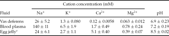
Cation concentrations and pH (mean ± standard deviation) in vas deferens fluid and blood plasma of Cynops pyrrhogaster

a Ukita et al. (Reference Ukita, Itoh, Watanabe and Onitake1999).
Sperm motility
Sperm were pre-treated with each VDCC blocker in RVDS for 3 min. They were suspended in JE or hJE that contained the blocker at the same concentration. For the controls, sperm were suspended in JE or RIS. Sperm motility was observed under a phase-contrast microscope (BX51, Olympus) equipped with a ×10 objective at room temperature and recorded for 10 min on a personal computer with a digital video camera (Moticam 2000; Shimadzu). Sperm with a waving undulating membrane were evaluated as ‘motile sperm’. Experiments were performed independently at least three times. More than 50 sperm were observed in each experiment, and the percentages of motile sperm were calculated. Significant differences were estimated by Student's t-test.
Results
SEM observation
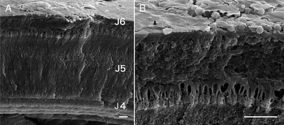
The egg jelly of C. pyrrhogaster is composed of six distinct layers (Okimura et al., Reference Okimura, Watanabe and Onitake2001). In the outermost layer, an ARIS-localizing sheet-like structure covers the outer surface and the SMIS-localizing granules are sequestered under this structure (Watanabe et al., Reference Watanabe, Kubo, Takeshima, Nakagawa, Ohta, Kamimura, Takayama-Watanabe, Watanabe and Onitake2010). These structural feature and localization of the molecules construct the AR-mediated initiation of sperm motility. In the present study, to estimate the structural conditions for the unique sperm–egg interaction in detail, specimens were obtained that maintained fine structures on the egg jelly surface and the detailed features of its components were observed. The sheet-like structure was 0.29 ± 0.16 μm (n = 8) thick and completely covered the outer surface of the egg jelly (Fig. 1). Granules 2.0 ± 0.44 μm (n = 30) in diameter were accumulated beneath the sheet-like structure and in direct contact with the structure (Fig. 2). Many small particles with diameters of 54 ± 14 nm (n = 20) were observed on the surface of the granules. The number of the granules differed throughout the egg jelly surface and a lack of granules was observed occasionally.
Scanning electron microscopy (SEM) observation of the surface of egg jelly of C. pyrrhogaster. (A) Cut surface of the egg jelly. J4–J6 indicate the outer layers. (B) High magnification view of (A). Occasionally, no granules were observed beneath the sheet-like structure (arrow). Bar: 10 μm.

Scanning electron microscopy observation of the sheet-like structure and granules in the surface of the egg jelly. (A–C) View of the egg jelly surface. A few layers of granules are observed beneath the sheet-like structure. (D,E) Field-emission scanning electron microscopy observation of granules. The granule is in direct contact with the sheet-like structure (D). Many particles are observed on the surface of the granules (D, E). Bar: 0.5 μm.

Measurement of ion concentration by ICP-OES
The cation concentrations of the egg jelly of C. pyrrhogaster were previously estimated as 24 mM Na+, 2.7 mM K+, 5.1 mM Ca2+, and 0.39 mM Mg2+ by ICP-OES and determined a pH of 8.5 (Ukita et al., Reference Ukita, Itoh, Watanabe and Onitake1999; Table 1). To determine whether the concentrations of some ions in the egg jelly are critically different from fluids from other tissues, the cation concentrations in blood plasma and vas deferens fluid were also estimated in the current study, as representatives of body fluid and the fluid from the sperm reservoir in the male reproductive tract, using ICP-OES. Vas deferens fluid contained 26 ± 5.2 mM Na+, 1.3 ± 0.080 mM K+, 0.12 ± 0.0058 mM Ca2+ and 0.063 ± 0.012 mM Mg2+; its pH was 6.9 ± 0.23 (Table 1). The cation concentration of the blood plasma was 140 ± 11 mM Na+, 6.5 ± 1.9 mM K+, 1.7 ± 0.49 mM Ca2+ and 0.78 ± 0.24 mM Mg2+ (Table 1). The pH of the cut surface of the liver was 7.2 ± 0.19. The Na+ concentration of samples that contributes to reproduction was lower than that of body fluids such as blood plasma. Notably, Ca2+ in the egg jelly was maintained specifically at a concentration between 3- and 40-fold higher than the vas deferens fluid and blood plasma, and the pH was also higher.
Effects of Ca2+ channel inhibitors on induction of AR
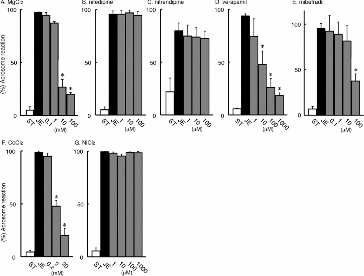
The involvement of the sperm Ca2+ channels in ARIS-induced AR in JE was investigated. Sperm that were pre-treated with 0.1–100 mM Mg2+ in RVDS and then suspended in JE exhibited a decrease in the percentage of sperm undergoing the AR in a dose-dependent manner (Fig. 3A ). However, 97 ± 0.35% of the sperm underwent the AR in JE with RVDS pre-treatment without Mg2+. When sperm that had been pre-treated with RVDS were suspended in RIS as the control, only 5.0 ± 3.1% of the sperm underwent the AR. These data suggest that a Ca2+ channel is involved in the induction of the AR.
Effect of voltage-dependent Ca2+ channel (VDCC) blockers on the induction of the acrosome reaction (AR) in jelly extract (JE). Sperm were pre-treated with a VDCC blocker and suspended in JE that contained the VDCC blocker. At 5 min, they were fixed in glutaraldehyde and observed by microscopy. The AR was evaluated by the absence of the acrosome in the tip of sperm head. Data are the mean ± standard error (SE). White and black columns show percentages of sperm undergoing the AR in RIS and JE, respectively, as controls. Grey columns show those in the JE that contained each VDCC blocker at the indicated concentration. (A) MgCl2. (B) Nifedipine. (C) Nitrendipine. (D) Verapamil. (E) Mibefradil. (F) CoCl2. (G) NiCl2.

When sperm were pre-treated with RVDS that contained 1–100 μM nifedipine and nitrendipine, blockers of the L-type VDCC, and then suspended in JE, most sperm underwent AR, as did the sperm pre-treated with RVDS and suspended in JE, at 96 ± 3.1% and 80 ± 7.4%, respectively (Fig. 3B ,C). Only 5.0 ± 2.6% and 22 ± 13% of the sperm underwent AR in the control, in which sperm were pre-treated with RVDS and suspended in RIS.
Sperm pre-treated with 1 μM–1 mM verapamil, which blocks L-type VDCCs with high affinity and T-type VDCCs at a high concentration (Arnoult et al., Reference Arnoult, Villaz and Florman1998), were suspended in JE. The amount of sperm undergoing AR was decreased in a dose-dependent manner (Fig. 3D ), whereas 94 ± 2.1% sperm underwent AR without the blocker. Only 5.8 ± 0.68% of the sperm underwent AR when the sperm pre-treated with RVDS were suspended in RIS as the control.
In regard to blockers that are effective against T-type VDCCs, when sperm were pre-treated with 0.1–100 μM mibefradil or 0.2–20 mM CoCl2 in RVDS and then suspended in JE, the percentage of sperm that underwent AR was decreased at 100 μM (mibefradil) or in a dose-dependent manner (Co2+) (Fig. 3E ,F), whereas 95 ± 5.5% (mibefradil) or 98 ± 1.2% (Co2+) of the sperm underwent AR without blockers. In the controls for mibefradil and Co2+, sperm were pre-treated with RVDS and then suspended in RIS, and only 6.8 ± 2.9% and 4.4 ± 1.5% of the sperm, respectively, underwent AR. These results suggest that T-type VDCC may be a major candidate of gated VDCCs relating the ARIS-induced AR. Ni2+, which blocks the α1 H subunit of T-type VDCCs (Son et al., Reference Son, Lee, Lee and Han2000), showed no inhibitory effect on AR induction at concentrations of 1 μM–1 mM (Fig. 3G ).
Effects of VDCC blockers on initiation of sperm motility
SMIS is inactivated in the egg jelly by association with other factor(s) contained in the egg jelly of C. pyrrhogaster (Mizuno et al., Reference Mizuno, Watanabe and Onitake1999; Watanabe et al., Reference Watanabe, Kubo, Takeshima, Nakagawa, Ohta, Kamimura, Takayama-Watanabe, Watanabe and Onitake2010). Although the activation process is unknown, SMIS activity appears when JE is heated (Mizuno et al., Reference Mizuno, Watanabe and Onitake1999).
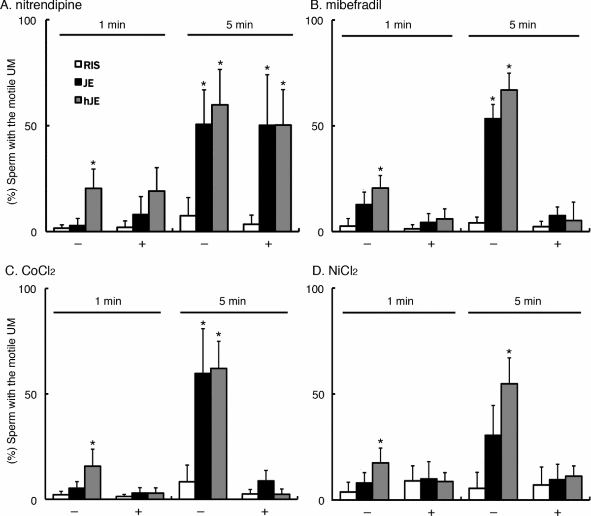
In sperm pre-treated with 100 μM nitrendipine, an L-type VDCC blocker, in RVDS, the percentage of motile sperm in JE, hJE or RIS was low at 1 min and increased significantly by 5 min (P < 0.01) (Fig. 4A ). In the control, when sperm were pre-treated with RVDS, the percentages of motile sperm in JE and hJE were also low at 1 min and increased significantly by 5 min (P < 0.01). These results suggest that L-type VDCCs do not contribute to the initiation of sperm motility by SMIS.
Effect of voltage-dependent Ca2+ channel (VDCC) blockers on initiation of sperm motility in the jelly extract (JE). Sperm were pre-treated with 100 μM nitrendipine (A); 100 μM mibefradil (B); 20 mM CoCl2 (C); or 1 mM NiCl2 (D); and suspended in JE that contained the same VDCC blocker. They were observed by microscopy at 1 and 5 min. Sperm with a waving undulating membrane (UM) were considered to be motile. Data are mean ± standard error (SE).

T-type VDCC blockers inhibited the initiation of sperm motility in both JE and hJE. In sperm pre-treated with 100 μM mibefradil (Fig. 4B ), 20 mM Co2+ (Fig. 4C ), or 100 μM Ni2+ (Fig. 4D ) in RVDS, the percentages of motile sperm in JE and hJE as well as RIS were low for 5 min. In the control, sperm pre-treated with RVDS, the percentages of motile sperm in JE and hJE were low at 1 min and increased significantly by 5 min (P < 0.01). These results suggest that the gated channel involved in the SMIS-induced initiation of sperm motility has a different specificity for Ni+ from that involved in the ARIS-induced AR.
Discussion
Based on the fine structure of the egg jelly surface on which ARIS and SMIS are localized in a specific pattern, it was suggested that initiation of sperm motility is mediated by AR in the internal fertilization of the newt C. pyrrhogaster (Watanabe et al., Reference Watanabe, Kubo, Takeshima, Nakagawa, Ohta, Kamimura, Takayama-Watanabe, Watanabe and Onitake2010). This unique feature is suggested to be specific to the internal fertilization of urodeles among amphibians (Takayama-Watanabe et al., Reference Takayama-Watanabe, Campanella, Kubo and Watanabe2012). More details of the morphology of the egg jelly surface and the contribution of calcium ions and Ca2+ channels with regard to the induction of AR and the initiation of sperm motility were investigated.
Scanning electron microscopy revealed the precise size of a sheet-like structure that covered the outer surface of egg jelly and that granules were attached under the sheet-like structure (Fig. 1). Small particles covered the surface of the granules, indicating that each granule is formed by aggregation of the particles. Because the granules maintained direct contact with the sheet-like structure (Fig. 2), the digestion of the sheet-like structure upon the induction of the AR simply exposes the SMIS in the granules to the inseminated sperm, and the SMIS triggers the subsequent initiation of sperm motility. Occasionally, no granules were observed under the sheet-like structure. In in vitro insemination with sperm collected from the vas deferens, 38% of the sperm did not initiate motility on the surface of the egg jelly (Watanabe unpublished data), whereas most sperm collected from the vas deferens responded to ARIS and SMIS (Ukita et al., Reference Ukita, Itoh, Watanabe and Onitake1999; Hiyoshi et al., Reference Hiyoshi, Sasaki, Takayama-Watanabe, Takai, Watanabe and Onitake2007). The phenomenon of many sperm failing to initiate motility on the surface of egg jelly may, in part, be ascribed to the inhomogeneous distribution of the granules on the surface of the egg jelly.
Extracellular Ca2+ concentrations of at least 5 mM and the presence of external Ca2+ are required for the induction of the AR by ARIS (Hiyoshi et al., Reference Hiyoshi, Sasaki, Takayama-Watanabe, Takai, Watanabe and Onitake2007) and the initiation of sperm motility by SMIS (Watanabe & Onitake, Reference Watanabe, Onitake and Sever2003; Watanabe et al., Reference Watanabe, Ito, Watanabe and Onitake2003), respectively, in the newt C. pyrrhogaster. A high pH is also prerequisite for both events (Watanabe & Onitake, Reference Watanabe, Onitake and Sever2003; Hiyoshi et al., Reference Hiyoshi, Sasaki, Takayama-Watanabe, Takai, Watanabe and Onitake2007). Thus, the 5 mM Ca2+ maintained in egg jelly, which is higher than the concentration in blood plasma and vas deferens fluid (Table 1), is sufficient for the induction of AR as well as the initiation of sperm motility in this species. In anuran amphibians, egg jelly is considered to be a source of Ca2+ for sperm–egg interaction at fertilization in freshwater in which the Ca2+ content is poor (Katagiri, Reference Katagiri1987; Omata, Reference Omata1993). Similarly to external fertilization species, egg jelly in C. pyrrhogaster, which contains a high concentration of Ca2+, enables sperm to interact with an egg at the beginning of internal fertilization. Furthermore, the pH of the egg jelly (Ukita et al., Reference Ukita, Itoh, Watanabe and Onitake1999), which is higher than that in the vas deferens fluid and body fluid (Table 1), may ensure sperm–egg interaction in C. pyrrhogaster. Because egg jelly becomes refractory to penetration by sperm shortly after eggs are spawned in freshwater (Matsuda & Onitake, Reference Matsuda and Onitake1984), sperm should immediately respond to ARIS and SMIS to initiate motility upon contact with the surface of the egg jelly in the cloaca, the site of fertilization in C. pyrrhogaster. The structural and physiological characteristics of the surface of the egg jelly (Figs. 1 and 2 and Table 1) are well suited to fulfil this requirement.
The present study showed that both the induction of the AR and initiation of sperm motility in the sperm of C. pyrrhogaster mostly depend on T-type VDCCs because both events were inhibited by T-type VDCC blockers, which are effective in some vertebrate species (Mishra & Hermsmeyer, Reference Mishra and Hermsmeyer1994; Bezprozvanny & Tsien, Reference Bezprozvanny and Tsien1995; Arnoult et al., Reference Arnoult, Villaz and Florman1998; Jiménez et al., Reference Jiménez, Bourinet, Leuranguer, Richard, Snutch and Nargeot2000; Son et al., Reference Son, Lee, Lee and Han2000; Díaz et al., Reference Díaz, Bartolo, Delgadillo, Higueldo and Gomora2005). Furthermore, it may be possible that CatSper mediates the induction of AR or the initiation of sperm motility in C. pyrrhogaster because mibefradil, which effectively blocks Ca2+ influx through CatSper in mammalian sperm (Strünker et al., Reference Strünker, Goodwin, Brenker, Kashikar, Weyand, Seifert and Kaupp2011), also blocks both events in the newt. Ca2+ influx through the CatSper channel is inevitable for hyperactivated motility and is responsible for the early phase of the increase of intracellular Ca2+ by zona pellucida proteins that can induce the AR (Xia & Ren, Reference Xia and Ren2009). These data suggest that sperm motility or AR may be induced by a signalling event that is homologous to the one that occurs in internal fertilization species. However, because genes for CatSper channel subunits are not found in the genome of the externally fertilizing anuran amphibian, Xenopus (Silurana) tropicalis (Cai & Clapham, Reference Cai and Clapham2008), it is worth examining whether urodele amphibians, most of which undergo internal fertilization, maintain CatSper genes in their genome.
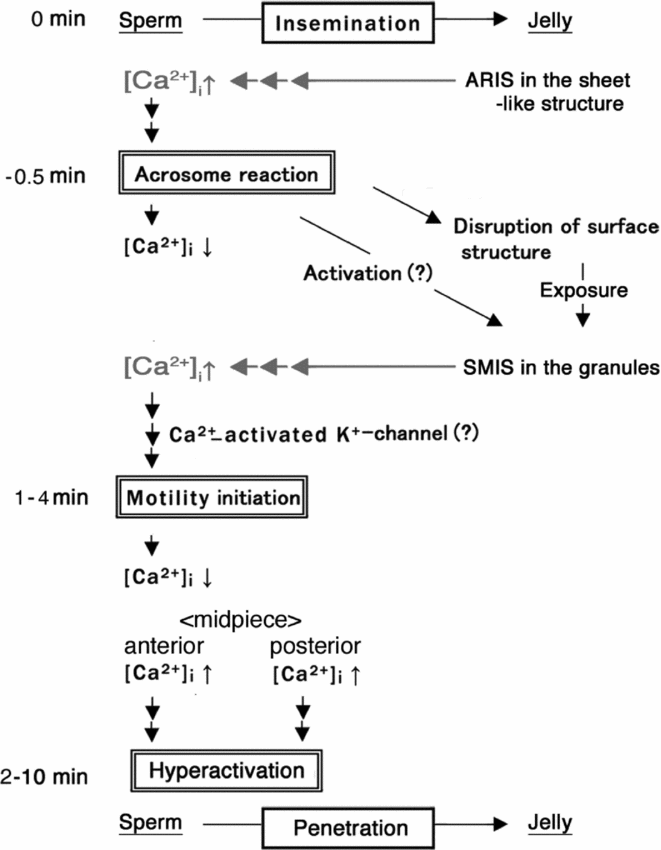
The results of the present study indicate that induction of AR and initiation of sperm motility depend on Ca2+ channels with different dependency on Ni+ (Figs. 3 and 4). The Ni+ is a selective blocker for the α1 H subunit of T-type VDCCs (Lee et al., Reference Lee, Gomora, Cribbs and Perez-Reyes1999; Son et al., Reference Son, Lee, Lee and Han2000), and indicated that sperm of C. pyrrhogaster sequentially gate distinct Ca2+ channels in the AR-mediated initiation of motility. Recently, it has been reported that two different types of VDCCs, possibly L-type and T-type, were gated in the sperm of C. pyrrhogaster to strengthen motility at the anterior and posterior midpiece, respectively, and to enable the sperm to penetrate the jelly matrix (Takahashi et al., Reference Takahashi, Kutsuzawa, Shiba, Takayama-Watanabe, Inaba and Watanabe2013). These data suggest that, during natural fertilization of C. pyrrhogaster, complex control of the intracellular Ca2+ level through multiple types of Ca2+ channels is needed for sperm stored in the female to participate in fertilization. Figure 5 shows the proposed signalling cascade for the initiation of sperm motility mediated by the AR and the subsequent strengthening of motility with reference to changes in the intracellular Ca2+ in the sperm. When sperm stored in the spermatheca are pushed onto the surface of egg jelly, ARIS on the sheet-like structure directs the sperm to gate a Ca2+ channel (Fig. 3). An influx of Ca2+ from egg jelly increases the intracellular Ca2+ level of the sperm, resulting in the induction of AR. After the AR, the intracellular Ca2+ level immediately decreases to basal levels (Watanabe et al., Reference Watanabe, Takayama-Watanabe, Vines and Cherr2011). The sheet-like structure on the surface of the egg jelly is then disrupted, possibly by acrosomal enzymes (Watanabe et al., Reference Watanabe, Kubo, Takeshima, Nakagawa, Ohta, Kamimura, Takayama-Watanabe, Watanabe and Onitake2010), and the SMIS in the granules is exposed and becomes accessible to the sperm. An activation step may be required for SMIS action because the SMIS in egg jelly is largely inactive (Mizuno et al., Reference Mizuno, Watanabe and Onitake1999). SMIS causes the sperm to activate another gated Ca2+ channel (Fig. 4). An influx of Ca2+ from the egg jelly again increases intracellular Ca2+ level of the sperm, resulting in initiation of sperm motility, which may be mediated by a Ca2+-activated K+-channel (Watanabe et al., Reference Watanabe, Ito, Watanabe and Onitake2003). In motile sperm, the intracellular Ca2+ level again immediately decreases to the basal level (Watanabe et al., Reference Watanabe, Takayama-Watanabe, Vines and Cherr2011). The motility of the sperm increases in a time-dependent fashion (Watanabe & Onitake, Reference Watanabe, Onitake and Sever2003). During this change in motility, Ca2+ channels of the sperm distinctly mediate the changes of the motility in the anterior and posterior midpiece, and the Ca2+ influx finally causes a hyperactivation–like higher motility state that allows sperm to penetrate the egg jelly matrix (Takahashi et al., Reference Takahashi, Kutsuzawa, Shiba, Takayama-Watanabe, Inaba and Watanabe2013). In bioassays using sperm collected from the vas deferens, the AR is induced within 30 s by ARIS (Hiyoshi et al., Reference Hiyoshi, Sasaki, Takayama-Watanabe, Takai, Watanabe and Onitake2007), but sperm motility is sporadically initiated within 1–4 min by SMIS (Watanabe et al., Reference Watanabe, Ito, Watanabe and Onitake2003; Watanabe et al, Reference Watanabe, Takayama-Watanabe, Vines and Cherr2011). As a result, the sperm exhibit a higher motility state within 2–10 min (Watanabe & Onitake, Reference Watanabe, Onitake and Sever2003; Takahashi et al., Reference Takahashi, Kutsuzawa, Shiba, Takayama-Watanabe, Inaba and Watanabe2013). The differences in the time course for the initiation and enhancement of motility among sperm may, in part, be ascribed to the control of intracellular Ca2+ levels through multiple channels. Further study of the control of intracellular Ca2+ in the sperm will provide a great deal of information about the mechanism by which sperm stored in the female are prepared for internal fertilization in C. pyrrhogaster.
A working model of sperm–egg interaction on the surface of the egg jelly of Cynops pyrrhogaster. Interaction of newt sperm with egg jelly is proposed based on previous reports. Data from the present study suggest that the acrosome reaction (AR) by AR-inducing substance (ARIS) and initiation of sperm motility by sperm motility-initiating substance (SMIS) (indicated by grey colour) are mediated by gating of distinct Ca2+ channels.

Acknowledgements
We express special thanks to Professor Fumitaka Yanagisawa, Department of Earth and Environmental Sciences, Yamagata University for technical support in performing ICP-OES. We also thank Dr Manabu Ishizaki of Yamagata University for technical support in field-emission SEM. This work was supported by a Grant-in-Aid for Scientific Research (C) 24570246 and (A) 24240062 from the Japan Society for the Promotion of Science and was performed in part as joint research in the Japanese Association for Marine Biology (JAMBIO).