I. INTRODUCTION
Since the discovery of the silicate bioactive glass (BG) designated 45S5 (Bioglass®) by Hench et al. in 1971,Reference Hench, Splinter, Allen and Greenlee1 45S5 and BGs based on the 45S5 composition have been widely researched and used for biomedical applications, such as drug delivery systems, implants for bone repair, and bone cements.Reference Hench2, Reference Hench, Andersson, Hench and Wilson3 Because of their brittleness, BG implants commonly lack the requisite mechanical properties and reliability for repairing load-bearing bones. Metals and alloys, such as stainless steel, cobalt–chromium, and titanium metallic alloys, are the most widely used implants for internal fixation and the repair of loaded bone. However, bacterial infection of these metallic implants is a serious concern,Reference Alta, Bechert, Steinrucke, Wagener and Seidel4 despite rigorous antiseptic surgical procedures and systemic antibiotics.
Previous reports show that the infection rate after implant surgery is in the range 0.5–3% and that chronic infection has been one of the major causes of periprosthetic implant failure.Reference Esposito, Hirsch, Lekholm and Thomsen5–Reference Schierholz, Morsczeck, Brenner, Konig, Yucel and Korenkov7 Currently, to inhibit bacterial activity after implant surgery, high doses of antibacterial agents and long-term antibiotic treatments are commonly used, which may lead to potentially severe side effects and to increasing prevalence of bacterial-resistant strains, such as methicillin-resistant Staphylococcus aureus (MRSA).Reference Eggimann and Pittet8, Reference Harbarth, Samore, Lichtenberg and Carmeli9 Alternative or improved bacterial inhibition techniques are needed to reduce the incidence of infection after implant-related surgery.
BG coatings have been researched and developed for improving the biocompatibility of metallic and ceramic implants and their bonding to bone.Reference Hench2, Reference Hench, Andersson, Hench and Wilson10–Reference Rahaman, Li, Bal and Huang19 Most of the attempts to coat metallic implants with BGs have had limited success, mainly due to poor adhesion of the coating to the metallic substrate.Reference Hench, Andersson, Hench and Wilson10 However, a borate BG glass with a low concentration of SiO2 (designated H12; composition: 8Na2O, 40CaO, 2Al2O3, 8SiO2, 40B2O3, 7.5SiO2, 2.5P2O5; mol%) was biocompatible and to form a strongly adherent coating on a Ti–6Al–4V alloy.Reference Peddi, Brow and Brown18
In addition to the H12 glass described above, several borate-based BGs have been developed and evaluated in recent years for biomedical applications.Reference Liang, Rahaman, Day, Marion, Riley and Mao20–Reference Han and Day23 The replacement of larger amounts of SiO2 in the 45S5 composition with B2O3 resulted in borate-based BG compositions with a higher conversion rate to HAReference Huang, Day, Kittiratanapiboon and Rahaman21 as well as a higher thermal expansion coefficient.Reference Lopez-Esteban, Saiz, Fujino, Oku, Suganuma and Tomsia24 By adjusting the composition, the borate-based BG can have an optimized bioactivity coupled with a thermal expansion coefficient similar to that of titanium. Some borate BGs have been shown to have a greater capability than silicate BGs (such as 45S5 and 13-93) to support bone formation in vivo,Reference Fu, Rahaman, Bal, Kuroki and Brown25 while the addition of SrO has been shown to enhance the osteoinductivity of some borate-based BGs used in the treatment of bone defects.Reference Pan, Zhao, Zhang, Zhang, Li, Li, Lam, Lu, Wang, Huang, Lin and Chang26
An advantage of BGs over other biomaterials is their compositional flexibility. In particular, borate BGs can be doped with elements such as Ag to impart antibacterial properties to the glass.Reference Adams, Santchi and Millencamp27–Reference Silver29 As the glass degrades, Ag+ ions are released at a biologically acceptable rate. Our previous work showed that the addition of 0.75–2 wt% Ag2O to a borate BG did not affect the in vitro bioactivity of the glass (conversion to hydroxyapatite, HA) upon immersion in a simulated body fluid (SBF).Reference Luo, Xiao, Wei, Jia, Zhang, Huang, Jin, Rahaman and Day30 Incubation of mouse MC3T3-E1 osteoblasts and L929 fibroblasts in media containing soluble extracts of the Ag-containing borate BG showed that the effects of the Ag on cell proliferation were concentration dependent. Extracts from the BGs containing 0.75 and 1.0 wt% AgO were not harmful to the cells; in comparison, extracts from the glass containing 2 wt% AgO resulted in a decrease in cell proliferation. However, the antibacterial properties of the Ag-containing borate BGs were not evaluated.
The objective of this work was to evaluate the ability of Ag-containing borate BG coatings to improve the biocompatibility and antibacterial properties of Ti implants in vitro and in vivo. The conversion of the coatings to HA and the release of Ag+ions was studied as a function of immersion time in SBF. The ability of BG-coated Ti implants to simultaneously support fracture fixation and eradication of MRSA-induced infection was evaluated in a rabbit tibial fracture model.
II. MATERIALS AND METHODS
A. Preparation of borate BG coating on Ti
Borate BG (composition: 6Na2O, 8K2O, 8MgO, 16CaO, 2P2O5, 6SrO, 36B2O3, 18SiO2; mol%) doped with 0, 0.75, or 1.0 wt% Ag2O was prepared by melting a mixture of analytical grade Na2CO3, K2CO3, MgCO3, CaCO3, NaH2PO4·2H2O, SrCO3, H3BO3, SiO2, and Ag2O (Sinopharm Chemical Reagent Co., Ltd., Shanghai, China) in a platinum crucible for ∼1 h at 1150 °C. After the melt was quenched between cold stainless steel plates, the glass was crushed and sieved to give particles of size <50 μm. A layer of glass particles with a thickness of ∼100 μm was deposited on medical grade Ti plates (4.0 × 0.5 × 0.2 cm) (TA3; Trauson Medical Instrument Co., Ltd, Jiangsu, China), and the system was placed directly into a furnace preheated to 900 °C. After 3 min, the BG-coated Ti was removed from the furnace and cooled slowly to room temperature in air.
B. Characterization of borate BG coating on Ti
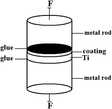
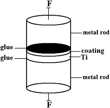
Cross sections of the BG-coated Ti were sputter coated with Au/Pd and examined in a scanning electron microscope (SEM, Hitachi, S2360N, Tokyo, Japan) at an accelerating voltage 15 kV and a working distance of 10 mm. The adhesive strength between the BG coating and the titanium plate was measured using an Instron testing machine (Grove City, PA). Metal rods were glued to the flat surfaces of BG-coated Ti disks (10 mm in diameter × 2 mm in thick) and used to apply a tensile load to the sample (Fig. 1) at an extension rate of 1 mm/min. The adhesive strength was determined as the force at delamination divided by the cross-sectional area of the interface. Six samples were tested for each BG coating, and the results were presented a mean ± standard deviation.
Sample geometry used to determine the interfacial adhesive strength of the BG coating on titanium.
C. Conversion of BG coating to HA in K2HPO4 solution
The BG-coated Ti substrates were immersed for varying times in aqueous K2HPO4 solution (0.02 M; starting pH = 9.0) at 60 °C, and, after drying, the conversion of the coatings to HA was characterized as a function of immersion time using SEM and x-ray diffraction (XRD). SEM (Hitachi S2360N) was used under the conditions described previously to examine the microstructure of the converted layer. For XRD, the converted layer was lightly scraped off the surface of the BG coating and ground into a powder. XRD (D/max 2550VB3+/PC; Rigaku, The Woodlands, TX) was performed using Cu Kα radiation (λ = 0.15406 nm) in a step-scan mode (10°/min) in the range of 10–70° 2θ. The concentration of Ag+ ions released from the glass coating into the K2HPO4solution was measured as a function of immersion time using inductively coupled optical emission spectroscopy (Optima 2100 DV, PerkinElmer, Wellesley, MA).
D. Implantation of BG-coated Ti substrates in a rabbit tibial fracture model
All surgical procedures were designed, approved, and performed by the Animal Care Committee of Shanghai Sixth People’s Hospital, Shanghai Jiaotong University, China, and were conducted in accordance with the NIH guide for care and use of laboratory animals and comply with the national laws on animal experiments. A rabbit tibial fracture model was used to evaluate the ability of the BG-coated Ti implants to support fracture fixation and eradicate MRSA-induced bone infection.
Forty adult female pathogen-free New Zealand White rabbits (average weight = 2.7 ± 0.2 kg) were randomly divided into four groups and used in the experiments. The tibia approximately 0.5 cm below the tibial tubercle was traversed after exposing one side of the tibia and implanted with the four groups of plates; each plate had four matching screws used for fixation. The four groups consisted of Ti without coating (Group 1), coated with BG containing 0 wt% Ag2O (Group 2, designated BG), 0.75 wt% Ag2O (Group 3, designated BG-0.75Ag), or 1.0 wt% Ag2O (Group 4, designated BG-1.0Ag). After the fixation step, osteomyelitis was induced by injecting 0.1 mL of MRSA suspension (1 × 10⁷CFU/mL).
Postimplantation, all animals were monitored daily for general conditions, including activity, body temperature, weight, and appearance of the implant site. Six weeks postimplantation, all the animals were pharmacologically euthanized for autopsy by injecting an intravenous overdose of pentobarbital sodium.
E. Radiographic and histological analyses
Radiographic images of the right tibia of the rabbits were taken before the implantation of the plates, and after implantation times of 1, 3, and 6 wk, to observe healing of the bone infection. At sacrifice (6 wk postimplantation), the excised tibial specimens were fixed in 10% formaldehyde, decalcified in ethylenediaminetetraacetic acid, dehydrated in a graded series of ethanol, and embedded in paraffin. Sections (∼5-μm thick) in the longitudinal direction of the tibia were stained with toluidine blue (TB) and observed using transmitted light microscopy.
F. Microbiological examination
Tissue samples from the infected tibial cavity were collected and kept in sterile tubes for microbiological examination. The samples were inoculated on blood agar (Oxoid Ltd., Basingstoke, U.K.) and incubated for 48 h at 37 °C. The existence of Staphylococcus aureus was identified using the coagulate tube test and the API Staph system (ATB 32 Staph; Bio-Merieux, Marcy-I’Etoile, France).
G. Quantitative microcomputed tomography analysis
The tibial fracture sites with surrounding bone and soft tissue were harvested after 6 wk and tomography images were acquired. The procedures were omitted for they were similar to those described somewhere else.Reference Morgan, Mason, Chien, Pfeiffer, Barnes, Einhorn and Gerstenfeld31, Reference Nyman, Munoz, Jadhav, Mansour, Yoshii, Mundy and Gutierrez32 The region of interest (ROI) was defined 0.5 cm above and below the fracture line. Total callus volume (TV), mineralized callus volume (BV), mineralized callus surface (BS), trabecular number (Tb. N), trabecular separation (Tb. Sp), and trabecular thickness (Tb. Th) were measured for the ROI of each sample.
H. Statistical analysis
Measurements (N = 10) for the ROI of each sample were expressed as a mean ± standard deviation. Analysis for differences between groups was performed using one-way analysis of variance with Tukey’s posthoc test; differences were considered significant for P < 0.05.
III. RESULTS
A. Characterization of borate BG coating on Ti
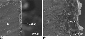
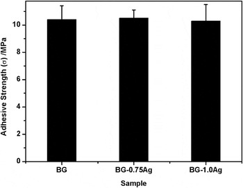
SEM images of the cross sections of BG coatings (1.0 wt% Ag) on Ti substrates showed that the coatings had a uniform thickness of ∼20 μm [Figs. 2(a) and 2(b)]. The coating appeared to be dense and well bonded to the Ti substrate [Fig. 2(b)]; however, there was some pullout of the coating material, which presumably resulted from the sectioning and polishing of the sample [Fig. 2(a)]. The interfacial adhesive strength between the BG coating (0 wt% Ag2O) and the Ti surface was 10 ± 1 MPa; the average adhesive strength did not change significantly with the presence of the Ag2O (0.75 and 1.0 wt%) in the glass (Fig. 3).
SEM images of the cross section of a borate BG coating on a titanium substrate: (a) lower magnification and (b) higher magnification.

The interfacial adhesive strength between titanium plates and borate BG coatings containing 0 wt% Ag2O (BG), 0.75 wt% Ag2O (BG-0.75Ag), and 1.0 wt% Ag2O (BG-1.0Ag). The differences among the groups are not significant.

B. Bioactivity of borate BG coating in vitro
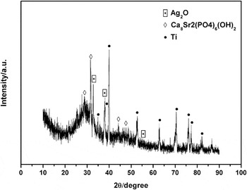
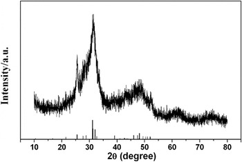
Upon immersion of the BG-coated Ti substrates in K2HPO4 solution, reaction and conversion of the BG coating occurred, starting at the surface of the BG coating and proceeding inward. For an immersion time of 2 wk, XRD of the reaction product showed that the main reflection peaks corresponded to those of a reference HA (JCPDS Card No. 34-0480) (Fig. 4). The broad width of the major peak (31.8° 2θ) is an indication that the HA crystals might be nanosized, partially crystallized, or a combination of both. After removal of the reacted layer, the major XRD peaks of the BG coating (1.0 wt% Ag2O) remaining on the Ti substrate corresponded to those of Ti and Ag2O; the peaks corresponding to a Sr-substituted HA presumably resulted from incomplete removal of the reaction product (Fig. 5) or from an interfacial reaction between the glass and the Ti during the coating step.
XRD pattern of the borate BG coating (1.0 wt% Ag2O) on the Ti substrate after reaction for 2 wk in K2HPO4solution and removal of the reacted layer.

XRD pattern of the reaction products of the borate BG coating (BG-1.0Ag) after immersion in K2HPO4solution (0.02 M) for 2 wk at 37 °C. The pattern of a reference HA (JCPDS Card No. 34-0380) is shown for reference.

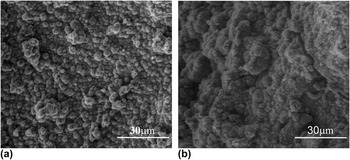
SEM images of the surface of the BG coating (1.0 wt% Ag2O) after immersion for 3 and 7 days in K2HPO4 solution showed that the converted layer consisted of fine particles [Figs. 6(a) and 6(b)], presumably HA as determined previously by XRD. The converted layer became thicker with increasing immersion time as well as more porous and rougher.
SEM images of the surface of the borate BG coating (1.0 wt% Ag2O) after immersion in K2HPO4solution for (a) 3 days and (b) 7 days.

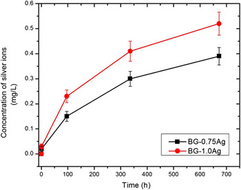
Figure 7 shows the concentration of Ag+ ions dissolved from the BG coating (0.75 and 1.0 wt% Ag2O) into the K2HPO4 solution as a function of immersion time. The two curves have similar shapes, but the Ag+ concentration at any immersion time is higher for the BG coating with the higher Ag2O content. For each glass, the Ag+ concentration increased more rapidly initially but then slowed. The release of Ag+ was still detected after 4 wk, when the experiment was stopped, showing the ability of the BG coating to provide sustained release of Ag+.
Concentration of silver ions released from borate BG coatings containing 0.75 wt% Ag2O (BG-0.75Ag) and 1.0 wt% Ag2O (BG-1.0Ag) into K2HPO4solution at 60 °C as a function of immersion time.

C. Bone repair
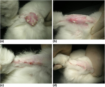
Postimplantation, the animals showed some differences depending on the implant groups (Fig. 8). Of the 10 animals implanted with Ti substrates with no BG coating (Group 1), one animal died at week 1, and radiographs taken at week 3 showed that one animal suffered from internal fixation failure. Six weeks postimplantation, sinuses were found in five animals, while the remaining three animals had significant swelling of the leg.
Photographs of the implant sites in the animals implanted with (a) Ti, (b) Ti coated with BG, (c) Ti coated with BG containing 0.75 wt% Ag2O (BG-0.75Ag), and (d) Ti coated with BG containing 1.0 wt% Ag2O (BG-1.0Ag) 6 wk postimplantation.

All 10 animals implanted with BG-coated Ti implants (BG; Group 2) (0 wt% Ag2O) survived the 6-wk implantation. Radiographs taken at week 3 showed internal fixation failure in one animal. At 6 wk, sinuses were found in four animals and two animals had poor wound healing and various degrees of soft tissue swelling. The remaining three animals recovered well and no sign of systemic complications were evident.
All 10 animals implanted with BG-0.75Ag implants (Group 3) or with BG-1.0Ag implants (Group 4) survived the 6-wk implantation with no internal fixation problems. Except for two animals in Group 3, which showed slightly poor wound healing, all animals in Group 3 and Group 4 showed no sign of systemic complications.
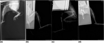
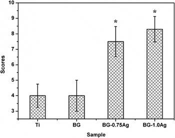
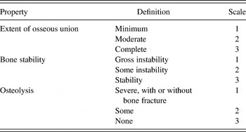
Examples of radiographs taken 1, 3, and 6 wk postimplantation are shown in Fig. 9; for the sake of brevity, the remaining radiographs are not shown. Based on the radiographs, the extent of osseous union, the stability of the bone, and the degree of osteolysis were each subjectively evaluated for each implant according to the scale given in Table I. The individual scores for osseous union, bone stability, and degree of osteolysis were combined and averaged for each implant group (Fig. 10). As shown, Group 1 (Ti) and Group 2 (BG) implants had approximately the same x-ray scores (∼4), indicating a low rate of osseous union, poor bone stability, and a large extent of osteolysis. In comparison, Group 3 (BG-0.75Ag) and Group 4 (BG-1.0Ag) implants had significantly higher x-ray scores (∼7.5–8), indicating a higher rate of osseous union, better bone stability, and less osteolysis.
Radiographs of rabbit tibiae implanted for 3 wk with (a) Ti (Group 1); (b) Ti coated with BG containing 0 wt% Ag2O (Group 2); (c) Ti coated with BG containing 0.75 wt% Ag2O (Group 3); and (d) Ti coated with BG containing 1.0 wt% Ag2O (Group 4).

Combined x-ray scores (osseous union, bone stability, and degree of osteolysis) for four groups of implants (6 wk postimplantation): Group 1: uncoated Ti; Group 2: Ti coated with BG containing 0 wt% Ag2O (BG); Group 3: Ti coated with BG containing 0.75 wt% Ag2O (BG-0.75Ag); Group 4: Ti coated with BG containing 1.0 wt% Ag2O (BG-1.0Ag). (*Significant difference compared to Ti and BG; P < 0.05.)

Scale used in grading radiographs for extent of osseous union, bone stability, and osteolysis in a rabbit tibial fracture model.

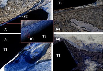
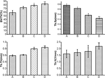
Transmitted light images of TB-stained sections of the implants in rabbit tibiae (implantation time = 6 wk) are shown in Fig. 11. Soft tissue was stained blue, while bone tissue was stained yellow. Under higher magnification of the interface between coating and bone tissue, many inflammatory cells were found in Group 1 and 2 [Figs. 11(a) and 11(b)], in the interface between coating and bone tissue. Animals in Group 4 also showed good contact between the implant and bone; bone trabeculae were arranged densely, and the implant was coated with bone callus. Osteoid tissues were formed on the surface of the implants for Groups 2–4 after the 6-wk implantation, but animals in Group 1 (Ti) showed more severe inflamed tissues, with a low density of bone trabeculae and no osteoid tissue. Accordingly, quantitative micro-computed tomography (micro-CT) analysis results (Fig. 13) also showed that animals in Group 3 and 4 possessed significantly higher BV/TV, Tb. Th, and Tb. N, while significantly lower Tb. Sp than Group 1 and 2, indicating fracture sites treated with Ag-containing BG coatings for 6 wk have a higher rate of osseous union, better bone stability, and less osteolysis.
Transmitted light images of TB-stained sections of rabbit tibiae, after implantation for 6 wk with (a) Ti (Group 1); (b) Ti coated with BG containing 0 wt% Ag2O (Group 2); (c) Ti coated with BG containing 0.75 wt% Ag2O (Group 3); and (d) Ti coated with BG containing 1.0 wt% Ag2O (Group 4); ST, soft tissue; IC, inflammatory cells.

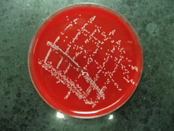
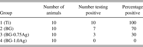
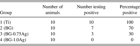
Microbiological investigations showed that all animals in Group 1 (Ti implants) tested positive for MRSA-induced infection (Fig. 12), while 70% of the animals in Group 2 (BG) tested positive. In comparison, only 30% of the animals in Group 3 (BG-0.75Ag) and none of the animals in Group 4 (BG-1.0Ag) tested positive, showing the effectiveness of the Ag-containing BG coatings for controlling MRSA-induced infection (Table II).
Cultivated MRSA from the secretion of a rabbit implanted with uncoated Ti plate (Group 1) for 6 wk.

Comparison of Ti implants coated with BG containing 0.75 wt% Ag2O (BG-0.75Ag) or 1.0 wt% Ag2O (BG-1.0Ag) for controlling MRSA-induced infection after implantation for 6 wk in a rabbit tibial fracture model. Ti implants coated with BG containing no Ag2O and the uncoated Ti implants were used as controls. The number of animals testing positive for MRSA-induced infection after the 6-wk implantation is shown.
IV. DISCUSSION
The Ag-containing borate BG coating used in this work provides an approach for imparting a combination of antibacterial activity and bioactivity to bioinert implants such as Ti used in orthopedic surgery. Titanium implants coated with borate BG containing an optimum amount of Ag (1 wt% Ag2O) showed better ability to support fracture fixation and cure MRSA-induced infection in a rabbit tibial model, when compared to uncoated Ti implants or Ti implants coated with borate BG without Ag.
A benefit of the present approach is the compositional flexibility of the coating material. Since the Ag is incorporated into the glass during the glass-forming step, the desired concentration of Ag can be readily incorporated into the glass. Another advantage is that the Ag is incorporated as ions into the glass structure, instead of a discreet second phase. As a result, the release rate of the Ag is controlled by the conversion rate of the borate BG to HA in the body fluid. Figure 7 shows that the release of Ag from the glass into an aqueous phosphate solution scaled as the initial concentration of Ag in the glass. In the present work, a single glass composition was used. However, our previous work showed the ability to control the conversion rate of borate-based BG by controlling the composition of the glass.Reference Gu, Xiao, Lu, Huang, Rahaman and Wang33 Consequently, by controlling the Ag content and the composition of the borate BG coating, implants with clinically desirable Ag release rates can be created.
The release kinetics of Ag from the coating is an important parameter for bacterial inhibition. The amount of Ag+ released into the K2HPO4 solution increased more rapidly during the early period and more slowly at longer times (Fig. 7). As severe infection often takes place soon after surgery, this rapid release of Ag+ during the early period could be a valuable benefit of the BG-coated implants. The recurrence of infection is most likely to occur within the first 2 wk of surgery.Reference Walenkamp34 After this period of higher risk, a sustained release of Ag ions at the requisite concentration levels makes the coating an ideal material for a bacteria-inhibiting implant surface.Reference Kumar and Munstedt35
The results showed that a dense adherent BG coating with a uniform thickness was formed on Ti plates by depositing a layer of BG particles on the Ti, followed by sintering (Figs. 2 and 3). During the sintering step, the layer of particles formed a dense coating by viscous flow and bonded with the Ti substrate. The adhesive strength of the BG coating on the Ti was 10 ± 1 MPa. Although the optimization of the adhesive strength was not the main focus of the present work, it is likely that higher interfacial strengths could be achieved by controlling the time–temperature schedule and the ambient atmosphere used in the sintering process. As oxidation of the Ti substrate in air can limit the interfacial strength, the sintering step in the present work was carried out rapidly (3 min at 900 °C) to limit oxidation. However, the use of an inert atmosphere (e.g., argon) coupled with an optimized sintering schedule might be expected to improve the adhesive strength. Furthermore, because of the ease of fabrication, the present method can be used to prepare coatings with varying thicknesses and to coat implants with simple or complex shapes.
XRD and SEM showed that the incorporation of Ag did not seriously affect the bioactive potential of the borate BG coating in vitro. The use of a borate BG coating can, in addition, provide attractive properties for implant applications. Because of its high bioactive potential, the borate BG converts rapidly to a HA-type material when immersed in an aqueous phosphate solution, such as the body fluid or K2HPO4 solution. Dissolution of the network modifiers and degradation of the glass network lead to the release of ions such as Ag+, Na+, K+, and BO33+ (or HBO33+) into the solution, while the reaction between Ca2+, Sr2+ ions released from the glass and PO43+ ions present in the solution leads to the formation of HA on the surface of glass.Reference Huang, Rahaman, Day and Li36 Na+, K+, Ca2+, and Sr2+ ions released from the coating are all present naturally in the body. The release of boron is of particular interest; an optimum concentration of boron has been shown to promote tissue healing, whereas above a certain concentration, boron can be toxic to cells.Reference Fu, Rahaman, Bal, Kuroki and Brown25, Reference McCoy, Kenney, Montgomery, Irwin, Williams and Orrell37, Reference Ning, Yao, Wang, Huang, Fu, Liu, Jiang and Zhang38 Our previous work showed that the parent borate BG used in the present investigation was biocompatible, bioactive, and had no adverse effect on cell proliferation in vitro.Reference Luo, Xiao, Wei, Jia, Zhang, Huang, Jin, Rahaman and Day30
The differences in rehabilitation conditions among the four groups of implants (Figs. 9, 10, and 13; Table II) indicate that the bacteriostasis property of the borate BG coating was enhanced by an increasing amount of Ag2O in the glass. Our previous work showed that the same borate BG doped with an optimum amount of Ag2O (0.75–1.0 wt%) enhanced the proliferation of bone marrow-derived stem cells (BMSCs) but a higher amount (2.0 wt%) inhibited proliferation.Reference Luo, Xiao, Wei, Jia, Zhang, Huang, Jin, Rahaman and Day30 Under these conditions, a design criterion for developing Ag-containing borate BG coatings with good antibacterial properties might be an optimum concentration of Ag2O, which will enhance the proliferation of BMSCs in vitro.
Quantitative micro-CT analysis results for four groups of implants (6 wk postimplantation): Group A: uncoated Ti; Group B: Ti coated with BG containing 0 wt% Ag2O (BG); Group C: Ti coated with BG containing 0.75 wt% Ag2O (BG-0.75Ag); Group D: Ti coated with BG containing 1.0 wt% Ag2O (BG-1.0Ag). (*Significant difference compared to Ti and BG; P < 0.05.)

While radiological, histological, and conventional bacterial culture results (Figs. 10, 11, and 13; Table II) supported the efficacy of the coating for infection control, three animals implanted with the BG-0.75Ag group were still detected with MRSA infection. However, the results for the BG-1.0Ag group showed that the borate BG coating containing 1.0 wt% Ag2O cured the infection and supported fracture fixation in all 10 animals within 6 wk of implantation, indicating that 1.0 wt% Ag2O is an optimum concentration for the borate glass used in this work.
V. CONCLUSIONS
Borate BG coatings (∼20-μm thick) containing 0.75 or 1.0 wt% Ag2O improved the biocompatibility and antibacterial properties of Ti implants. The coating adhered strongly to the Ti substrate, with an adhesive strength of 10 ± 1 MPa. Immersion of BG-coated Ti substrates in an aqueous (K2HPO4) resulted in the formation of a HA surface layer within 3 days and sustained release of Ag+ ions for over 4 wk. Titanium implants coated with BG containing 1.0 wt% Ag2O (designated BG-1.0Ag) were most effective for simultaneously supporting fracture fixation and eradicating MRSA-induced infection within 6 wk in a rabbit tibial model. None of the 10 animals implanted with BG-1.0Ag tested positive for MRSA-induced infection; in comparison, all 100% of the animals implanted with uncoated Ti and 70% implanted with BG-coated Ti (without Ag2O) tested positive for the infection. Implants coated with borate BG coatings containing an optimum concentration of Ag could provide an effective approach for reducing implant-related bone infection.
ACKNOWLEDGMENT
This work was supported by the National Natural Science Foundation of China through the project 51072133 and 81201377 and the Shanghai Science Committee through the project 08441900500. Disclosures: Everyone in the author list has no disclosure for this article.