Introduction
The genus Paralecithodendrium Travassos, 1921 (Lecithodendriidae) is cosmopolitan, and its representatives parasitize bats (Chiroptera) worldwide. Among them, Paralecithodendrium longiforme (Bhalerao, Reference Bhalerao1926) and Paralecithodendrium cryptolechitum (Zdzitowiecki, Reference Zdzitowiecki1969) have similar morphological characteristics and can co-occur in European bats, making identification of these digenean species difficult.
Paralecithodendrium longiforme was described by Bhalerao (Reference Bhalerao1926) as Lecithodendrium longiforme from the northern free-tailed bat Mops jobensis (Miller, 1902) in Myanmar (former Burma). This lecithodendriid species is widespread in the Palearctic and has a wide host range among bats (Sharpilo and Iskova Reference Sharpilo and Iskova1989). Paralecithodendrium longiforme is a common parasite of bats, found in many European countries (Cosson and Albalat Reference Cosson and Albalat2007; Dubois Reference Dubois1956; Horvat et al. Reference Horvat, Cabrilo, Paunovic, Karapandza, Jovanovic, Budinski and Bjelic Cabrilo2015, Reference Horvat, Cabrilo, Paunovic, Karapandza, Jovanovic, Budinski and BjelicCabrilo2017; Hurkova Reference Hurkova1963; Kochseder Reference Kochseder1969; Lanza Reference Lanza1999; Matskasi Reference Matskasi1967; Odening Reference Odening1968; Rysavy Reference Rysavy1956; Sharpilo and Iskova Reference Sharpilo and Iskova1989; Shimalov et al. Reference Shimalov, Demyanchik and Demyanchik2002, Reference Shimalov, Demyanchik and Demyanchik2011; Shimalov Reference Shimalov2021; Skvortsov Reference Skvortsov1969; Soltys Reference Soltys1959; Sumer and Yildirimhan Reference Sumer and Yildirimhan2017, Reference Sumer and Yildirimhan2018, Reference Sumer and Yildirimhan2019; Timon-David Reference Timon-David1964; Yanchev and Stoykova Reference Yanchev and Stoykova1973; Zdzitowiecki Reference Zdzitowiecki1969). In Russia, this species has been recorded in bats in the Samara region (Demidova and Vekhnik Reference Demidova and Vekhnik2004; Kirillova et al. Reference Kirillova, Kirillov and Vekhnik2007, Reference Kirillova, Kirillov and Vekhnik2023a, Reference Kirillova, Kirillov, Vekhnik and Ruchin2023b; Kirillov et al. Reference Kirillov, Kirillova and Chikhlyaev2012a, Reference Kirillov, Kirillova and Vekhnik2012b, Reference Kirillova and Kirillov2017; Kirillova and Kirillov Reference Kirillova and Kirillov2017, Reference Kirillova and Kirillov2024), Mordovia (Kirillova et al. Reference Kirillova, Kirillov, Vekhnik and Ruchin2023b; Kirillova and Kirillov Reference Kirillova, Kirillov, Shchenkov, Knyazev and Vekhnik2024; Ruchin et al. Reference Ruchin, Kirillov, Chikhlyaev and Kirillova2016), and the Republic of Karelia (Lebedeva et al. Reference Lebedeva, Belkin, Stanyukovich, Bespyatova and Bugmyrin2020).
Paralecithodendrium cryptolecithum was first described by Zdzitowiecki (Reference Zdzitowiecki1969) as Prosthodendrium cryptolecithum from the pond bat Myotis dasycneme (Boie, 1825) in Poland. However, P. cryptolecithum has not been recorded in Europe since then. Rysavy (Reference Rysavy1956) described Paralecithodendrium magnum ex Rhinolophus ferrumequinum (Schreber, 1774) as Prosthodendrium magnum, which some authors consider a synonym of P. longiforme (Hurkova Reference Hurkova1963; Matskasi Reference Matskasi1967; Sharpilo and Iskova Reference Sharpilo and Iskova1989; Skvortsov Reference Skvortsov1969; Timon-David Reference Timon-David1964). Since only P. longiforme is usually reported for European bat hosts, we assume that such records also include P. cryptolecithum.
In this research, we studied P. cryptolecithum and P. longiforme from bats of the Samara region (Middle Volga region, European Russia). The aim of our study was to provide reliable identification of these two Paralecithodendrium species using morphological and morphometric analyses, to present new molecular phylogenetic data on P. cryptolecithum, and to confirm the validity of this species.
Materials and methods
Trematode collection and examination
Adult Paralecithodendrium trematodes were collected from dead bats in the vicinity of Shiryaevo village (Samara region, European Russia) in December 2023. A total of 10 bats belonging to four species were examined: three individuals each of Myotis dasycneme and Eptesicus nilssoni Keyserling & Blasius, 1839 and two individuals each of Myotis daubentonii (Kuhl, 1817) and Plecotus auritus (Linnaeus), 1758. No animals were intentionally killed for the study; bats died naturally while hibernating in abandoned mine workings.
Only alive, motile, and sexually mature trematodes were collected for the study. Abandoned adits were inspected every 5–6 days for dead bats. Microclimatic conditions in the adits, such as year-round temperatures from +2 to +5°C and high air humidity, contribute to good preservation of both bat corpses and their helminths. For morphological examination, digeneans were recovered from the bat intestines and killed by heating in water. The samples were then stained with aceto-carmine, dehydrated in a series of ethanol (70–96%), cleared in clove oil, and mounted in Canada balsam (Anikanova et al. Reference Anikanova, Bugmyrin and Ieshko2007; Ivashkin et al. Reference Ivashkin, Kontrimavichus and Nazarova1971). For molecular phylogenetic analysis, Paralecithodendrium specimens were fixed in 96% ethanol and stored at +4°C. A total of 49 trematode individuals were studied: 15 specimens of P. cryptolechitum and 34 specimens of P. longiforme.
The drawings were made using a Levenhuk M500 BASE Digital Camera and an RA-7 drawing tube attached to a Pzo-Biolar light microscope. The following morphometric characteristics of trematodes were studied: length and width of the body, oral and ventral suckers, pharynx, pseudocirrus sac, sinistral and dextral testes, ovary and eggs, as well as the ratio of body length to width, width of the oral sucker to ventral suckers, and width of the oral sucker to the pharynx. Egg sizes were calculated based on 5–7 eggs from the distal part of the uterus of each trematode specimen. All measurements are given in millimeters. For comparative analysis, data from previously published works containing drawings and morphometric parameters of P. cryptolechitum and P. longiforme were used. The parasites were identified according to Zdzietowiecki (1969) and Sharpilo and Iskova (Reference Sharpilo and Iskova1989). Voucher specimens of trematodes are stored at the Institute of Ecology of the Volga River Basin of the Russian Academy of Sciences (Togliatti, Russia).
DNA extraction, amplification, sequencing, and phylogenetic analysis
Total DNA was isolated from individual specimens using Chelex-100 with Proteinase-K solution. Primers ‘28sy/28sz’ (Hillis and Dixon Reference Hillis and Dixon1991) and ‘3S/ITS2.2’ (Cribb et al. Reference Cribb, Adlard and Bray1998; Morgan and Blair Reference Morgan and Blair1995) were used to amplify partial 28S rDNA and the ITS2 region, respectively. PCR parameters for the ‘28sy/28sz’ primer pair were used after Kirillova et al. (Reference Kirillova, Kirillov, Shchenkov, Knyazev and Vekhnik2024). For the ITS2 region, the PCR parameters were as follows: initial denaturation at 95°C, 3 min; denaturation 95°C, 30 sec; annealing 60.2°C, 20 sec; extention 72°C, 35 sec; final elongation 72°C, 5 min; 35 cycles at 25 μl in total. Primers JB3 (5`-TTT TTT GGG CAT CCT GAG GTTTAT-3`) and JB4.5 (5` -TAA AGA AAG AAC ATA ATG AAA ATG-3`) (Bowles et al. Reference Bowles, Blair and McManus1992) were used to amplify the partial cox1 gene. The thermal cycler parameters were as follows: initial denaturation at 95°C, 3 min; 35 cycles of denaturation 20s, 95°C; annealing 20s at 48.9°C; elongation 50s at 72°C; final elongation 72°C, 5 min. Amplicons were sequenced by ‘Evrogen’ company (Moscow, Russia).
To estimate the phylogenetic position of Paralecithodendrium specimens, the datasets were processed using a pipeline implemented in the ‘R’ programming language (R core team 2022). The code is available at the GitHub repository: ‘https://github.com/Shchenkov/dirty_seqs’. Multiple sequence alignment, trimming, and model testing are automated in the script using the following libraries: ‘BiocManager’ (Morgan and Ramos Reference Morgan and Ramos2024), ‘BiocGenerics’ (Huber et. al. Reference Huber, Carey, Gentleman, Anders, Carlson, Carvalho, Bravo, Davis, Gatto, Girke, Gottardo, Hahne, Hansen, Irizarry, Lawrence, Love, MacDonald, Obenchain, Ole’s, Pag’es, Reyes, Shannon, Smyth, Tenenbaum, Waldron and Morgan2015), ‘msa’ (Bodenhofer et. al. Reference Bodenhofer, Bonatesta, Horejs-Kainrath and Hochreiter2015), ‘seqinr’ (Charif and Lobry Reference Charif, Lobry, Bastolla, Porto, Roman and Vendruscolo2007), ‘ape’ (Paradis and Schliep Reference Paradis and Schliep2019), ‘Biostrings’ (Pagès et. al. Reference Pagès, Aboyoun, Gentleman and DebRoy2024), ‘ade4’ (Dray and Dufour Reference Dray and Dufour2007), ‘readr’ (Wickham et. al. Reference Wickham, Hester and Bryan2024), microseq (https://rscholar.com/pkg/microseq), ‘phangorn’ (Schliep Reference Schliep2011), ‘catGenes’ (Cardoso and Cavalcante Reference Cardoso and Cavalcante2024). Bayesian Inference analysis was performed with Mr. Bayes 3.2.7a on a local workstation. Trees were run as four separate chains (default heating parameters) for 15 million generations. The list of sequences used in the phylogenetic analysis is given in Table 1.
List of species incorporated into molecular phylogenetic analysis

The final length of alignment was 869 bp for the 28S rDNA dataset, 434 bp for the ITS2 region, and 323 bp for the partial coI mtDNA gene. We included Plagiorchis koreanus in the 28S rDNA dataset, Maritrema afanassjewi in the ITS2 region dataset, and Plagiorchis multiglandularis in the partial coI mtDNA gene dataset as outgroups in our analysis. Quality of the chains was estimated using the built-in MrBayes tools. Based on the estimates, the first 5,000 generations were discarded for burn-in in the 28S rDNA and ITS2 datasets. Pairwise distances (p-distances) were calculated based on partial coI mtDNA gene sequences with MEGA11 software (Tamura et al. Reference Tamura, Stecher and Kumar2021) under standard parameters. The heatmap was generated with ‘ComplexHeatmap’ library (Gu Reference Gu2022) in the ‘R’ programming language using a native clustering algorithm.
Results
Molecular phylogenetic analysis
In our analysis based on the 28S rDNA dataset (Figure 1), Ochoterenatrema and Lecithodendrium species appear to form a well-supported clade related to Pycnoporus spp. + Paralecithodendrium hurkovaae. Together, these clades branch with Paralecithodendrium species. In the BI analysis, there are three fully supported separate species-level clades: P. chilostomum and P. longiforme + P. cryptolecithum. Within the P. longiforme subclade, there is no correlation between individual trematode specimens and their hosts. Paralecithodendrium parvouterus forms a basal clade for all species listed above, with full support. Next, Ophiosacculus mehelyi, Limathulum fulvum and a separate clade of three pleurogenid species, Macyella postgonoporus + Leyogonimus polyoon and Gyrabascus oppositus, branch subsequently.
Results of molecular phylogenetic analysis (BI) based on partial 28S rDNA sequences. Supports below 0.95 are not shown; supports above 0.95 are marked with “*” . Newly obtained sequences are underlined.

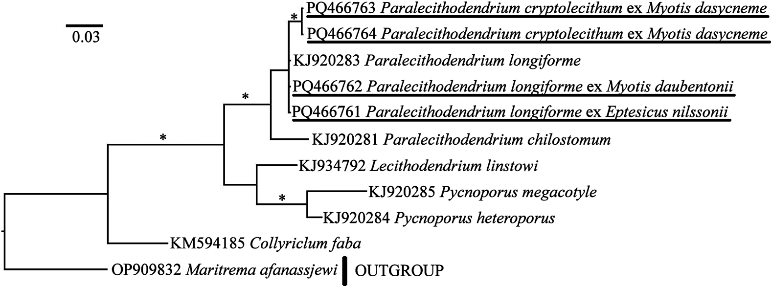
According to the phylogenetic analysis of the ITS2 dataset (Figure 2), P. cryptolecithum forms a separate clade, while the sequences of P. longiforme are almost identical to each other. Paralecithoendrium chilostomum appears to branch basally to the species above. Three lecithodendriid species (L. linstowi, Pycnoporus megacotyle, and P. heteroporus) form a sister clade to the genus Paralecithodendrium. Collyriclum faba branches basally to the other species. The results of pairwise distances analysis between partial coI mtDNA sequences of P. longiforme and P. cryptolecithum revealed an interspecific difference (Figure 3) of 13%–14%. There are no obvious differences between sequences within the corresponding species (maximum is 1%).
Results of molecular phylogenetic analysis (BI) based on partial sequence of ITS2 region. Supports below 0.95 are not shown; supports above 0.95 are marked with “*”. Newly obtained sequences are underlined.

Pairvised distances based on partial coI mtDNA sequences of studied species represented as a heatmap.

Systematics and morphological characteristics of Paralecithodendrium spp.
Superfamily Microphalloidea Ward, 1901
Family Lecithodendriidae Lühe, 1901
Genus Paralecithodendrium Travassos, 1921
Paralecithodendrium cryptolechitum (Zdzitowiecki, Reference Zdzitowiecki1969) (Figure 4)
Paralecithodendrium cryptolecithum ex Myotis dasycneme, general view, (a, b, c). Scale bar 0.5 mm.

Host: Myotis dasycneme.
Geographical locality: Shiryaevo village (Samara region, Russia).
Availability: GenBank No PQ466759, PQ466760 - 28S rDNA; PQ466763, PQ466764 - ITS2; PQ579978, PQ579979 - coI mtDNA.
Accession numbers in collection of IEVB RAS: No 2311, 2326, 2328.
General description of Paralecithodendrium cryptolecithum (based on 15 specimens). Body oviform or oval, not flattened dorsoventrally. Tegument unarmed. Oral sucker oval, subterminal. Ventral sucker round, preequatorial, smaller than oral sucker. Pharynx large, round. Esophagus very short, often not observed. Caeca short and wide. Posterior ends of intestinal branches reach anterior margins of testes (Figure 4). Testes round, opposite, approximately equal, located laterally at ventral sucker level and partially overlapped by it. Pseudocirrus sac rounded, with convoluted seminal vesicle, lies submedially between ventral sucker and caecal bifurcation. In some individuals, pseudocirrus sac more or less overlapped by ventral sucker; in others, it is located at a distance from ventral sucker (Figure 4). Genital pore submedian. Ovary round, lies to the left or right of ventral sucker on its anterior or anterolateral edge and more or less overlapped by it (Figure 4). Ootype with Mehlis’ gland located at posterior or posterolateral margin of ovary. Vitellaria lie laterally and extend from oral sucker to anterior margins of testes. Vitellarium consists of numerous small follicles of irregular shape, which form two groups. From both groups of vitelline follicles, vitelline ducts extend dorsally and merge at level of ventral sucker, forming vitelline reservoir. Vitelline groups not connected medially, but individual follicles may reach body midline dorsally (Figure 4). Numerous uterine loops occupy entire space behind testes. Metratherm morphologically indistinct. Eggs numerous, operculated. Excretory pore terminal. Excretory vesicle V-shaped.
Paralecithodendrium longiforme (Bhalerao, Reference Bhalerao1926) (Figure 5)
Paralecithodendrium longiforme ex Myotis daubentonii, general view (a); Paralecithodendrium longiforme ex Eptesicus nilssoni, general view (b); Paralecithodendrium longiforme ex Plecotus auritus, general view (c). Scale bars 0.5 mm.

Host: Myotis daubentonii, Eptesicus nilssoni, Plecotus auritus.
Geographical locality: Shiryaevo village (Samara region, Russia).
Availability: GenBank No PQ466755–PQ466758 – 28S rDNA; PQ466761, PQ466762 – ITS2; PQ579974–PQ579977 – coI mtDNA.
Accession numbers in collection of IEVB RAS: No 2313, 2314, 2329, 2334, 2335, 2337, 2339.
General description of Paralecithodendrium longiforme (based on 34 specimens). Body lanceolate, flattened dorsoventrally. Tegument unarmed. Oral sucker oval, subterminal. Ventral sucker round, preequatorial, slightly smaller than oral sucker. Pharynx round. Esophagus very short, often not observed. Intestinal branches short and wide, and can reach anterior margins of testes. Testes oval, one always slightly larger than another, lie laterally at ventral sucker level, and may be partially overlapped by it (Figure 5). Pseudocirrus sac slightly transversely elongated, with a convoluted seminal vesicle, lies medially or submedially between ventral sucker and caeca bifurcation. Pseudocirrus sac can reach anterior margin of ventral sucker (Figure 5b) or be more or less overlapped by it (Figure 5a, 5c). Genital pore median or submedian. Ovary oval, located at level of anterolateral, posterolateral or posterior edges of ventral sucker (Figure 5), always covered by it. Mehlis’ complex lies at posterior edge of ovary. Seminal receptacle large, oval. Vitellarium consists of large but few follicles, which form two groups. Vitelline follicle groups lie laterally between oral sucker and anterior margins of testes. Follicle groups can reach anterior margin of testes, at least on one side (Figures 5a and 5c), or vitellaria not reach level of testes (Figure 5b). Vitellaria always merge dorsally. Vitelline ducts extend dorsally from both vitelline follicle groups and merge at level of posterior margin of ovary, forming vitelline reservoir. Numerous uterine loops occupy entire space behind testes. Metratherm morphologically indistinct. Eggs numerous, operculated. Excretory pore terminal. Excretory vesicle V-shaped.
Discussion
We studied the morphological and morphometric features of two species of Paralecithodendrium parasitizing bats in the Middle Volga region (European Russia) and provided new molecular phylogenetic data on these trematodes. We presented a complete description of P. cryptolechitum from M. dasycneme with morphometric characteristics and confirmed the validity of this species. All examined specimens of P. longiforme and P. cryptolechitum correspond well to their previous descriptions (Tables 2, S1, and S2). Characteristic features of these two species, such as body shape, body length to width ratio, size and number of vitelline follicles, were always present.
Measurements of the studied Paralecithodendrium specimens

Note: mean values are given in parentheses; 1 – number of Paralecithodendrium specimens examined, OS – oral sucker, VS – ventral sucker.
The results of our molecular phylogenetic analysis are generally consistent with previous data. The phylogenetic relationships between representatives of Lecithodendriidae and Pleurogenidae are the same as those shown in recent papers by Fernandes et al. (Reference Fernandes, Dos Santos, de Vasconcelos Melo, Achatz, McAllister, Bonilla and Tkach2022), Kirillova et al. (Reference Kirillova, Kirillov, Shchenkov, Knyazev and Vekhnik2024), Sokolov et al. (Reference Sokolov, Shchenkov, Kalmykov and Smirnova2020), and Tkach et al. (Reference Tkach, Gasperetti, Fernandes, Carrion-Bonilla, Cook and Achatz2024). In our analysis of the 28S rDNA dataset, P. cryptolecithum was found to be a separate species along with P. longiforme and P. chilostomum. Phylogenetic analysis based on ITS2 region sequences also confirmed that P. cryptolecithum is a separate species.
At the same time, we have revealed variability in the topology of some internal organs and morphometric characteristics of the studied trematodes. This concerns Paralecithodendrium specimens both from different host species and from conspecific bat individuals. Thus, the largest specimens of P. longiforme were found in P. auritus (Figure 5c). Trematode individuals ex M. daubentonii and E. nilssoni were smaller (Figures 5a and 5b). Accordingly, the largest sizes of suckers and internal organs were registered in trematodes from P. auritus. Variability in the sizes of the body and internal organs, as well as the body length to width ratio, was more pronounced in trematodes from M. daubentonii (Table 2).
Both trematode species are characterized by high variability in body length and width, as well as in the sizes of suckers and reproductive organs (Table 2). Less pronounced variability was observed in the size of the pharynx, the ratio of body length to width, the ratio of the width of the oral and ventral suckers, and the ratio of the width of the oral sucker to the pharynx. The size of the eggs is relatively constant in both species (Table 2).
When identifying related trematode species, it is necessary to take into account their intraspecific morphological and morphometric variability. Paralecithodendrium longiforme and P. cryptolechitum are morphologically very similar. Thus, Zdzitowiecki (Reference Zdzitowiecki1969) points out that Hurkova (Reference Hurkova1963) identified trematodes ex M. dasycneme as P. longiforme, but they were identical to P. cryptolechitum. We critically reviewed our materials on bat trematodes from the Middle Volga region and found that trematodes ex M. dasycneme identified as P. longiforme correspond to the description of P. cryptolechitum (Kirillov et al. Reference Kirillov, Kirillova and Chikhlyaev2012a, Reference Kirillov, Kirillova and Vekhnik2012b).
Our study showed that the sizes of oral and ventral suckers, reproductive organs, and body may be similar in P. longiforme and P. cryptolechitum. Therefore, the use of morphometric features for the identification of these two species is insufficient. Zdzitowiecki (Reference Zdzitowiecki1969) indicates that the main diagnostic features of these species are body shape, the ratio of body length and width, and the size and number of vitelline follicles. We came to the same conclusion in our study.
Thus, for reliable identification of P. longiforme and P. cryptolechitum, the following distinguishing features should be taken into account:
-
1. Body size. Paralecithodendrium longiforme has a lanceolate body shape, while P. cryptolechitum has an ovoid body shape;
-
2. Body length to width ratio. In P. longiforme, the body length is always more than twice the width, while in P. cryptolechitum, it is always less than twice the width;
-
3. Size and number of vitelline follicles. In P. longiforme, the vitelline follicles are relatively large and few in number. Paralecithodendrium cryptolechitum has numerous small follicles.
In addition, in P. longiforme, the vitelline follicles are always merged dorsally, whereas in P. cryptolechitum, the vitelline fields are not connected dorsally, and only individual follicles may reach the midline of the body.
Hurkova (Reference Hurkova1959) described Paralecithodendrium carolinum (as Prosthodendrium carolinum) ex M. daubenronii from the territory of the Czech Republic. The main characters of this species, distinguishing it from P. longiforme, are the following: the pseudocirrus sac is located immediately below the caeca bifurcation, not reaching the anterior margin of the ventral sucker; the ovary lies behind the ventral sucker postacetobularly; the vitellarium occupies the space from the oral sucker to the anterior margin of the pseudocirrus sac.
Paralecithodendrium carolinum and P. longiforme do not differ in their main morphometric features. In our study, we showed that P. longiforme is characterized by wide morphological variability. Thus, the pseudocirrus sac can be located to the left or right of the midline of the body, or lie medially (Figure 4). It can reach the anterior margin of the ventral sucker or be overlapped by it to a greater or lesser extent.
The ovary in P. longiforme can be located at the level of the anterolateral, posterolateral, or posterior margins of the ventral sucker. The vitelline follicles can either reach only the level of the pseudocirrus sac or reach the anterior margin of the testes (Figure 4). Such variability in the topology of the internal organs of P. longiforme was also noted by Zdzitowiecki (Reference Zdzitowiecki1969) and Sharpilo and Iskova (Reference Sharpilo and Iskova1989).
Since its first description, P. carolinum has been reported in Rhinolophus ferrumequinum by Mituch (Reference Mituch1964) in Slovakia and by Timon-David (Reference Timon-David1964) in France, in M. daubentonii by Matskasi (Reference Matskasi1967) in Hungary, and in Myotis mystacinus Kuhl, 1817 and Eptesicus serotinus Schreber, 1774 by Skvortsov (Reference Skvortsov1969) in Moldova. Along with typical specimens of P. carolinum, Timon-David (Reference Timon-David1964) found trematodes that differed in the location of the ovary relative to the level of the ventral sucker. Matskasi (Reference Matskasi1967) found that in his specimens of P. carolinum the vitellarium reached the level of the ventral sucker. Skvortsov (Reference Skvortsov1969) also noted that in some individuals of P. Carolinum, the ovary was located at the level of the ventral sucker.
It follows from the above that the listed morphological characters of P. carolinum cannot be used as diagnostic because of their variability. Judging by the morphological descriptions and drawings presented in the works of Hurkova (Reference Hurkova1959, Reference Hurkova1963), Timon-David (Reference Timon-David1964), Matskasi (Reference Matskasi1967), and Skvortsov (Reference Skvortsov1969), we believe that the authors were dealing with morphological variability of P. longiforme individuals. Sharpilo and Iskova (Reference Sharpilo and Iskova1989) synonymized P. carolinum with P. longiforme. We agree with the latter authors and consider P. carolinum to be a synonym of P. longiforme until proven otherwise.
Analysis of our previous and literature data showed that P. longiforme and P. cryptolechitum have different host specificities. Thus, the main host for P. cryptolechitum is the pond bat M. dasycneme (Kirillova and Kirillov Reference Kirillova, Kirillov, Shchenkov, Knyazev and Vekhnik2024; Zdzitowiecki Reference Zdzitowiecki1969). We also found P. cryptolechitum in the Brandt’s bat Myotis brandtii Eversmann, 1845 (Kirillova et al. Reference Kirillova, Shchenkov, Kirillov and Ruchin2022, 2023a, 2023b; Kirillova and Kirillov Reference Kirillova, Kirillov, Shchenkov, Knyazev and Vekhnik2024). In contrast, P. longiforme parasitizes a wide range of European bats (Sharpilo and Iskova Reference Sharpilo and Iskova1989).
Thus, the combined use of molecular and morphological methods in our study allowed us to identify closely related species – P. longiforme and P. cryptolechitum. For the first time, we provide complete morphological descriptions of these two species, as well as molecular data for P. cryptolechitum.
Supplementary material
The supplementary material for this article can be found at http://doi.org/10.1017/S0022149X24001019.
Financial support
The research was supported by the Russian Science Foundation, grant number 23-24-10021, https://rscf.ru/en/project/23-24-10021/.
Competing interest
The authors declare that they have no conflict of interest.
Ethical standard
The authors assert that all procedures contributing to this work comply with the ethical standards of the relevant national and institutional guides on the use, care, and dissection of animals.