




No CrossRef data available.
A ten year review of atrioventricular septal defects
Published online by Cambridge University Press: 20 January 2005
Abstract
Objectives: To review all cases of atrioventricular septal defects in Northern Ireland from January 1990 to February 1999, examining clinical and morphological features, management, and outcome. Methods: A retrospective case note analysis of 106 subjects with comparisons between subgroups. Results: An atrioventricular septal defect was part of a more complex abnormality in 50 of the patients (47%). Down's syndrome was present in 57 (54%). Cardiac surgery was performed in 81%. The defects were unrestrictive in 69 patients (65%), 45 of whom had Down's syndrome. Complex associated abnormalities existed in 36 patients, and 10 of these died without cardiac surgery. Operative mortality was 9.5% for those with co-existing Down's syndrome group, and 14.3% for the chromosomally normal patients. The ventricular components of the septal defect were restrictive in 23 patients (22%), with 9 having Down's syndrome. Spontaneous closure occurred in more than half of these patients. Mortality was zero. The septal defect was exclusively at atrial level in 14 patients (“primum” defects – 13%), and 3 of these had Down's syndrome. Operative mortality was again zero. Median duration of postoperative follow-up was 3 and a half years. Overall, moderate to severe left atrioventricular valvar regurgitation was observed postoperatively in 23% at follow-up. Conclusions: Mortality was highest in the atrioventricular septal defects with an unrestrictive ventricular component. Uncomplicated cases had good outcomes. Patients without Down's syndrome tended to have more associated cardiac abnormalities, and to have more postoperative arrhythmias. Approximately half of the defects with restrictive ventricular components closed spontaneously. Moderate postoperative left atrioventricular valvar regurgitation was commonest in patients with the defect exclusively at atrial level.
Information
- Type
- Original Article
- Information
- Copyright
- © 2004 Cambridge University Press
Atrioventricular septal defects account for about one-twentieth of all congenital cardiac malformations. Craig and colleagues1 had previously reviewed surgical management of atrioventricular septal defects in Northern Ireland for the period from 1987 through 1991. We wished to update that series, and obtain details of those cases where surgery was not performed.
The atrioventricular separating structures in the normal heart have two components, a muscular part inferiorly formed from the overlapping areas of atrial and ventricular musculature, and a membranous component separating the right atrium from the subaortic outflow tract. The aortic root is then located between the left atrioventricular valve and the atrioventricular septum. Both of these components are absent in the setting of an atrioventricular septal defect with common atrioventricular junction, the aortic valve being anterior to the common atrioventricular junction, and with lengthening and narrowing of the left ventricular outlet. Defects unified in this fashion vary anatomically, particularly in regard to the atrioventricular valvar morphology, the potential for shunting through the defect, and the ventricular balance. It is the attachments of the bridging leaflets of the common atrioventricular junction that determine the level of shunting.
Atrioventricular septal defects may occur in association with other anomalies, including tetralogy of Fallot, double outlet right ventricle, and isomerism of the atrial appendages.2 There is a strong association with Down's syndrome. In a prospective screening study of patients with Down's syndrome in Northern Ireland, about one-sixth had atrioventricular septal defects.3 Over the period of our study, 319 babies born in Northern Ireland had karyotypically confirmed Down's syndrome, and 57 (18%) of these had atrioventricular septal defects.
Management of patients with atrioventricular septal defects has become more aggressive over the last 15–20 years in an attempt to reduce the early effects of the complication of cardiac failure, and the later one of irreversible pulmonary hypertension. More primary repairs are being undertaken, and more patients with chromosomal abnormalities are being considered fit for surgery. Our study details recent practice in one unit, with the majority of surgery being undertaken by one surgeon. The findings reflect the benefits of early diagnosis and surgery.
Materials and methods
We used a database of all patients assessed by paediatric cardiologists in Northern Ireland from January 1990 to February 1999 to identify all cases of atrioventricular septal defects with common atrioventricular junction diagnosed within that period. Relevant data were then collected retrospectively from the case notes of the identified patients. For inclusion, the patient had to have shunting through the septal defect at atrial level, and thus we excluded any patients with common atrioventricular junction but with shunting exclusively at ventricular level. Morphological details were obtained from echocardiographic, catheterisation, operative, and post-mortem findings.
Since November 1987, all babies born in Northern Ireland with Down's syndrome have been screened by echocardiography in early infancy for detection of congenital cardiac disease. Cases were classified as those associated with Down's syndrome or those with normal chromosomes, and as being complicated by major associated abnormalities or uncomplicated. Cases were considered uncomplicated when having no other major medical or cardiac abnormalities apart from mild to severe atrioventricular valvar regurgitation, patency of the arterial duct, a septal deficiency within the oval fossa, a bicuspid aortic valve, or mild stenosis of either the right or left pulmonary artery. Atrioventricular valvar regurgitation was categorized as trivial, mild, moderate, or severe according to the echocardiographic findings. Trivial atrioventricular valvar regurgitation was not included in our analyses.
Further classification into three subgroups was performed based on findings at diagnosis. Thus, we divided the patients into those with an unrestrictive ventricular component to the defect; those with a restrictive component identified on echocardiography with a gradient of greater than 30 mmHg using Doppler echocardiography, and those with shunting confined exclusively at atrial level.
Over 90% of the operations were performed by the same surgeon. In the majority of cases, correction was achieved using a single patch, but occasionally two patches were used, especially when the ventricular component was confined beneath one of the bridging leaflets, and it was possible to close the defect without incising the leaflet. The zone of apposition between the left ventricular components of the bridging leaflets, the so-called “cleft”, was not routinely closed unless it appeared necessary because the reconstructed valve was regurgitant on intra-operative testing by distension using cold saline. Intraoperative transoesophageal echocardiography was not routinely used. In cases with unbalanced ventricles, the decision on management was based on the subjective assessment made by the surgeon of adequacy or otherwise of the underdeveloped ventricle. In general, if there was any doubt over the adequacy of a ventricle, an initial palliative procedure was carried out, usually banding of the pulmonary trunk. In marginal cases, primary repair was more likely to be undertaken when the right rather than the left ventricle was small. Operative mortality was defined as death within thirty days of surgery, or beyond that interval, but prior to discharge from hospital.
Cardiac failure was defined as impairment of cardiac function requiring treatment with diuretics and/or digoxin. Balanced ventricles were defined as those cases where the common atrioventricular junction was committed equally to the right and left ventricles. Respiratory postoperative complications were defined as clinical evidence of respiratory tract infection, pneumothorax, or pleural effusion with accompanying radiological abnormality. Postoperative complications of cardiac rhythm were defined as bradyarrhythmias or tachyarrhythmias persisting following transfer from the operating room.
To allow for variations in weight by age and gender, values were transformed to standard deviation scores. The standard deviation score relates each value to the normal population following the equation {measured weight – mean weight for age and gender}/weight standard deviation at this age, but with additional allowance made for lack of normality in the distribution of weight. For chromosomally normal patients, the computerised 1990 British Growth Standard was used.4 For patients with Down's syndrome, we used data appropriate for this syndrome.5
The statistical computer packages “Statview” and “SPSS” were used to analyse the results. Descriptive statistics and Kaplan Meier survival curves were employed. Comparison of Kaplan Meier curves for Down's syndrome and chromosomally normal patients were made using the log rank test.
Results
Atrioventricular septal defects which included shunting at atrial level were found in 106 patients, and 86 (81%) of these have had surgery. An additional 5 patients (5%) are awaiting surgery, while 13 (12%) were considered unsuitable for surgery because of severe associated cardiac or medical problems with critical illness in 9 (8.5%), or because the defects were so small that no clinical compromise was demonstrable and spontaneous closure was expected in 4 (3.5%). Two patients died while on the waiting list for surgery. Cardiac catheterisation was performed in 26 patients (25%). Of the overall group, 58 patients were male, and 50 (47%) were deemed to be complicated within the definitions given above. There was one case with a significant anomaly of the atrioventricular valve in the form of an absent mural leaflet. This patient had Down's syndrome. There were no cases with double orifice of the atrioventricular valve.
Table 1 shows the median gestation, weight at birth, and age at diagnosis of those with and without Down's syndrome.
Table 1. Median gestation, birth weight and age at diagnosis of 106 patients with atrioventricular septal defects, 57 of whom had Down's syndrome.

We lost 3 patients to follow-up, one shortly after prophylactic banding of the pulmonary trunk and ligation of a patent arterial duct at the age of eight months, another one year and eight months after primary repair, and the third four years after primary repair.
Atrioventricular septal defects with an unrestrictive ventricular component
This morphology was found in 69 patients (Table 2). Their median age at diagnosis was 4 days, with a range from 1 to 2342 days, and cardiac failure was present in 35 (51%). Down's syndrome occurred in 45 (65%), with 18 (40%) of these being complicated. Of the chromosomally normal patients, 18 (75%) had complications, and 8 died without undergoing cardiac surgery, 5 having severe associated medical problems, specifically necrotizing enterocolitis, and VATER, Edward's, Noonan's, and Goldenhar syndromes. The first two died following episodes of sepsis, Edward's syndrome is life-limiting, the patients with Noonan's syndrome died from probable hypertrophic cardiomyopathy, and the patient with Goldenhar syndrome died from renal failure subsequent to gastrointestinal surgery. The other 3 patients who died had multiple cardiac abnormalities. The first had a common arterial trunk along with totally anomalous pulmonary venous connection. The second had a right-sided heart with a right aortic arch, hypoplasia of the ascending aorta, and both great vessels arising from an anterior chamber. The third patient also had complex malformations including a right-sided heart, double outlet right ventricle, infradiaphragmatic totally anomalous pulmonary venous connection, and a deformed right component of the common atrioventricular valve. In addition, there was asplenia, bilateral trilobed lungs, and pulmonary congestion. These three neonates were critically unwell, and it was considered that they would not survive surgery. Ventilation was electively discontinued on the third, fourth, and first days of life, respectively.
Table 2. Preoperative features of 106 patients with atrioventricular septal defects.

Two patients with Down's syndrome died preoperatively. One had a tracheo-oesophageal fistula, and died at the age of 310 days of respiratory syncytial virus positive bronchiolitis. The other died at home at the age of 11 months having previously suffered a near miss cot death with global cerebral damage at the age of five months.
Unbalanced ventricles were much commoner in the chromosomally normal patients, being found in 42% as opposed to only 6.7% of those with trisomy 21. No patients had severe left atrioventricular valvar regurgitation.
Obstruction of the left ventricular outflow tract was present in two patients, neither of whom had Down's syndrome. The first, described already, had a right-sided heart and a right aortic arch, hypoplasia of the ascending aorta, and both great vessels arising from an anterior chamber. This patient died preoperatively. The other had a hypoplastic left ventricle and coarctation of the aorta, but these obstructions were relieved following surgery. None of these defects closed spontaneously.
Surgery was performed in 42 patients with Down's syndrome, 15 of whom were complicated (Tables 3 and 4). Of the operations, 7 were palliative or involved incomplete repair, being undertaken at a median age of 77 days, with a range from 26 to 256 days. The other 35 procedures involved primary complete repair at a median of 163 days, with a range from 20 to 335 days. The median weight at operation was 4.7 kg, with a range from 2.7 to 9.2 kg. The 7 palliative or incomplete repair procedures were banding of the pulmonary trunk and ligation of a patent arterial duct in three; banding of the pulmonary trunk in isolation in one; construction of a modified left Blalock-Taussig shunt in one; incomplete repair with banding of the pulmonary trunk and ligation of a patent duct in one, and incomplete repair with banding of the pulmonary trunk in the seventh.
Table 3. Operative courses of 49 patients with uncomplicated atrioventricular septal defects.

Table 4. Operative courses of 37 patients with complicated atrioventricular septal defects.

Surgery was performed in 14 chromosomally normal patients, 10 of whom were complicated (Tables 3 and 4). Palliative procedures had been performed in 5, at a median age of 31 days, with a range from 1 to 483 days, one of the patients being over one year of age at the time of the surgery. The pulmonary trunk was banded in two patients, totally anomalous pulmonary venous connection relieved in one, supracardiac totally anomalous pulmonary venous connection corrected with ligation of a patent duct in another, the right pulmonary artery reconstructed as part of a bilateral bidirectional cavopulmonary anastomosis in another combined with ligation of a patent duct, and the pulmonary trunk banded along with repair of aortic coarctation in the fifth. Primary repair was carried out in the other nine patients at a median age of 103 days, with a range from 40 to 837 days, two patients being older than one year at the time of surgery. The median weight at operation was 3.90 kg, with a range from 2.2 to 11.5 kg. Three patients await surgery.
The commonest postoperative complication in this group was respiratory, involving 59.5% of the patients with Down's syndrome, and half of the chromosomally normal patients. Ventilation was required for a mean of 4.3 days, with a standard deviation of 3.84, and a standard error of the mean of 0.51. Arrhythmias, only one episode of which required treatment, occurred in 5 (12%) of the patients with Down's syndrome. The treated patient, who had a complicating muscular ventricular septal defect, developed atrial fibrillation and received digoxin. Of the chromosomally normal patients, 3 (21%) developed arrhythmias, and two needed treatment. One patient without other complications developed a junctional tachycardia, and was treated with digoxin and subsequently, two and a half years postoperatively, developed atrial flutter and needed anticoagulation. The second patient had a degree of hypoplasia of the left ventricle and developed a nodal rhythm, which was treated with digoxin and amiodarone.
Operative mortality amongst these patients was higher (14.3%) in those who were chromosomally normal, with two patients dying. One of these patients had a “Swiss cheese” type of muscular ventricular septum, while the other had complicating totally anomalous pulmonary venous connection, abnormally related great arterial trunks, and a small left ventricle. Palliation was attempted in the latter.
Operative mortality was 9.5% in the patients with Down's syndrome. One patient died with acute renal failure and a chest infection at the age of 4 months, this occurring 17 days after banding of the pulmonary trunk and ligation of a patent duct. He had been born at 27 weeks gestation and had necrotizing enterocolitis with moderate preoperative atrioventricular valvar regurgitation. Another patient died 18 days postoperatively from respiratory complications. He was 324 days old at the time of surgery, and had mild preoperative atrioventricular valvar regurgitation. He had associated duodenal atresia, congenital hypothyroidism, and gastro-oesophageal reflux. A third patient died of respiratory failure 19 days after surgery at 4 months of age. He had trivial preoperative atrioventricular valvar regurgitation. The fourth patient died of respiratory failure 39 days after repair of the atrioventricular septal defect repair at almost 5 years of age. He had a smaller left ventricle and complete absence of the atrial septum, and had previously undergone banding of the pulmonary trunk at 77 days of age. He did not have any atrioventricular valvar regurgitation preoperatively.
In patients without other complications, the operative mortality was zero for those who were chromosomally normal, and 4.0% for those with Down's syndrome.
There was one late postoperative death in a chromosomally normal patient. This was due to congestive cardiac failure precipitated by varicella zoster infection in a girl with moderate left ventricular hypoplasia who had been palliated 79 days previously.
In the patients with Down's syndrome, there were two late deaths following primary repair. One patient known to have pulmonary hypertension developed septicaemia, while the other died from congestive cardiac failure and a respiratory tract infection 100 days after repair at 6 months of age. Post-mortem examination of the latter revealed evidence of severe pulmonary hypertension and an abnormal thymus.
Postoperative follow up for this group was for a median of 48 months, with a range from 2 to 95 months. Symptoms persisted in 8 patients, 5 with Down's syndrome. Of these, 3 were diagnosed as having asthma. Of the chromosomally normal patients, 42.9% had mild and 21.4% had moderate to severe left atrioventricular valvar regurgitation. Of the patients with Down's syndrome, 28.6% had mild, 21.4% had moderate, and none had severe left atrioventricular valvar regurgitation. One chromosomally normal patient with a small left ventricle required replacement of the left atrioventricular valve 439 days after repair, which had been undertaken at 166 days of age.
Of the whole group, only one patient had congestive cardiac failure, but 3 had residual septal defects. A second operation was required in 7, and a third operation in one. One uncomplicated and chromosomally normal patient had residual trivial obstruction of the left ventricular outflow tract, albeit with no evidence of encroachment of the patch on the outflow tract.
In the chromosomally normal patients, weight standard deviation scores improved from a mean of −3.675, with a standard deviation of 1.63, at operation to −1.486 with a standard deviation of 1.65 at follow up. In the patients with Down's syndrome, the change was from a mean of −1.33, with standard deviation of 1.01, at operation to −0.62 with standard deviation of 1.17 at follow-up.
We have analysed the survival for the patients with unrestrictive ventricular components categorized as having Down's syndrome or being chromosomally normal, and as complicated or uncomplicated whether or not surgery was performed, in Kaplan Meier survival curves (Figs 1–3).
Survival was better for the patients with Down's syndrome (Fig. 1), with cumulative survival at one year of 0.861, and 95% confidence intervals from 0.741 to 0.981, compared to 0.542, with 95% confidence intervals from 0.342 to 0.742, for the chromosomally normal patients. This difference was statistically significant by the log rank test (p = 0.0036).

Figure 1. Kaplan Meier survival curves for all the patients having an unrestrictive ventricular component to their atrioventricular septal defect. The Y-axis shows survival, while the X-axis shows age in years. The data for the patients with Down's syndrome is shown by the dashed line, while the solid line shows the data for the chromosomally normal patients. The filled circles represent censored patients.

Figure 2. The Kaplan Meier survival curves for all patients with an unrestrictive ventricular component to the septal defect, and in the absence of any complicating features. The format for the graph is as shown in Figure 1.

Figure 3. Kaplan Meier survival curves for all patients with an unrestrictive ventricular component to the atrioventricular septal defects, but in the setting of associated complications. The format is again as for Figure 1.
Complications had a marked impact on survival (Figs. 2 and 3), with one year cumulative survival for complicated and chromosomally normal patients being 0.611, with 95% confidence intervals from 0.381 to 0.841, compared to 0.826, with 95% confidence intervals from 0.183 to 1, for the patients with Down's syndrome. This difference was again statistically significant as assessed by the log rank test (p = 0.0069).
Atrioventricular septal defects with a restrictive ventricular component
This combination was found in 23 patients (Table 2). Their median age in days at diagnosis was 25, with a range from 1 to 1341, and cardiac failure was present in 8 (35%). Down's syndrome occurred in 9 (39%), with 4 (44%) of these having additional complications. Of the chromosomally normal patients, 7 (50%) were complicated. No patients died.
Spontaneous closure of the ventricular component occurred in 5 (56%) of the patients with Down's syndrome, and in 8 (57%) of the chromosomally normal patients.
Surgical repair was performed in 6 patients with Down's syndrome, three of whom were complicated (Tables 3 and 4). This occurred at a median age of 366 days, with a range from 181 to 1262 days. The median weight at operation was 6.74 kg, with a range from 3.82 to 12.5 kg. Surgery was undertaken in 13 chromosomally normal patients, 7 of whom had additional complications (Tables 3 and 4). The operation in one was palliative ligation of a patent arterial duct. Preoperative echocardiography did not detect an aortic coarctation in one of the 13 chromosomally normal patients undergoing surgery. A tight coarctation was present just distal to the left subclavian artery, and there was a small distal aortic arch. The arterial duct had become ligamentous.
Of the 12 chromosomally normal patients undergoing complete repair, the median weight at the time of the operation was 8.8 kg, with a range from 3.64 to 18.0 kg. One patient awaits surgery. The median age at surgery was 424 days, with a range from 22 to 1817 days.
The main clinical difference between the patients with and without Down's syndrome was that preoperative atrioventricular valvar regurgitation was much commoner in the chromosomally normal patients, being seen in half as opposed to only 11% of those with Down's syndrome.
Respiratory and arrhythmic problems were the most frequent complications encountered postoperatively. Respiratory problems occurred in half of the patients with Down's syndrome, and arrhythmias in 16.7%. Respiratory complications occurred in one-quarter of the chromosomally normal patients, and arrhythmias in almost two-fifths. One of the chromosomally normal patients developed 2 to 1 atrioventricular block, then sinus rhythm with first degree block. Later sinus rhythm alternated with a nodal rhythm of a satisfactory rate. No treatment was necessary. The patient had Alagille's syndrome. All of the other arrhythmias resolved spontaneously.
None of these patients died, nor were there any late postoperative deaths.
The patients have been followed postoperatively for a median of 29 months, with a range from 0.25 to 77.0 months. Of the patients, 2, one with Down's syndrome, had persisting symptoms, and one of these was diagnosed as having asthma. Of the patients without Down's syndrome, half had mild and a quarter had moderate left atrioventricular valvar regurgitation, compared to one-third and one-sixth respectively of the patients with Down's syndrome. None had severe left atrioventricular valvar regurgitation, and none had required replacement of the valve.
No patients had congestive cardiac failure, but two had residual septal defects.
One had required a second operation, but none needed a third operation. Residual obstruction of the left ventricular outflow tract was present in 2, with gradients of 13 and 14 mmHg.
In those who were chromosomally normal, weight standard deviation scores improved from a mean of −2.130, with standard deviation of 1.34, at operation to −0.842, with standard deviation of 1.42, at follow-up. In those with Down's syndrome, the changes were from a mean of −1.75, with standard deviation of 1.09, at operation to −1.58, with standard deviation of 0.82, at follow-up.
Shunting exclusively at atrial level
We found “primum” atrial septal defects, with no associated ventricular defect, in 14 patients (Table 2). The median age at diagnosis was 185 days, with a range from 2 to 1856 days, and cardiac failure was present in four (28.6%). Down's syndrome was present in 3 patients, and none of these had further complications. Of the chromosomally normal patients, three (27%) had additional complications. One of these had a hypoplastic left heart and a patent arterial duct and died preoperatively. The pathologist commented that “rather unusually the atrial septal lesion was of an atrioventricular canal type with deformity of the tricuspid valve”. Another of the chromosomally normal patients had fibroelastosis on the epicardial surface of the left ventricular outflow tract. Postoperatively, he had a residual moderate degree of obstruction within the left ventricular outflow tract, with a maximum gradient of 20 mmHg.
One patient awaits surgery, and one has a defect that does not require surgery. Primary repair was performed in 11 patients, two of whom had Down's syndrome and were uncomplicated (Tables 3 and 4). The surgery was performed at a median age in days of 529, with a range from 67 to 2222 days. The median weight at operation was 10.0 kg, with a range from 3.42 to 18.0 kg.
Of the patients proceeding to surgery, 44% of the chromosomally normal patients had mild to moderate atrioventricular valvar regurgitation preoperatively, while no patients with Down's syndrome had preoperative atrioventricular valvar regurgitation.
The commonest postoperative problem was respiratory, occurring in two-thirds, of the chromosomally normal patients, and in half of those with Down's syndrome. No patient in this group suffered a postoperative arrhythmia, and no patients died either during surgery or subsequently.
The patients have been followed up for a median period of 42 months, with a range from 4 to 90 months. All patients were asymptomatic at follow-up. Of the patients without Down's syndrome, 44% had mild, 22% had moderate, and none had severe left atrioventricular valvar regurgitation. Of the two patients with Down's syndrome undergoing surgery, one had moderate left atrioventricular valvar regurgitation. None required valvar replacement, but one had a residual obstruction in the left ventricular outflow tract, with a gradient of 40 mmHg. None had residual septal defects or required further surgery.
In the chromosomally normal patients, the weight standard deviation scores improved from a mean of −1.783, with a standard deviation of 1.57, at the time of operation to −0.631, with a standard deviation of 1.16, at follow-up. In those with Down's syndrome, the change was from a mean of −1.93, with standard deviation of 0.88 at operation, to −0.59, with standard deviation of 1.37, at follow-up.
Discussion
Our review demonstrates that uncomplicated cases of atrioventricular septal defects, including those with exclusively atrial shunting, the so-called “primum atrial septal defect”, have good outcomes from primary repair irrespective of whether or not the patients are chromosomally normal. We did not attempt to include patients with exclusively ventricular shunting in the setting of a common atrioventricular junction, as complete case ascertainment would have been impossible retrospectively.
Operative mortality in uncomplicated patients with unrestrictive ventricular components to their atrioventricular septal defects is 3.2%. This finding concurs with that of Brando et al.,6 who showed an operative mortality of 3%, two out of sixty-seven, for patients with atrioventricular septal defects with common atrioventricular valve and unrestrictive ventricular shunting but without complicating coexisting cardiac anomalies who underwent primary repair from 1990 onwards. Preoperative uncontrolled congestive cardiac failure is the major problem encountered in managing this group of patients, with failure to thrive being the consequence of the cardiac failure. The risk of a chest infection is increased by pulmonary venous congestion through left atrioventricular valvar regurgitation. Pulmonary hypertension is a well-recognised risk of delayed surgery. We advocate surgery before six months of age, and have demonstrated good results with babies weighing 2.2 kg or more at the time of surgery. Our findings are in keeping with those of Stellin et al.,7 who have demonstrated a lower operative mortality, at 3.4%, for patients with uncomplicated unrestrictive atrioventricular septal defects less than three months old at surgery than for those greater than three months old where it was 15%. Primary repair, however, was not possible in all of our patients. Palliation had to be employed in 12 patients with unrestrictive defects, 10 of whom were complicated, and in one patient with a restrictive defect who also had additional complications. We included complicated patients with major associated medical or cardiac abnormalities in our review as these patients are particularly difficult to manage, and we deemed scrutiny of their treatment and outcome to be important. Early diagnosis is vital, and cardiological screening of infants with Down's syndrome as soon as possible after birth is recommended so that early medical and surgical treatment can be instituted.3, 8 The ensuing absence of congestive cardiac failure permits effective developmental and physiotherapy support.
The main differences between the patients with and without Down's syndrome were that more of the chromosomally normal patients had complicated lesions, and that preoperative atrioventricular valvar regurgitation was also commoner in these chromosomally normal patients. An explanation for this latter finding comes from the fact that the left mural leaflet of the common atrioventricular valve is generally smaller in the chromosomally normal patients than in those with Down's syndrome. Very few of our patients developed pulmonary hypertension, or had obstruction of the left ventricular outflow tract.
We found that eight patients required reoperation, with one needing replacement of the left atrioventricular valve. Of these eight patients, only one was without severe associated medical or cardiac abnormalities.
The severity of preoperative atrioventricular valvar incompetence did not affect the mortality of our group of patients, unlike the experience of Studer et al.,9 who studied 310 patients from the period of 1967 through 1982. They graded atrioventricular valvar regurgitation from 0 to 5, and found that survival at 12.5 years postoperatively was 95% when preoperative valvar incompetence was absent or mild, as opposed to 88% when it was more severe. The difference compared to our findings may have been because none of our patients had severe atrioventricular valvar regurgitation preoperatively, possibly because they underwent surgery at a younger age. Only one of our patients needed replacement of the left atrioventricular valve, confirming the prediction of Studer and his colleagues in 19829 that fewer valvar replacements would be required for future generations of children with atrioventricular septal defects. Longer follow-up of our patients may reveal others who will require valvar replacement. We found that residual moderate left atrioventricular valvar regurgitation was commonest in patients with exclusively atrial shunting, at 27.3% versus 21.3% in those with ventricular shunting at the time of diagnosis (Table 2).
A notable finding was that spontaneous closure of the ventricular component of the atrioventricular septal defect occurred in approximately half of patients who were both chromosomally normal and with Down's syndrome when the defect was restrictive at the time of diagnosis. To our knowledge, the frequency of spontaneous closure of the ventricular component of the septal defect has not previously been highlighted in the literature. It is important to be aware of this possibility when planning surgery and counselling families.
Survival is particularly good for those patients with restrictive ventricular components or exclusively atrial shunting (Tables 3 and 4).
Long-term follow up of patients with atrioventricular septal defects, whether surgery has been performed or not, is critical to minimise morbidity.
Although our study is retrospective, it does give an overall view of a decade in which referral of patients with atrioventricular septal defects was standardised and early surgical intervention was the normal practice. The numbers, although not large, are sufficient to allow major differences in the anatomical subgroups to be ascertained. Our series of patients, furthermore, demonstrates that early diagnosis and careful monitoring, with appropriate timing of surgery, lead to good outcomes in the majority of patients with atrioventricular septal defects, irrespective of whether or not the patients were chromosomally normal.
Acknowledgements
We thank Jayne Johnston and Lorraine Davidson for performance of echocardiograms, and Chris Patterson for assistance with the statistical analysis.
References
Craig B, Mulholland C, Gladstone D, O'Kane H, Cleland J. Surgical management of complete atrioventricular septal defect. Ir J Med Sc 1992; 161: 146.Google Scholar
Najm HK, Coles JG, Endo M, et al. Complete atrioventricular septal defects. Results of repair, risk factors and freedom from reoperation. Circulation 1997; 96 (S11): 311–315.Google Scholar
Tubman TRJ, Shields MD, Craig BG, Mulholland HC, Nevin NC. Congenital heart disease in Down's syndrome: two year prospective early screening study. BMJ 1991; 302: 1425–1427.Google Scholar
Freeman JV, Cole TJ, Chinn S, Jones PRM, White EM, Preece MA. Cross-sectional stature and weight reference curves for the UK, 1990. Arch Dis Child 1995; 73: 17–24.Google Scholar
Styles ME, Cole TJ, Dennis J, Preece MA. New cross sectional stature, weight and head circumference references for Down's syndrome in the UK and Republic of Ireland. Arch Dis Child 2002; 87: 104–108. L,M,S data copyright of the Down's Syndrome Medical Interest Group.Google Scholar
Bando K, Turrentine MW, Sun K, et al. Surgical management of complete atrioventricular septal defects. A twenty-year experience. J Thorac Cardiovasc Surg 1995; 110: 1543–1554.Google Scholar
Stellin G, Vida VL, Milanesi O, et al. Surgical treatment of complete A-V canal defects in children before 3 months of age. Eur J Cardiothorac Surg 2003; 23: 187–193.Google Scholar
Amark K, Sunnegardh J. The effect of changing attitudes to Down's syndrome in the management of complete atrioventricular septal defects. Arch Dis Child 1999; 81: 151–154.Google Scholar
Studer M, Blackstone E, Kirklin John W, et al. Determinants of early and late results of repair of atrioventricular septal (canal) defects. J Thorac Cardiovasc Surg 1982; 84: 523–542.Google Scholar
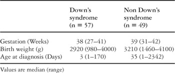
Table 1.
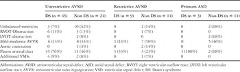
Table 2.
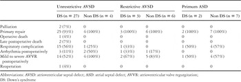
Table 3.
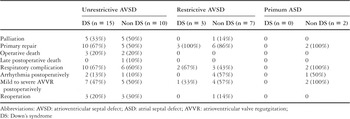
Table 4.

Kaplan Meier survival curves for all the patients having an unrestrictive ventricular component to their atrioventricular septal defect. The Y-axis shows survival, while the X-axis shows age in years. The data for the patients with Down's syndrome is shown by the dashed line, while the solid line shows the data for the chromosomally normal patients. The filled circles represent censored patients.

The Kaplan Meier survival curves for all patients with an unrestrictive ventricular component to the septal defect, and in the absence of any complicating features. The format for the graph is as shown in Figure 1.

Kaplan Meier survival curves for all patients with an unrestrictive ventricular component to the atrioventricular septal defects, but in the setting of associated complications. The format is again as for Figure 1.
 You have
Access
- 18
- Cited by