Social media synopsis
Persistent hypoxemia in neonates despite improved respiratory distress and increased supplemental oxygen needs echocardiography in evaluating Chiari network.
# persistent hypoxemia
# Chiari network
During fetal development, the right valve of the sinus venosus segregates all systemic venous blood exiting the superior vena cava, inferior vena cava, and coronary sinus from the tricuspid valve and shunts oxygen-rich blood into the left atrium. Reference Allen1 The Chiari network is an embryologic remnant of the right sinus venous valve; it is thin, fenestrated, and mobile and attached to multiple lesions within the right atrium. The rate of identification of the Chiari network on transesophageal echocardiography was reportedly 2%. Reference Schneider, Hofmann, Justen and Meinertz2 The Chiari network is often diagnosed incidentally by echocardiography. Neonates with the Chiari network may rarely present with hypoxemia due to restricted blood flow from the right atrium to the right ventricle and right-to-left shunting across the foramen ovale. Reference Aljemmali, Bokowski, Morales and Abdulla3 The Chiari network can impede blood flow to various degrees and necessitate surgical intervention. Reference Aljemmali, Bokowski, Morales and Abdulla3 Therefore, it is crucial to diagnose the cause precisely in prolonged hypoxemic infants. We report the case of a neonate with hypoxemia and a Chiari network that was diagnosed with real-time telemedicine.
Case report
A primiparous mother with no underlying medical conditions delivered a female neonate spontaneously at 38 weeks of gestation, with an uneventful perinatal history. At birth, the newborn had a weight of 3236 g, height of 50.0 cm, heart rate of 160/min, blood pressure of 79/44 mmHg, respiratory rate of 60/min, clear respiratory sounds, no heart murmurs, and no external anomaly. She presented with grunting. Arterial oxygen saturation by pulse oximetry (SpO2) was 88–92% in room air. Arterial blood gas analysis showed no respiratory acidosis or hypercapnia. Complete blood counts and biochemical markers were within normal limits. Electrocardiography findings were normal, and a chest X-ray film showed a cardiothoracic ratio of 0.58 and mild prominence of the pulmonary vasculature.
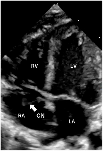
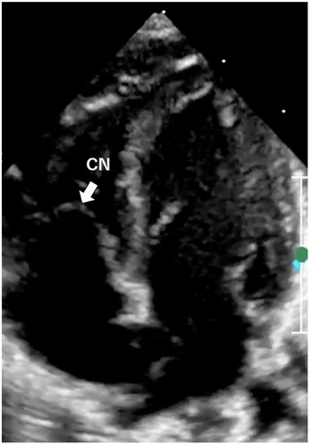
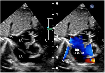
At 3 hours of life, the SpO2 was low, measuring 92–95% even after treatment with 30% oxygen, although other clinical signs of respiratory distress resolved. Even after increasing the supplemental oxygen to 40%, the SpO2 remained unchanged significantly and measured 96%. Echocardiography revealed a thin membranous structure in the right atrium attached to the right atrial wall, protruding into the right ventricle during diastole, and moving in and out through the orifice of the tricuspid valve (Figs 1 and 2). The flow through the inferior vena cava was guided by a right-to-left flow across the patent foramen ovale (Fig 3). The tricuspid valve and the ring of the pulmonary valve had diameters of 10.2 mm (92% of normal) and 7.1 mm (82% of normal), respectively. No other abnormalities were identified; good biventricular function was noted. We initially suspected tricuspid valve dysplasia; however, this was not confirmed. Therefore, we consulted a tertiary care facility by real-time telemedical echocardiography. The structure was identified as a Chiari network by a paediatric cardiologist. We expected spontaneous improvement of the hypoxemia with a natural improvement in the pulmonary vascular resistance; hence, surgery was not performed. The patient was followed up conservatively. At 5 months of age, her development and growth were normal, right-to-left shunting had decreased, and the SpO2 was 95% without oxygen.
The apical four chambers view shows the Chiari network in the right atrium. CN: Chiari network, RA: right atrium, RV: right ventricle, LA: left atrium, LV: left ventricle.

Apical four chambers view in diastole shows the Chiari network protruded into the right ventricle.

Subcostal sagittal view shows right-to-left flow shunting across the patent foramen ovale due to a large Chiari network. IVC: inferior vena cava, RA: right atrium, LA: left atrium, RL shunt: right-to-left flow shunt.

Discussion
Our neonate showed a low SpO2 and right-to-left flow shunting across the patent foramen ovale due to a large Chiari network. A narrow ring of the pulmonary valve indicated that the Chiari network reduced the flow from the right ventricle to the pulmonary artery before birth.
Schneider et al. described that right-to-left shunting occurred more often in patients with Chiari networks than in the general population due to a high right atrial pressure, large patent foramen ovale, or streaming effect. Reference Schneider, Hofmann, Justen and Meinertz2 Aljemmali et al. reported a Chiari network with cyanosis due to a right-to-left shunt across a patent foramen ovale in patients with structurally normal hearts and reviewed five similar cases. Reference Aljemmali, Bokowski, Morales and Abdulla3 Surgical removal was performed in one patient, and cyanosis improved in other patients spontaneously within the first few days of life. In our case, hypoxemia gradually resolved with only a temporary oxygen supply.
The Chiari network is regarded as a normal anatomic variant with negligible clinical importance; however, it has been associated with fetal hydrops Reference Bendadi, van Tijn, Pistorius and Freund4 and right heart hypoplasia Reference Trento, Zuberbuhler, Anderson, Park and Siewers5 in previous studies. These reports suggested that a Chiari network causes varying degrees of right ventricular inflow obstruction, which may cause CHD, necessitating surgical intervention. Various other complications such as thrombus formation and arrhythmia Reference Schneider, Hofmann, Justen and Meinertz2 have been reported associated with the Chiari network. However, no such complications were observed in our case.
In our case, low SpO2 persisted even after her tachypnea and grunting improved, and SpO2 remained unchanged with adequate supplementation of oxygen; therefore, we suspected cyanotic CHD and performed echocardiography. Excessive supplementation of oxygen facilitates a decrease in pulmonary vascular resistance and closure of the ductus arteriosus, which may deteriorate the CHD condition. If hypoxemia does not improve with adequate oxygenation, oxygen should be discontinued, and echocardiography should be performed to diagnose cyanotic CHDs. Colour echocardiography or contrast echo test would be very useful to detect right-to-left flow shunting across the patent foramen ovale due to cyanotic CHDs. The Chiari network does not have a high pulmonary blood flow and is not ductal-dependent; oxygen was administered to achieve normal SpO2.
Our echocardiography findings revealed an abnormal structure in the right atrium and right-to-left shunting across the patent foramen ovale. Echocardiography is considered most helpful in diagnosing a Chiari network; however, the Chiari network is an uncommon diagnosis, which must be recognised appropriately to prevent misdiagnosis. General paediatricians in our region are inexperienced in diagnosing rare CHD. Reference Sotodate, Oyama and Saiki6 For general paediatricians, telemedicine facilitates a quicker and accurate diagnosis Reference Randolph, Hagler and Khandheria7 and significantly decreases the need to transport infants with mild or no heart disease, Reference Webb, Waugh and Grigsby8 as in our case.
In conclusion, although the Chiari network is a benign structure, it may cause a decrease in SpO2 due to reduced right ventricle inflow. If low SpO2 persists even after improvement in respiratory distress symptoms, echocardiography should be performed immediately to evaluate cyanotic CHD accurately. When abnormal structures in the right atrium and right-to-left shunting across the patent foramen ovale are identified, the Chiari network needs to be differentiated. A real-time telemedical echocardiography system helps differentiate rare CHDs at a primary care centre.
Acknowledgements
We thank infant and parents, doctors, and medical staff involved in this study.
Financial support
This research received no specific grant from any funding agency, commercial, or not-for-profit sectors.
Conflicts of interest
None.
Ethical standards
Informed consent was obtained from the parents for the publication of this case report. The authors assert that all procedures contributing to this work comply with the ethical standards of the relevant national guidelines on human experimentation (Japan) and with the Helsinki Declaration of 1975, as revised in 2008, and have been approved by the institutional committees (Japan).