Introduction
Squamous cell carcinoma (SCC) of the tympanic membrane is an extremely rare entity, with only two cases previously reported in the literature.Reference Gisselsson1, Reference Somers, Vercruysse, Goovaerts, Govaerts and Offeciers2 The diagnosis may prove elusive, as biopsies of the external auditory meatus will exclude a malignant process.
We present a recent patient with SCC of the tympanic membrane. We also discuss the investigation, diagnosis, natural history and management of this rare condition, together with the staging and management of tumours of the temporal bone, and the differences between these closely related but prognostically different entities.
Case history
A 42-year-old woman with a history of type one diabetes mellitus presented to the ENT department with left-sided otalgia and otorrhoea. She was treated conservatively and her symptoms settled. However, she developed a nasal blockage, and nasal polyps were subsequently diagnosed on follow up. She underwent a septoplasty and functional endoscopic sinus surgery.
The patient later developed recurrent left-sided otorrhoea. On examination, she was found to have granulation tissue on the postero-superior aspect of the tympanic membrane. She was treated conservatively with topical antibiotics and aural toilet.
However, within six months the patient developed a blood-stained aural discharge. A granular mass was seen to be arising from the floor of the external auditory canal. This was biopsied three times under general anaesthesia. Histological analysis found squamous dysplasia each time.
The patient underwent computed tomography (CT), which demonstrated a slightly thickened left tympanic membrane but a normal middle ear and mastoid, and no evidence of malignant otitis externa.
The patient's ear continued to discharge. As a result, she underwent a cortical mastoidectomy and tympanoplasty. During the procedure, the inferior half of the tympanic membrane was noted to have an abnormal, granular, scarred appearance and was partially excised. The middle ear was normal and the ossicular chain was intact.
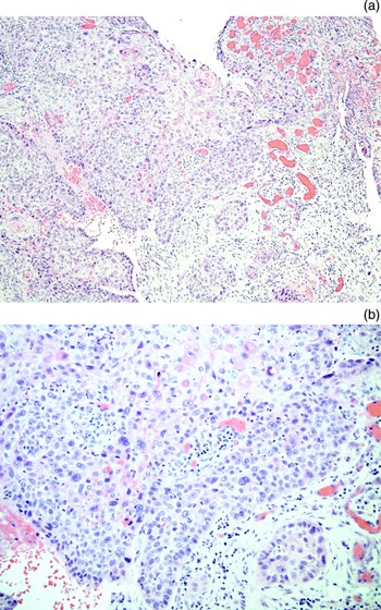
Histological analysis of the tympanic membrane established a diagnosis of SCC (Figure 1).
Photomicrographs of tympanic membrane: (a) ×10; (b) ×20. (H&E)

The patient underwent full staging and discussion at the regional multidisciplinary meeting. Staging CT and magnetic resonance imaging (MRI) scans of the temporal bone and neck showed no evidence of residual or metastatic disease.
The patient underwent a revision radical mastoidectomy. Frozen sections were taken from the residual tympanic membrane and the skin of the external auditory canal; these showed no evidence of malignancy. The external auditory canal skin, remaining tympanic membrane, fibrous annulus, posterior canal wall, and malleus and incus were removed en bloc; post-operative analysis showed no evidence of residual tumour.
At the time of writing, the patient was due to undergo middle-ear reconstruction, ossiculoplasty and mastoid obliteration.
Discussion
Squamous cell carcinomas arising from the tympanic membrane are extremely rare. The present case represents the third published report of SCC of the tympanic membrane. The first case was reported by Gisselsson in Sweden in 1951.Reference Gisselsson1
The aetiology of this particular entity remains somewhat of a mystery due to its rarity. In all reported cases, the presentation was initially one of chronic otitis externa with poor response to conservative management.Reference Gisselsson1, Reference Somers, Vercruysse, Goovaerts, Govaerts and Offeciers2 Once the acute phase had settled, otoscopic examination in all three reported cases demonstrated the presence of granulations arising from the tympanic membrane. The presence of these granulations led in each case to biopsy of the tympanic membrane and exploration of the middle ear.
Demographic factors again provide no correlation. One of three reported patients in the literature was diabetic, and none of the patients smoked tobacco. Unlike our female patient, the other patients were male, aged 65 and 70 years. In addition, there does not appear to be any consistency as regards the anatomical location of the tumour on the tympanic membrane, although this is hardly surprising given its rarity.
In all three published cases, simple biopsy was suspicious for malignancy but did not enable a definitive diagnosis. In each case, the resection specimen itself yielded the conclusive histological diagnosis of SCC.
Computed tomography has become the radiological investigation of choice in assessing the anatomy of the temporal bone and ossicular chain. Magnetic resonance imaging yields additional information to the surgeon regarding the nodal status of the head and neck, and supplies higher resolution evidence of soft tissue involvement.
As the intraparotid lymph and retropharyngeal nodes are a first-echelon drainage site for cancers of the external auditory canal, special attention should be paid to the ipsilateral parotid gland and lymph nodes. The lymphatic drainage of the middle ear runs to the mastoid and deep cervical nodes. The specific lymphatic drainage of the tympanic membrane is not well described in the literature. The denotation of nodal status is identical to the current UK tumour-node-metastasis (TNM) system used for other cancers of the head and neck.Reference Argiris, Karamouzis, Raben and Ferris3
Although SCC arising from and confined to the tympanic membrane is exceptionally rare, such cancers arising from the pinna and external auditory canal are a well documented entity. These tumours can involve the tympanic membrane and the middle ear, and are staged and managed in the same way as SCCs of the temporal bone. Therefore, it seems prudent to discuss the management of tympanic membrane SCC under the broader umbrella of temporal bone SCC.
The accepted staging system for temporal bone carcinoma is the modified Pittsburgh system based on the TNM classification.Reference Moody, Hirsch and Myers4 This system includes those tumours arising from the external auditory canal and middle ear. The staging system is based on clinical, radiological and pathological findings. Within the TNM staging system for temporal bone SCC arising from the external auditory canal, the T component is staged as follows: T1 = tumour limited to the external auditory canal, without bony erosion or soft tissue involvement; T2 = tumour with external auditory canal bone erosion; T3 = tumour eroding the osseous external auditory canal (full thickness), with limited (<0.5 cm) soft tissue involvement or tumour involving the middle ear, mastoid, or both; and T4 = tumour eroding the cochlea, petrous apex, medial wall of the middle ear, carotid canal, jugular foramen or dura, or with extensive soft tissue involvement (>0.5 cm) (e.g. involvement of the temporomandibular joint or stylomastoid foramen) or evidence of facial paresis.
Surgery is the treatment modality of choice. En bloc resection is often performed, as positive margins are associated with poor survival rates. Irradiation is ineffective as a primary treatment modality.Reference Moody, Hirsch and Myers4, Reference Moffat, Wagstaff and Hardy5 Chemoradiation may be offered to those unfit for surgery, as palliative therapy.Reference Moffat, Wagstaff and Hardy5 As an adjunct, most authors advocate a full course of post-operative irradiation to patients with stage T3 and T4 tumours.Reference Moody, Hirsch and Myers4–Reference Kunst, Lavieille and Marres6
The relevant operative procedures have previously been described in detail.Reference Hirsch, Chang and Myers7 Surgical indications are based on the location and stage of the tumour.Reference Hirsch, Chang and Myers7 Medina et al. have described several modifications of the lateral temporal bone resection technique based on the location of disease.Reference Medina, Park, Neely and Britton8
In patients such as ours, in whom tumour was isolated to the tympanic membrane, en bloc dissection and multiple frozen sections should be performed to demonstrate not only that clear margins have been obtained but also that there is no local spread of disease. However, previous case reports have supplied no operative detail to indicate whether this took place or not.
Lateral temporal bone resection is appropriate for T1 and T2 tumours of the pinna or external auditory canal, and includes resection of the external auditory canal, tympanic membrane, malleus and incus. Tumour stage three disease requires a subtotal temporal bone resection, and for T4 disease a total temporal bone resection can be used.Reference Hirsch, Chang and Myers7, Reference Medina, Park, Neely and Britton8
Complete resection may include the petrous apex, with the internal carotid artery preserved or resected. It may also be necessary to sacrifice other critical structures, such as the sigmoid sinus, internal jugular vein, dura and cranial nerves, in order to provide a surgical cure. Therefore, many T4 tumours are considered inoperable. Clearly, significant morbidity is incurred with a resection of this nature. Poor survival rates coupled with significant treatment morbidity irrespective of treatment type make the management of large tumours controversial.Reference Hirsch, Chang and Myers7
Metastases to the neck are uncommon; however, selective neck dissections are performed in some centres. As one would expect, the presence of neck metastasis is associated with a poor prognosis; however, neck dissection performed as a prophylactic measure has not been shown to improve survival rates.Reference Moody, Hirsch and Myers4, Reference Moffat, Wagstaff and Hardy5
• True squamous cell carcinoma isolated to the tympanic membrane is extremely rare
• Persistent otorrhoea with the presence of chronic inflammation or granulations adjacent to the tympanic membrane should raise the possibility of malignancy
• Biopsy alone may not yield a definitive histological diagnosis
Treatment complications depend on the extent of the resection and the use of radiation. Irradiation has established complications, including fibrosis of soft tissues, osteoradionecrosis, and fibrosis or injury to the salivary glands if included in the radiotherapy field.
Survival rates for early T1 and T2 tumours are good, with most series reporting an 80–100 per cent survival at five years. Advanced lesions have a poor prognosis regardless of radical management, with two-year survival rates of 0–40 per cent.Reference Moody, Hirsch and Myers4, Reference Moffat, Wagstaff and Hardy5
The limited number of cases of temporal bone malignancies at each individual institution precludes definitive conclusions regarding the optimum management protocol. The future of improved treatment strategies lies in a multi-institutional effort to adopt a staging system and to develop prospective trials.
The two other reported cases of tympanic membrane SCC had no extension beyond the tympanic membrane and were cured with resection of the membrane alone. It is our recommendation that subsequently identified cases of SCC of the tympanic membrane be staged and managed as for temporal bone SCC at other sites. The three reported cases of tympanic membrane SCC have all been T1 N0 M0 tumours which were cured by surgical resection of the primary tumour alone. In our case, frozen section analysis of local structures was performed to demonstrate clear margins, after CT and MRI demonstrated no evidence of spread.
Conclusion
Tympanic membrane SCC is rare but can be treated by primary surgery alone if the diagnosis is made early.
Acknowledgement
The authors would like to acknowledge Dr R Swarmy for his expertise in processing and analysing the histological specimens.