Introduction
Kjer's disease is a hereditary autosomal dominant condition causing optic atrophy. It was first described by Batten in 1896, and named Kjer's optic neuropathy in 1959.Reference Votruba, Fitzke, Holder, Carter, Bhattacharya and Moore1, Reference Kjer2 It involves slowly progressive, bilateral atrophy of the optic nerves, which is characterised by loss of visual acuity, scotomas, optic disc pallor and abnormalities of colour vision.Reference Votruba, Fitzke, Holder, Carter, Bhattacharya and Moore1, Reference Jaeger3, Reference Hoyt4 Autosomal dominant optic atrophy is the most common form of hereditary optic neuropathy.Reference Kjer, Eiberg, Kjer and Rosenberg5 The majority of cases are associated with mutations in the OPA1 gene.Reference Bette, Zimmermann, Wissinger and Knipper6, Reference Delettre, Lenaers, Pelloquin, Belenguer and Hamel7 A prevalence of between 1: 10 000 in Denmark and 1: 50 000 elsewhere has been reported.Reference Kjer, Eiberg, Kjer and Rosenberg5, Reference Lyle8
In some cases of autosomal dominant optic atrophy, the optic pathology is accompanied by other clinical findings, such as hearing impairment, which can range from subclinical to severe and congenital.Reference Hoyt4, Reference Berlin, Hood, Morlet, Wilensky, Li and Mattingly9–Reference Delettre-Cribaillet, Hamel, Lenaers, Pagon, Bird, Dolan and Stephens12 Other neurological and neuromuscular symptoms have also been described, such as ataxia and sensory-motor polyneuropathy.Reference Amati-Bonneau, Guichet, Olichon, Chevrollier, Viala and Miot13, Reference Amati-Bonneau, Milea, Bonneau, Chevrollier, Ferre and Guillet14 Pure tone audiometry may show high- or low-frequency sensorineural hearing loss. Auditory brainstem responses may be absent or severely desynchronised; however, otoacoustic emissions may be completely normal.Reference Delettre-Cribaillet, Hamel, Lenaers, Pagon, Bird, Dolan and Stephens12, Reference Amati-Bonneau, Guichet, Olichon, Chevrollier, Viala and Miot13, Reference Amati-Bonneau, Odent, Derrien, Pasquier, Malthiery and Reynier15
These clinical and investigative data agree with the diagnostic criteria for auditory neuropathy spectrum disorder.16 This condition encompasses a range of disorders which affect auditory neural function but spare cochlear outer hair cell function.Reference Berlin, Morlet and Hood17, Reference Starr, Picton, Sininger, Hood and Berlin18 Auditory neuropathy spectrum disorder may occur in the absence of any apparent medical problem, or in combination with a variety of other symptoms such as anoxia, hyperbilirubinaemia, infectious processes and immune disorders.Reference Kraus, Sininger and Starr19 The condition can be associated with several genetic diseases and syndromes, including Freidrich's ataxia, Charcot–Marie–Tooth syndrome, mitochondrial disorders and optic nerve atrophy.Reference Berlin, Hood, Morlet, Wilensky, Li and Mattingly9, Reference Kraus, Sininger and Starr19
In cases with auditory neuropathy, the neural response has a diminished ability to follow fast temporal changes in the stimulus. The primary lesion is presumably located in the inner hair cells, in the synapses between hair cells and the auditory nerve, or in the auditory nerve fibres themselves.Reference Starr, Sininger and Pratt20 This lesion affects the normal synchronous activity in the auditory nerve, which explains the absence of a normal brainstem response. Patients with auditory neuropathy spectrum disorder complain of poor speech recognition. This is a result of impairment in temporal processing.Reference Zeng, Oba, Garde, Sininger and Starr21
Case report
A 30-year-old, Caucasian woman presented to the otorhinolaryngology department of our tertiary referral hospital for evaluation. She had already been diagnosed with Kjer's disease one year previously. She complained of hearing difficulties especially in noisy situations. Her family history included at least four generations suffering from optic atrophy combined with hearing loss. The patient had no complaints of tinnitus or vertigo, although she sometimes felt slight instability when walking.
Genetic evaluation had already been undertaken, and had demonstrated a mutation of the OPA1 gene (R445H in the guanosine triphosphatase domain).
Neurological evaluation identified peripheral neuropathy, dysarthria and ataxia.
Methods
Otorhinolaryngological evaluation was carried out, followed by audiological assessment. Pure tone audiometry, speech audiometry (with the Dutch consonant-vowel-consonant syllables) and speech-in-noise audiometry (with the Plomp test) were performed using an Interacoustics AC 40 audiometer (Interacoustics, Assens, Denmark) and headphones TDH39 (Telephonics Corporation, New York, USA).Reference Bosman and Smoorenburg22, Reference Plomp and Mimpen23
Middle-ear immitance and reflex measurements were assessed using Interacoustics GSI 33 equipment.
Transient evoked otoacoustic emissions were assessed using a non-linear click stimulus of 75 dB peSPL.
Auditory brainstem responses were evoked with an alternating click stimulus of 11.1 clicks/second, using Interacoustics EP15 equipment. Response signals were filtered in a pass band of 50–3000 Hz. The stimuli were applied using E-A-R tone 3A insert earphones (E-A-R Auditory Systems, Indianapolis, Indiana, USA). (Standard headphones are not appropriate in cases of auditory neuropathy spectrum disorder because of possible stimulus artefacts which can hinder the identification of cochlear microphonics.) Reversed click polarity was used to identify the presence of cochlear microphonics.Reference Berlin, Bordelon, St John, Wilensky, Hurley and Kluka24 Separate traces of rarefaction and condensation stimulus runs were plotted in an overlaid view.
Results
In this patient, complete otorhinolaryngological evaluation showed no abnormal findings.
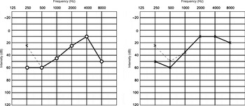
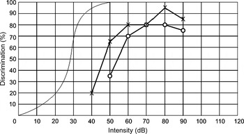
Pure tone threshold audiometry demonstrated thresholds of 60 dB nHL at 250 and 500 Hz, with a rising slope up to 10 dB nHL at 4000 Hz at both ears (Figure 1). Speech audiometry at the right ear showed a speech reception threshold shift of 28 dB and a discrimination loss of 20 per cent; at the left ear, a threshold shift of 20 dB and a discrimination loss of 10 per cent were found. Both ears showed a slight regression at 90 dB nHL (Figure 2). The determination of reliable thresholds was compromised because the responses of the patient were often slow and inconsistent. Although both tone and speech audiometry results raised the suspicion of possible aggravation (i.e. some exaggeration of the actual hearing loss), the outcomes of two repeated tests were consistent. This prompted a full audiometric assessment.
Pure tone audiometry results.

Speech audiometry results.

Speech in noise thresholds were obtained. We observed elevated signal to noise ratios of + 6.8 dB for the right ear and +6.4 dB for the left ear (normal range: −8 to −3 dB ).Reference Plomp and Mimpen23
Tympanometry showed a type A tympanogram in both ears, according to the visual admittance classification system of Liden and Jerger.Reference Liden25, Reference Jerger26
Middle-ear muscle acoustic reflexes were absent at all frequencies, at the maximum stimulus level of 110 dB, at both ears.
Clear otoacoustic emissions were present in both ears, with a whole-wave reproducibility of 97 per cent at the right ear and 98 per cent at the left.
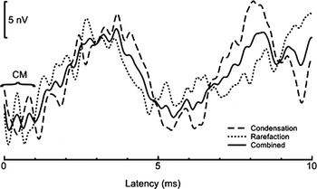
No clear auditory brainstem responses were observed in either ear, up to 100 dB nHL. However, both ears showed cochlear microphonics (Figure 3). The possibility of stimulus artefact was excluded by performing a no-stimulus control run with the insert earphone tubing clipped.
Cochlear microphonics results.

Hearing aids did not improve the patient's speech reception ability, and were rejected by the patient. A trial with an assistant listening device was offered in order to facilitate listening in noisy environments.
Discussion
The presented case displayed a combination of audiological outcomes which is generally considered to constitute evidence of auditory neuropathy spectrum disorder, according to international guidelines, i.e. (1) speech audiometry results apparently in disagreement with tone audiometry results; (2) detectable otoacoustic emissions; and (3) no clear auditory brainstem response, other than cochlear microphonics in the first milliseconds.16
This patient's audiometric results showed an inconsistency between pure-tone and speech audiograms, but results were consistent in longitudinal analysis. Middle-ear muscle acoustic reflexes were absent at all frequencies, at the maximum stimulus level of 110 dB, at both ears. This is in line with observations in other cases of auditory neuropathy, in which no normal reflexes were found.Reference Berlin, Hood, Morlet, Wilensky, Li and Mattingly9 Clear otoacoustic emissions were present in both ears, providing proof of normal outer hair cell function.Reference Kemp27 Normal outer hair cell function was also confirmed by cochlear microphonics, in the absence of clear auditory brainstem responses.Reference Berlin, Morlet and Hood17, Reference Starr, Picton, Sininger, Hood and Berlin18
Auditory neuropathy spectrum disorder is an uncommon symptom in adults. Our patient had been previously diagnosed with Kjer's disease, a condition associated with optic neuropathy. The combination of optic and auditory neuropathy suggests that both may be based on a common underlying factor, possibly related to a mitochondrial defect associated with the OPA1 genetic mutation.Reference Bette, Zimmermann, Wissinger and Knipper6, Reference Amati-Bonneau, Odent, Derrien, Pasquier, Malthiery and Reynier15
The OPA1 gene encodes a dynamin-related mitochondrial guanosine triphosphatase.Reference Amati-Bonneau, Milea, Bonneau, Chevrollier, Ferre and Guillet14, Reference Delettre, Lenaers, Griffoin, Gigarel, Lorenzo and Belenguer28, Reference Alexander, Votruba, Pesch, Thiselton, Mayer and Moore29 The OPA1 protein is involved in mitochondrial processes, and is widely expressed in the retina and brain.Reference Bette, Zimmermann, Wissinger and Knipper6, Reference Amati-Bonneau, Milea, Bonneau, Chevrollier, Ferre and Guillet14, Reference Delettre, Lenaers, Griffoin, Gigarel, Lorenzo and Belenguer28–Reference Zeviani30 An abnormality of the OPA1 protein may cause mitochondrial dysfunction, leading to insufficient energy support, which can result in neurodegenerative diseases.Reference Bette, Zimmermann, Wissinger and Knipper6, Reference Amati-Bonneau, Milea, Bonneau, Chevrollier, Ferre and Guillet14, Reference Matsunaga, Kaga and Starr31 Slower recovery of neurons due to reduced cell energy may disrupt the synchrony of neural firing, which could explain the observed abnormal auditory brainstem response.Reference Manchaiah, Zhao, Danesh and Duprey32 Presumably, the slow recovery of neurons could affect both the auditory and visual system. This may explain the combination of auditory and visual neuropathy present in our patient.
During testing, we were initially suspicious of aggravation of hearing loss. This was prompted by the inconsistency of the tone and speech audiograms, and by the patient's delayed test responses. However, two factors caused us to question our initial suspicions. Firstly, repeated tone and speech audiometry tests gave consistent outcomes, despite the patient's unusually delayed responses. Secondly, the low-frequency hearing loss indicated by the tone audiogram did not match the generally flat hearing loss that is more typical in cases of aggravated or simulated hearing loss. Subsequent results for brainstem responses and emissions led to the diagnosis of auditory neuropathy. We believe that the patient's inconsistent, delayed responses were probably a consequence of impaired auditory processing.
• The presented patient showed a rare combination of Kjer's disease and auditory neuropathy spectrum disorder
• Otorhinolaryngologists should be aware that optic atrophy can be accompanied by hearing loss
• Additional audiological tests may help diagnose auditory neuropathy spectrum disorder when clinical symptoms are unclear
• In such cases, initial, inconsistent audiological results may raise suspicion of simulated or aggravated hearing loss
This case shows that the combination of unexplained hearing loss and apparently inconsistent audiometric outcomes may be associated with auditory neuropathy spectrum disorder. However, such unexpected hearing evaluation outcomes can be related to neurological conditions, such as Kjer's disease. In the presented case, the combination of auditory neuropathy and Kjer's optic neuropathy may be based on a common mechanism, possibly related to slow neuron recovery due to an abnormality of the OPA1 protein.