

In “The New Accelerator,” a short story first published in 1901, by H.G. Wells, the physiologist Dr. Gibberne seeks to invent a drug that will accelerate the life of the body two or three times. Gibberne is a specialist on the action of drugs on the nervous system; the aim of the New Accelerator is to act on the body at a level more basic than that of the organ – not to make something that “gets at the brain champagne fashion and does nothing good for the solar plexus,” but instead to make a drug that “is a stimulant that stimulates all round, that wakes you up for a time from the crown of your head to the tip of your great toe, and makes you go two – or even three to everybody else's one” (Wells Reference Wells1979, 340).
Gibberne surprises even himself, inventing a potion that makes the body think, move, and work thousands of times faster. He and the narrator of the story – a writer who is the chemist's neighbor – take a dose of the drug and explore its effects. Gibberne warns the narrator that even though he will feel normal, “you will be going several thousand times faster than you ever did before, heart, lungs, muscles, brain – everything.” Part of the strangeness of the drug is the paradox of relative speeds, such that the effect of accelerating one's body is to make the rest of the world seem as though it is going thousands of times slower than it went before.
Gibberne and the narrator walk in amazement through this apparently slowed world, looking at “the end of the curtain, frozen, as it were, corner high, in the act of flapping briskly in the breeze” (ibid., 345). While they are going “fast all over,” hearts beating a thousand times a second, they watch a bee “sliding down the air with wings flapping slowly and at the speed of an exceptionally languid snail” (ibid., 347). They walk past a band playing, “though all the sound it made for us was a low-pitched, wheezy rattle, a sort of prolonged last sigh that passed at times into a sound like the slow muffled ticking of some monstrous clock” while around them “frozen people stood erect; strange, silent, self-conscious-looking dummies hung unstably in mid-stride” (ibid.).
This story, penned as it was at the turn of the twentieth century, is clearly responding to new experiences of transport and photography, a story of time when time itself seemed to be changing in nature (Doane Reference Doane2002; Kern Reference Kern1983). The narrator describes a physiology of expressions that suggests Edison's popular kinetescope record of a sneeze, or Georges Demeny's chronophotographic study of faces laughing or pronouncing “Vive la France!”: “A wink, studied with such leisurely deliberation as we could afford, is an unattractive thing . . . one remarks that the winking eye does not completely close, that under its dropping lid appears the lower edge of an eyeball and a line of white” (Wells Reference Wells1979, 347; Braun Reference Braun1994, 176–177). At the outset of the story, the narrator asks disbelievingly if such a thing as the new accelerator could even be possible: “‘As possible,’ said Gibberne, and glanced at something that went throbbing by the window, ‘as a motor-bus'” (Wells Reference Wells1979, 341). The motor-bus is even built into the temporal structure of the sentence; the glance, the bus, and the realism of the possibility are all compactly contained in one moment.
There is no reason to expect that changes in senses of the possible that we characterize as modernism should manifest in art, literature, and music, and not also appear in areas of human endeavor such as biology, particularly when historical actors such as Wells cross between the cultural domains of biology and literature (Pauly Reference Pauly and Ross1991). If we may speak of a biological modernism, it concerns in part a change in the materiality and fixedness of time in relation to biological matter; it is perhaps not a surprise to find biologists reading Wells’ story and seeing themselves in it. Writing in 1937 in a work entitled Biological Time, the biophysicist and popular philosopher Pierre LeComte du Noüy wrote an account of time-lapse microcinematography of cultured somatic cells as analogous to the potion in “The New Accelerator”:
Not only can we contemplate [cells] alive and magnified thousands of times on the screen, but we dispose, thanks to the cinematographic technique, of the apparently superhuman power of contracting their time in comparison to ours. Like the character in a remarkable story by Wells, we can accelerate the rhythm of the life of our constitutive elements. (LeComte du Noüy Reference Lecomte du Noüy1937, 103)
In time-lapse microcinematography of living cells, images are taken at regular intervals, and then the film is projected. An apparatus set up to take one frame per second, for example, produces 3,600 frames per hour, and those 3,600 images can be watched in 3.7 minutes at a projection rate of 16 frames per second (Michaelis Reference Michaelis1956, 36). The effect of having such a difference between the speed of capture of the images and the speed of projection of the images is to make it seem as if the cells are moving quickly. Watched through a microscope without such intervention, cells other than those of single-celled microorganisms generally move or change shape so slowly that it occurs below the threshold of perception of the observer. A cultured fibroblast, for example, moves at about 1 μm per minute, or its own length in one hour (Abercrombie Reference Abercrombie1980).
For du Noüy, microcinematography was akin to ingesting a drug that accelerated the body by acting on all its elements – cinema worked to accelerate cells in relation to the rest of the world. No matter that the cells were not actually any longer inside a body, but explanted into the glass and fluid vessels of tissue culture; they were inside an extended technological body to which the drug of cinema could be applied. Microcinematography is used by Du Noüy as the demonstration of the tangibility and measurability of biological time, a kind of time that is not the same as clock time. Du Noüy, who studied the healing times of wounds suffered by French soldiers on the front during World War I under the tutelage of surgeon Alexis Carrel, found that the younger the soldier, the faster the time of wound healing. Physiological time was a Bergsonian quantity that had to do with the physical accumulation of duration, which existed apart from clock time or the time of physics (Carrel Reference Carrel1931). The body was composed of heterochronies, as it was entirely possible that different tissues in different organs would move through biological time at distinctive rates according to their form and function. We who live in clock time can access these multiple teeming temporal worlds within with the help of cinema.
I begin with Wells’ story, and du Noüy's reading of it to capture the elements out of which this essay's arguments are constructed. First, cinema is a technological portal to another world of time. The word world is used by makers and watchers of microcinematography with the nonchalant confidence of a term assumed to be the right one, and it is a marker of the paradoxically inside-out nature of an exteriorized and seemingly autonomous temporal world of interior life. Jean Comandon, for example, early twentieth-century biological filmmaker and the first theorist of the microcinematograph, saw himself as a kind of impresario standing at the threshold of another world:
Scrutinizing that infinity in which he is immersed, the scientist discovers new worlds as teeming, as varied as our own. It is into one of these worlds, that of cells and microbes, that I am going to conduct you in a moment.Footnote 1
The world that lies through the portal is not elsewhere, not in outer space or under the sea. It is in and of the everyday macroscopic world, located in places such as “the interstices of time,” and unknowable without the intermediation of something – a moving image, an animation – that pulls a phenomenon “into human time” (Myers Reference Myers2006, 15).
Second, “The New Accelerator” highlights the importance of the affective elements of observation. The experience of moving through the portal – like the narrator's experience of speed being felt as everything else slowing – is counter to the intellectual understanding of what is happening. In microcinematography cells seem to move frenetically, agitatedly, even though the viewer “knows” that they are “in fact” moving extremely slowly in relation to human perceptual abilities. The feeling or affect of experiencing the microcinematographic film is similar: “a kind of shock to the retina,” that seems strange and “mad” (Wells Reference Wells1979, 344–346). One can ask what microcinematographers could see (or, observe, quantify, or identify) that they otherwise couldn't, but I want to pose the question more broadly, and inflected by cinema studies: What kind of spectators, experiencing what kind of perception, were the scientists using moving images to study cells in the twentieth century?
These scientists were not passive recipients of moving images; they constructed the very machines that generated the images. Because “few such instruments exist commercially. . . most research workers who wanted to use them for their work had to invent, design, and construct them for themselves” (Michaelis 1965, 71). The history of the microcinematographic portal is analyzed here in corresponding technical detail. The chronometers, incubators, cell cultures, micropuncture lasers and lathe beds are not here for their own sake, but as constitutive elements of distinctive portals to distinctive worlds, in other words, it is an understanding of technology as a distinctive form of scientific knowledge. This essay has learned from arguments for different shades of histories of technologies and technological methods – “materialized epistemology,” “instrument epistemology,” “thing knowledge,” or the study of scientific methodology – the imaging instruments and things of the laboratory, and how their use was debated and tinkered with in “methods talk,” can be productively interrogated epistemologically alongside more classical understandings of scientific knowledge as thought and text (Wise Reference Wise2006; Baird Reference Baird2004; Schickore Reference Schickore2007).
It is a commonly held view, with a history that originates in the eighteenth century, that instruments such as microscopes extend human vision (Schickore Reference Schickore2007). Or as Don Ihde has argued, “technologies, particularly instruments . . . should be regarded as means by which our perceptions and our wider experience are modified and transformed” (Ihde Reference Ihde1998, 1). Thus it follows that the microcinematograph extends our perception into the world of the very slow, the very small, and thus modifies our experience of cellular life and mediates the very possibility of its study. The reverse proposition is explored less often, perhaps because it seems less intuitive: that experience and human perception may be regarded as a means by which our technologies are modified and transformed, including “technologies of living substance” (Landecker Reference Landecker2007).
In the category of instruments transformed by experience and human perception I include cells, which as part of microcinematography become instruments for the investigation of their own lives and deaths. “What is extended, perhaps, is not the observer's sense but the living process of the body studied, and the epistemological domain of the apparatus in the generation of ‘life'” (Cartwright Reference Cartwright1995, 27). In this living machine, images of cellular life iteratively change the very means for living visibly in vitro; culture vessels and microscopes begin to move, become coupled to engines and gears, the fluids and surfaces of in vitro life shift, and the images generated again and again feed back into the apparatus for culturing and image-making. Cells are not outside this apparatus, as the “object” of study; as with other (macroscopic) filmic objects, cells “participate in the photographic presence of themselves . . . they are essential in the making of their own appearances” (Cavell Reference Cavell1979, xvi). “Cellular life” as a scientific object takes on a specific character with the physical embedding of cells in an incubator at the heart of the microcinemotographic machine where they participate in their own appearance (Latour Reference Latour and Daston2000).
What cinema does to biology and what biology does to cinema in the twentieth century is an important subspecies of the broader question of “cinema and the invention of modern life”; as such, it is a point of articulation with film theory and history more broadly (Charney and Schwartz Reference Charney and Schwartz1995). The question of the specificity of the film medium, the automation of image-making and observation, the relation of film and photographic technology to the spectatorial experience of a projected world, how viewers are placed subjectively in relation to the filmic depiction of physical reality, and film's “confrontations with time and time's passing,” have been considered at length and in depth in film studies (Rodowick Reference Rodowick2007, 73). While the “scientific cinema, its technical spectator and its filmed bodies make up an aspect of the cinematic apparatus that breaks in crucial ways with paradigms of cinematic technology and spectatorship generated in film scholarship around narrative and pictorial film texts and genres,” the relationship of macro to microcinema may also be understood to be at productive odds (Cartwright Reference Cartwright1995, 8). Despite differences – or perhaps because of them – it is particularly provocative to read theories generated for films of the macroscopic world in relation to films of the microscopic world. Both historical and philosophical considerations of scientific film can be radically and usefully opened to the larger world of cinema by close engagement with film theory; at the same time, perhaps the peculiarities of cellular films and biological times will inflect those theories in interesting new ways.
Longstanding philosophical issues are marked by the keywords “world” and “experience.” Here I limit the many possible discursions into phenomenology by using David Rodowick's rereading of Stanley Cavell's classic The World Viewed: Reflections on an Ontology of Film to think about films of the microscopic world (Cavell Reference Cavell1979; Rodowick Reference Rodowick2007). Why not just use Cavell (or other philosophers, for that matter)? The suitability of Rodowick's reading, poised as it is “between the questions what was cinema?” and “what will digital cinema become?” is due in part to the contemporary rediscovery of the power of time-lapse moving images in biology with the explosion of live cell imaging (Rodowick Reference Rodowick2007, 84). We are currently in the midst of a period of intense technical innovation in imaging molecules in living cells: vital fluorescent probes added or engineered into living cells, photomultipliers for detecting the faint fluorescent signals, motorized microscopes, digital recording devices, and computational software used to process, enhance, and reconstruct an image on a screen from the dataset gathered by the signal detectors. The “film and bright lights” era that is the subject of this essay is decisively over in biology, but paradoxically the value of interrogating it seems more urgent (Roux et al. Reference Roux, Münter, Frischknecht, Herbomel and Shorte2004, 334). Not that historians of science or practicing biologists ever asked “what is cinema” with such obsessive fervor as scholars of film apparently have, but they nonetheless may still participate in the feeling that “as film disappears into the electronic and virtual realm of numerical manipulation we are suddenly aware that something was cinema” (Rodowick Reference Rodowick2007, 31).
What was cinema to biology? Or, put slightly differently: what was the specificity of the film medium for cell biology, and thus what difference did it make to use moving images to study cellular life and not other media? Rodowick argues that a renewed fascination with film is related to thinking about the “new automatisms and ontologies” we may confront in the emergence of digital media. This essay, certainly, is motivated in part by thinking about how the cinematic life produced in the twentieth century both structures and is displaced by the new fluorescent molecular worlds of the living cell of contemporary digital practices (Landecker Reference Landecker2011).Footnote 2 The question of whether biology done using moving images was different from biology done without has both historical and contemporary import; in fact it reaches into the near future.
Here I tie the specificity of the microcinematographic machines and time-lapse films generated in them to a very specific form of production of a “microscopic world.” The notion of a microscopic world exists well before microcinematography, but the introduction of time-lapse mechanisms and practices significantly transforms the quality of that world from one that is spatial in dimension to one that is another world in time. The first part of this analysis focuses on the production of the microscopic world-in-time specific to microcinematography; the next three parts raise the question of what these practices do for biology that is different from other methods of visualization and analysis? What difference does cinema make in cell science?
The difference even raising this question makes to historical scholarship will perhaps be evident immediately in that the body of this essay concerns scientists to whom scant to no historical attention has ever been paid – Michael Abercrombie, a British embryologist and “pioneering ethologist of the cell”; Warren Lewis, and American embryologist and anatomist; Charles Pomerat, an American cell biologist and toxicologist; Wilton Earle, an American cancer biologist; and Marcel Bessis, a French haematologist and specialist in the study of leukemia (Medawar Reference Medawar1980, 11). Their work corresponds roughly to three areas of cellular life that proceed in the present continuous tense: creeping (cellular locomotion), drinking (pinocytosis or endocytosis), and dying (apoptosis).
I investigate these figures as the builders of machines for projecting the microworld, and in turn understand those worlds as worlds viewed – screened, experienced worlds that “place us subjectively with respect to a depicted physical reality” (Rodowick Reference Rodowick2007, 46). These aspects, I argue, are essential to any understanding of the basic question of the specificity of the film medium for cell science. In an ethnographically informed approach, I have sought to reconstruct aspects of experience and affect that were part of these figures’ work with science and film, out of the movies and documents they have left. This immediately raises the problem of capturing the perhaps ineffable in either textual inscriptions or the remaining film record, experience that may lurk in the interstices between possible modes of recordable expression and thus might be better accessed ethnographically in the present with practicing scientists, as anthropologist Natasha Myers has done so well with protein crystallographers (Myers Reference Myers2006; idem Reference Myers2008).
This caveat is offered on the way into going ahead and asserting that at least some important aspects of how it felt to work with film are nonetheless potentially extractable from the historical record, from two main sources: the way microcinematographic instruments were built and rebuilt, and from written descriptions of the practice. A sense of a distinctive world of cellular time is – like Wells’ compact containment of possibility, the glance, and the motor-bus in one narrative moment – built into the very sentence structure and grammar of papers written about cells studied using film. Elsewhere, I have discussed how microcinematographic films were narrated to and met by public audiences; but here I am particularly interested in the building of laboratory microcinematographic apparatuses, the making and viewing of time-lapse films as scientific work, and the affective and intellectual effects of working with/in the resulting cinematic microscopic world (Landecker Reference Landecker2005).
Part 1. Worlds and Times: The Microworld as a World Viewed
Cellular creeping behaviors were seen through the microscope well before cinema. Amoeba, protozoa, and white blood cells were observed moving around and behaving in response to stimuli well before the twentieth century (Schloegel and Schmidgen Reference Schloegel and Schmidgen2002). The act of looking at another scale of magnification has for several centuries been linked to the idea that things microscopic can be described as residing together in a spatial entity that can be called a world, as Catherine Wilson illustrates in her able description of early microscopy (Wilson Reference Wilson1995). Indeed, the “microscopic world” is a phrase that sounds entirely unremarkable even though most people would be hard pressed to define the location, edges, or definition of such a world. That microscropic world is assumed to be connected to this world, in fact to constitute it. This is often expressed as the microscopic being inside the macroscopic; in the case of living matter from the human or animal body, it is often depicted as an “inner world,” despite the manifestly obvious fact that microscopy is seldom endoscopy, and things generally have to be extracted from the body to be made visible.
At least since Thomas Huxley's Sunday-evening lecture of 1868 on the subject of his Scottish lay audience's protoplasmic basis and entirely cellular composition, it has been a popular pastime for biologists to spread the message that persons, by virtue of their constitutive biology, are not singular, individual, unitary or alone, but divisible, partable, and in their fundamental constitution multiple. Huxley told his audience that all one had to do to see that one's body was an aggregation of cells was to prick a finger, and look through a microscope at the “innumerable multitude of little, circular, discoidal bodies” that were the red blood cells, and at the white blood cells that “will be seen to exhibit a marvelous activity, changing their forms with great rapidity, drawing in and thrusting out prolongations of their substance, and creeping about as if they were independent organisms” (Huxley Reference Huxley1869, 133).
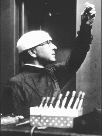
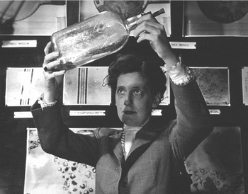
In other words, the technique of looking inside oneself to see the innumerable was to detach a bit of oneself and magnify it. In order to look inside oneself, one had to look outside oneself, and through a microscope. This gesture of extraction and presentation was most often done by the scientist such as Huxley, who stood between the screen or image and the audience, and said of the image: “this is you” (figs. 1a and 1b). The very fact that you could take a part of you and put it in front of you so that you could look at it, carried the full force of being multiple.
The gesture of extraction and presentation. Cells and representations of cells were demonstrated before the viewer's body to induce an experience of the inner world of the body. (1a) shows Alexis Carrel, a Franco-American surgeon and avid proponent of tissue culture, holding up a tube of cells to the light (undated, courtesy of Georgetown University Special Collections).

(1b) shows a scientist holding up a bottle of cultured cells in front of a wall of micrographs of cultured cells (undated, courtesy of the National Library of Medicine).

The philosopher Henri Bergson said of this partability of bodies in 1907 that “individuality . . . harbours its enemy at home.” As he saw it, reproduction, a basic characteristic of living beings, opposed any tendency to individuation.
For individuality to be perfect, it would be necessary that no detached part of the organism could live separately. But then reproduction would be impossible. For what is reproduction, but the building up of a new organism with a detached fragment of the old? Individuality therefore harbours its enemy at home. Its very need of perpetuating itself in time condemns it never to be complete in space. (Bergson Reference Bergson1911, 13)
Like the vision of life which hangs before Hans Castorp in Thomas Mann's Magic Mountain, the externalized vision of internal multiplicity could be both impressive and frightening simultaneously: “This body, then, which hovered before him, this individual and living I, was a monstrous multiplicity of breathing and self-nourishing individuals” (Mann Reference Mann1966, 277). Well before microcinematography then, elements of the microscopic world as a spatially distinct location residing inside bodies, which when externalized seemed surprisingly autonomous of the human, were part of the visual and conceptual understanding of cellularity.
What changed with microcinematography at the beginning of the twentieth century was the constitution of the microworld as a distinctive temporal domain. And this transformation, a fundamentally cinematic one, was not minor: “To see at a distance in space was commonplace by the nineteenth century. But to see at a distance in time was so confounding that it took nearly a hundred years to comprehend it” (Rodowick Reference Rodowick2007, 64). What would it mean for biology to be suddenly able to “expand or contract the time scale at will”? (Michaelis Reference Michaelis1956, 36). To understand the nature and significance of this shift in the microworld – to a world in time – and its implications for knowledge of the living, we need to understand the cinematic specificity of this twentieth-century microworld: the microworld as a world viewed (Cavell Reference Cavell1979).
Consider this description by Ronald Canti, read to the 1927 Tenth International Zoological Congress in Budapest, before screening The Cultivation of Living Tissue (Canti Reference Canti1928). These “außerordentlich schönen Filme” were, according to the editors’ note in the conference proceedings, shown on several occasions during this congress, and Dame Honor Fell, director of the Strangeways Laboratory where Canti filmed in this period, reminisced in the 1950s that she would “never forget the sensation that his film of migration and mitosis created when he showed it” in this venue (Fell Reference Fell1958, 490). Canti's lecture, however, began not with the film but with an extremely detailed account of the microcinematographic apparatus:
The trigger mechanism which determined the taking of a photograph and the changing of a photographic film was provided by a suitably modified electric clock which could be arranged to deliver electric impulses at the required intervals. A single impulse from the electric clock was led to a relay switch which closed the electric circuit actuating a small electric motor fitted with a suitable resistance in series for slow running. This motor was fitted with a warm gear and slowly revolved a drum carrying two cam wheels and four projecting arms for making mercury dip contacts. The function of the cam wheels was to pull upon wires running in a flexible spiral wire tube . . . and to actuate at a distance the two photographic shutters, the one for taking the microphotograph and the other for photographing the watch on the back of the film. (Canti Reference Canti1928, 88)
The apparatus was suspended between two clocks; one triggered the shutters; one was photographed when the shutters opened; and the image of the cell was merged with the image of the watch face with each exposure of the film.
The four projecting arms for making mercury dip contacts had four distinct functions. The first mercury contact short-circuited the resistance in series attached to the motor to make it run slowly, to facilitate the starting of the motor when the current reached it. The second turned on an electric light that illuminated the watch. The third sent a single impulse back to the relay switch to break the circuit to the motor and make it come to rest after having caused a single revolution of the drum. The fourth set a current going to a second electric motor. The second motor produced one revolution of a shaft, which caused one revolution of the gear of the camera film-driving mechanism and placed a new portion of film in position for taking the next photograph.
The microcinematograph thus took the automaticity of the photographic process to new heights, or built into it even further distances from the human hand: “Photographs are not hand-made; they are manufactured. And what is manufactured is an image of the world. The inescapable fact of mechanism or automatism in the making of these images is the feature Bazin points to as ‘[satisfying], once and for all and in its very essence, our obsession with realism'” (Cavell Reference Cavell1979, 20). Realism in scientific photography of course has its own terminology – “mechanical objectivity” might capture it best, a discourse of the mechanical replacement of the subjectivity of the observer that has its own distinct history in scientific representation (Daston and Galison Reference Daston and Galison2007). Another microcinematographic setup, built by Jean Comandon in France around the same period, was admiringly described as a “little factory of great precision,” which neatly expresses some of the satisfaction with this instrument's capacities to automate observation (Kazeef Reference Kazeef1938).
For philosopher Stanley Cavell, the removal of the human agent from the task of photographic reproduction was of interest not so much in terms of what photographs “represent, picture or mean,” but in terms of “how they place us subjectively” (Rodowick Reference Rodowick2007, 63). And because of its automaticities, “photography maintains the presentness of the world by accepting our absence from it. The reality in a photograph is present to me while I am not present to it” (Cavell Reference Cavell1979, 23). That reality present to the viewer is cropped from an indefinitely larger field: “A camera is an opening in a box: that is the best emblem of the fact that a camera holding on an object is holding the rest of the world away” (ibid., 24). Every explicit section of reality depicted thus carries with it the implied presence of the rest of the world, and its explicit rejection.
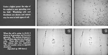
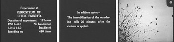
This sense of the presentness of a cropped section of the world is very vivid in the making and viewing of microcinematographic films, partly because the thing being viewed – a slide of living somatic cells – is already understood to be extracted from a body and more generally from the stream of biological time. In Canti's film, for example, scale is indicated on the film by showing the explant in the middle of a microscope slide being held by a hand. Then magnification is slightly increased by showing the middle of the slide and the fragment of tissue – the hand of course disappears as the viewer is drawn by the camera into the microscopic scale. Then the film shows the explant being photographed under a 50 mm objective lens, and then a 20 mm objective. The explant at this last magnification was itself outside of the field of view, positioned so that the viewers would see the edge of the growth sweeping across the screen, suggesting the “indefinitely extendible and contractive” frame, a frame extended in the direction of the very small in microcinematography by the addition of the microscope: “A close-up is of a part of the body, or of one object or small set of objects, supported by and reverberating the whole frame of nature” (fig. 2a) (Cavell Reference Cavell1979, 25).
Early scenes in Ronald Canti's Reference Canti1928The Cultivation of Living Tissue emphasized the constant growth, movement, and reproduction of cultured cells as they swept across the field of view. Intertitles narrated the scenes at different scales of magnification, instructing viewers to watch the wandering actions of the incessantly moving cells. Tissue culture itself was a technique for “cropping” complex living cells from the body and growing them isolated in glass vessels in the laboratory. Cells were extracted from the animal body and grown long-term in vitro; in this case the cells were originally derived from rat tumor or chick embryo tissue. Care had to be taken to keep the cells steady at body temperature so they would survive being filmed, and the apparatus included an incubator and stringent measures to restrict the amount of light shone on the sample.

The temporal microworld accessed through the microcinematographic apparatus does not share with macrophotography a sense of shock to human historical time – the time manipulated here was perceived to be within the endless repetitive rhythm of biological life. One recording of cell division or migration was considered to be pretty much the same as any other recording of cells, where the cell division or migration itself was unaffected by the course of human affairs even if the lighting or magnification or resolution of the image of cell division was changed by technical developments. What was photographed with these tools was, in Comandon's terms, a condensation of an extracted piece of teeming infinity; it had a fundamentally non-human otherness, even if it was considered an externalized view into “life within.” Both the rest of the body and the rest of the other scales of possible time were held away but also implied by the microcinematographic frame reverberating with “the whole frame of nature.” This apartness was accentuated by the need for the technical portal between the time of the observer and the time of the cell.
Rhetorically, Canti was pitiless in his depiction of the massiveness and singularity of the microcinematographic set-up that held the rest of the world away. The sense of dividing this space off from the macroworld in order to mechanically depict the microworld was enhanced by detailed attention given even to the embedding of the apparatus in concrete – any relative movements between the objective lens and the object being examined would cause blurring, so the whole thing was seated in a mass of concrete poured beneath the floor of the laboratory. Four metal posts embedded in the concrete passed up through holes in the floor; on the top of these posts was cast a slab of concrete and resting on this slab were alternate layers of concrete and rubber sponge. This layer also had four posts embedded in it; on these rested a wooden shelf on which sat the camera, pointing down above the microscope, which sat on the lower concrete layer surrounded by an incubator. These arrangements were described to the audience in relentless detail before the film was screened.
It has been argued that in the opening years of cinema it was “the technology which provides the immediate interest: what is promoted and sold is the experience of the machine” (Heath 1981, 221). The experience of the film and projector technology itself, which may have become commonplace for other kinds of moviegoers in the late 1920s, does not fade in relation to the film's content in the same way in scientific uses of film. For it continues to be mandatory to explain one's methods, and a continuously renewing “machine interest” is built into the discursive demands to foreground one's means of objectively representing the real and legitimating it as a means of registering life phenomena. Thus the opening technical description was a necessary part of experiencing the film and the apparatus at the same time. And when the film begins to roll, there is never a moment when that technical portal to the experience of cellular life becomes invisible (fig. 2b).
Dramatic scenes of cell division in the cultured tissues were accentuated by a switch in the third reel of The Cultivation of Living Tissue (1928) from direct light microscopy to dark field microscopy, in which the background was dark and the specimen was lit from the side. Note the ever-present chronometer hovering in the upper right hand corner of the screen, which indicated the contrast between the time unfolding in the experiment itself, and the accelerated time of viewing.

For Cavell, the automatisms of the photographic basis of film are one part of understanding cinema as “a succession of world projections.” The projector and screen are equally important components in generating the “relative specificity of cinematic experience as the projection of an ‘autonomous’ world” (Rodowick Reference Rodowick2007, 54). In this case, the screen was three meters high, and the magnification of the cells 45,000 to 78,000 diameters. It was a time-lapse film of embryonic chick cells and Jensen's Rat sarcoma cells in culture dividing and dying from radiation exposure, narrated by the filmmaker to his scientific audience as follows:
The chromosomes are seen forming an equatorial belt and as they migrate rapidly to the poles of the cell it seems as if they were pulling away from one another as in a “tug-of-war.” During this time bubbles of protoplasm are repeatedly shot out from the surface and withdrawn, and the constriction appears in the middle of the cells. This constriction deepens slowly at first then moves rapidly until finally there is only a narrow isthmus remaining. This persists for some time and then is finally broken away sometimes with a snap as the two newly formed daughter cells wander away from one another. (Canti Reference Canti1928, 92)
As Canti described what was happening on the screen he depicted a frenetic and violent scene of action and change, while noting that the scene was shown at 160 times its actual speed. He also used terms such as “snap” and “tug-of-war,” even as he cautioned his viewers in the very same sentence that “in watching these pictures there is always a desire to translate the movements into actions of every day life.” Then, radium is applied to the field of cells.
The field is seen as a seething mass of cells of both the motile and fibroblastic types . . . [a] cell may be seen in prophase at the top right hand corner just before the radium is applied at 6 o'clock. This cell, thus, subjected to irradiation while in mitosis, is prevented from completing division. At 6:20, i.e. twenty minutes after the emanation was placed in position, all the cells in the field are rendered motionless with an almost dramatic suddenness. (Ibid., 96)
Watching these films, the language of constant and violent form of movement becomes more comprehensible. Though one might think that it would be more accurate to marvel at the slowness and smallness of these changes upon seeing how much time elapses so quickly in order to bring the cellular movements to a scale of human perception, the image of the clock face on each frame of the film actually contributes to the sensation of speed; this is not just a recording of time elapsed, it is an image of time. The ever-present chronomoter or clock face in the corner of the picture hovers with its hands twirling like a frantic parody of time-keeping, so that the spinning of clock hands just adds to the impression of rapid and unceasing motion. Movement is all over this imposing screen as well, as the insides of cells are full of cytoplasmic streaming and the movements of organelles as the cells themselves change shape and move across the field of view. This is only accentuated by the spinning of the reels of the projector and the flicker of the projected film (or it would have been, before watching such a thing on Quicktime on one's computer, which is the situation of today's spectator).
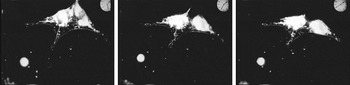
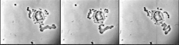
And then comes death. It is indeed an “almost dramatic suddenness,” in fact it is a surprisingly shocking cessation of movement, as the film rolls on and the clock hands twirl on, and the cells, just moments before a paroxysm of activity, become inert (fig. 2c). The screen becomes a moving image of stillness; the textual description of the scene shifts, its sentences shorten and stop seething and continuously undulating, and the image blurs.
Again twenty minutes after irradiation has commenced the cells are rendered motionless. This is quickly followed by contraction into the spherical form and by the breaking down process. . . . The cells throw out bubbles over their surface but these bubbles do not [show] the same active motion as was seen in the breaking down Sarcoma cells in the first reel. Further as time proceeds the outlines of the cell become frayed out and ragged. A rounded cell near the bottom of the field which is quite sharply defined at first is seen suddenly to commence disintegration at 3:45. (Ibid., 96)
The audience – the zoologists meeting in Budapest – experienced what was by all accounts a viscerally moving experience, the screening of life and death, but this should not be mistaken for some sense of “really being there,” which of course in this case would mean also being irradiated.
The depth of the automatism of photography is to be read not alone in its mechanical production of an image of reality, but in its mechanical defeat of our presence to that reality. The audience in a theater can be defined as those to whom the actors are present while they are not present to the actors. But movies allow the audience to be mechanically absent. The fact that I am invisible and inaudible to the actors, and fixed in position, no longer needs accounting for. . . . In viewing a movie my helplessness is mechanically assured: I am present not at something happening, which I must confirm, but at something that has happened, which I absorb (like a memory). (Cavell Reference Cavell1979, 25)
Canti asks the viewers of the scene of stilled dead cells to overlay this scene with a memory from just minutes before: “If the mind is now carried back to the commencement of the experiment the striking differences will at once become more apparent; the cells have ceased their movements, changed their shape and have commenced to break down” (Canti Reference Canti1928, 96). In Cavell's analysis, the screen that holds a projection “light as light” acts to both screen the viewer from the world and the world from the viewer. For Cavell, these “phenomenological facts of viewing” were “coextensive with the subjective condition of modernity, imagined here as a filmic way of encountering the world.” That is, that film embodied a philosophical situation – an ontology of “a self divided from the world by the window of perception” – a moving image of skepticism (Rodowick Reference Rodowick2007, 66–68).
A series of scenes at the end of The Cultivation of Living Tissue (1928) showed what happened when cultured cells were exposed to radiation. The rapid onset of stillness and death in the visual field after radium is applied is shown repeatedly, with each experiment's technical parameters indicated before the cells are shown and viewers directed to what they should see. These stills cannot capture the dramatic and paradoxical sensation of the transition from a moving image depicting constant movement to a moving image of stillness. The dead cells still move occasionally as if in the currents of the liquid they are in, but this movement is of an entirely different quality than those of the living cells.

For the 1920s biologist this subjective condition of modernity held no angst, quite to the contrary, it appeared to be a quite superb investigative position. The automatism, the distancing, the removal of the human hand, the extendible and contractible frame on the world, the positioning as the observer “to whom the actors are present while they are not present to the actors,” that excellent modernist viewpoint of invisible anonymity, all of these were reasons why, as Canti put it, film was a technique that had been chosen to address problems “which for various reasons could not be solved by direct visual observation”:
It was thought that by elaborating a cinema technique it would be possible to obtain records over long periods of time, which could be examined in a much shorter period of time, and further, which could be reexamined on as many occasions as might be required for their interpretation. Again it would be possible to run the film backwards and observe the events in the reverse order of time and thus trace to their origin any changes which might have taken place. In short it was decided that a technique which could afford opportunities such as these would constitute an ideal method for biological investigation. (Canti Reference Canti1928, 87)
This biologist did not want to be present to the cells at exactly the point they were present to him. There was little control to being there, in the moment, so to speak. Rather, the desire was, as du Nöuy put it, to be able to re-examine, run backwards, to disengage the observer's presence into parts that could be manipulated in relation to one another, specifically manipulating the time of observation in relation to the time of experiment. This would allow one to “accelerate the rhythm of our constitutive elements” – to dodge into the interstices of time and observe unsuspected from there this other temporal world.
Canti's narrative accentuated the automatic nature of the device. The shutter was not triggered by a human hand, but by an electric clock. The emission of light, the sequential movement of the film, the recording of time – all were automated. The human observer was even further distanced by the need to shield bodies from the radium source; the incubator was built with 2-inch lead walls. Further, the experiment, the work of the coupled microscope-incubator-film camera, was not accessible except through projection of the sequential images triggered by and including the various time-pieces. These are not just technical details: they constitute the world viewed.
Canti's films apparently caused astonishment in audiences from fellow biologists to Prince Albert, H.R.H. the Duke of York, to whom the films were shown in 1933 (Wilson Reference Wilson2005; Anon. 1933). They were taken as visual evidence of the possible efficacy of radiation treatment for cancer, since the radium exposure seemed to act to stop cells in the act of division, which supported the idea that tumors with actively dividing cells would be damaged more than the surrounding normal tissues. What I am interested in here, however, is not necessarily Canti himself, or the significance of either this particular film or the findings abstracted from it. Instead, I have staged the scene of this premiere of The Cultivation of Living Tissue to the scientific community and read it against Stanley Cavell's analysis of cinema as a succession of automated world-views in order to return to the question of worlds, and the ease with which microscopic and very slow things and their images are spoken of as other worlds.
In general, to get to another world, one needs a technology of one sort or another, even if it is simply a portal or trap door. This can be seen in a number of literary works, themselves portals of the narrative kind. H.G. Wells employed a Time Machine to get to the future in The Time Machine, a neuroactive potion to get to the interstices between seconds in the short story “The New Accelerator” discussed above, and a spaceship to get to the moon. In the case of cells, one world is connected to another by a portal in the form of the microcinematographic apparatus. Understanding this temporal microworld as specific to cinema helps explain the inside-out spectatorial experience of the extractive gesture: seeing a world of life that is internal to the body externalized, that is at once deeply foreign and startling and yet uncannily evocative of the “actions of everyday life.” The things on the screen are animate and are easily anthropomorphized, but at the same time are understood to exist in a world apart – the technical portal of cinema generates the experience of an autonomous world. Thus this temporal microworld is held at a distance by the very technology that makes it accessible; the apparatus and the face of relative time is as present as the cellular movements themselves in the experience of the projected film. This particular microworld is not just accessed by some entrance way or another, it is actually defined by that door. Given the necessity of the technological portal in scenarios science fictional and scientific, one could even arrive at a working definition of another world as that which has such a portal restricting access to it, determining what may be on the other side of it.
Part 2. The Microworld Viewed: Creeping, Drinking, Dying
What then did biologists do with this door to another world? The whole reason to return to the question of the specificity of the film medium in biology was to pose the question of what was distinctive to this spectatorial experience in terms of scientific knowledge production. In this section of the paper, I examine the work of some little-known but fascinating figures from twentieth-century biology, and argue that the apparently straightforward fact that they shared a technical interest in using and expanding the reach of cinematic techniques in biology can be analyzed as a shared style of encounter with the cell. This is not exactly the same as a “thought style”; but if, following Gaston Bachelard and Hans-Jörg Rheinberger, one understands the techniques, objects, and practices of representation in the laboratory as the “thinking machinery” of modern science, perhaps one may speak of a machine style in relation to certain genealogies of research (Fleck Reference Fleck1981; Rheinberger Reference Rheinberger1992, idem Reference Rheinberger1997).
As mentioned briefly above, time-lapse microcinematography began in earnest in the early twentieth century in the work of Parisian doctor, biological researcher and filmmaker Jean Comandon. Comandon has received extensive attention from historians of science and film, including myself, and so I will not linger on his contribution here, except to point to the fact that he was the first to explicitly raise the question this essay is pursuing, that of the specificity of the film medium for the production of biological knowledge.Footnote 3 In Comandon's view, time-lapse microcinematography was emblematic of the emergence of microbiology from what he called “the anatomical era” of fixatives and stains to the “physiological era.” The physiological era was characterized by the living subject: “one observes them anew in the living state, one can follow their modifications, their evolution; one experiments on the action of diverse chemical reagents or physical events that change their milieu” (Comandon Reference Comandon and Fescourt1932, 319; emphasis in the original).
It perhaps seems a simple point, that whether one's observations are of a dead subject or a living subject makes a difference – but it is deceptively so. There was something particular to cinema's “field of view,” so to speak, that repeatedly over the twentieth century drew biological film-makers to study cells as relational beings, in quite stark contrast to those who studied what one might call the more cell-intrinsic aspects of life, particularly the cellular structures involved in mitosis and meiosis (cell division). The artifactuality of histological methods of the anatomical period was, in Comandon's oeuvre, depicted as two simultaneous forms of representational violence: ripping the cell from its context of interacting elements, and from its world of time, the one proper to it. Comandon's classic films were dramas that showed the meeting of bacteria and white blood cells, the penetration of cells by parasites, the battle between nematodes and nematode-trapping fungi. In short, he was not so much interested in things as in the worlds of things; there was no necessity for those worlds to be natural, in fact, watching the cells react in time to the experimental manipulation of chemical reagents and physical forces in the milieu was one of the perceived benefits of the technique.
2a. Creeping
In order to explore the question of the specificity of the temporal microworld for cell biology, I turn here to the work of Michael Abercrombie. Abercrombie was an embryologist and cell biologist, who early in his career was deeply influenced by Ronald Canti, whose cinematographic apparatus was detailed above. Abercrombie spent a short postgraduate stint in 1934 working for C. H. Waddington at Strangeways Laboratory, where Canti did his initial cinematographic tinkering. Abercrombie is perhaps best known today as the person who coined the term “contact inhibition” in 1953 to describe how normal cells stop moving when in close contact to one another (in contrast to malignant cells, which are not stopped by such proximity). After Strangeways, Abercrombie moved to several other institutions, including University College London, before eventually returning as director of the Strangeways Laboratory at the end of his career, but this early exposure shaped the core of his practice for thirty-five years: film as a tool for the observation and quantification of cellular movement.
Peter Medawar called Abercombie a “pioneering ethologist of cells,” likening his study of the behavior of individual cells on film to the work of animal ethologists – who of course used film extensively in their studies of animal behavior (Mitman Reference Mitman1999). Writing that Abercrombie favored traditional methods of small-scale tissue culture over the mass tissue culture that became the norm later, Medawar observed that “if this had been the prevailing style of culture in Michael's day we would have learnt as much about the behaviour of individual cells as K. Lorenz and N. Tinbergen would have learnt about the behaviour of geese and ducks if they had confined their attention exclusively to large flocks or large populations instead of studying individual birds and their interactions with other birds” (Medawar Reference Medawar1980, 11).
It is in Abercrombie's work that the long-term results for biological knowledge of being startled by film begin to show. The originality of Abercrombie's thought and practice lies in his fundamental assertion that cell locomotion was a basic biological process, and thus a mechanism that required both empirical elaboration and theoretical synthesis across different kinds of organisms and across apparently different biological events, such as embryonic development and wound healing. Abercrombie saw cell locomotion as being not just overlooked as a fundamental morphogenetic phenomenon over the twentieth century, but as being both actively resisted and unconsciously suppressed by what he called “mitotic infatuation”: the notion that cellular movements in embryogenesis, wound healing, and tumor formation could be explained by localized cell multiplication, “an assumption that went strangely unquestioned, and hence was difficult to displace” (Abercrombie Reference Abercrombie1977, 337).
Abercrombie observed that E. B. Wilson's famous book The Cell in Development and Heredity conveys the impact of the discovery of mitosis, “that began the line of research to modern genetics,” but does not once mention cell locomotion. This revelation of mitosis, “this marvelously intricate process, closely linked with the deep human interests in generation and heredity, and of vast potential theoretical importance, made a profound impression which it seems to me has been a profound nuisance ever since to those concerned with the rightful place of cell locomotion” (ibid., 339). He demurred on a cause for this “temporary blind spot” in twentieth century biology, writing, “it is rash enough to suggest such an unconscious obstruction by a basic assumption has existed. It would be even rasher to try to explain it” (ibid., 343).
I will practice no such restraint. Abercrombie's insinuation of the blindspots produced by a fascination with the nucleus, mitosis, and then genes and DNA, to the exclusion of biological phenomena of growth and movement, gives us some insight into the intellectual and practical distinctiveness – and marginality – of using film as a research tool. Abercrombie's research was defined by his dual interest in cell locomotion and cell behavior – in movement, and movement in relation to an environment. In both cases he was using film to generate an empirical basis for a theory most other biologists didn't even know they needed. His vision on both counts was synthetic. On locomotion, he wrote: “The mechanism of locomotion of metazoan cells when they are crawling on a solid substrate can fairly be said to be wholly unknown” (Abercrombie, Heaysman, and Pegrum Reference Abercrombie, Heaysman and Pegrum1970, 393). He generated a set of hypotheses based on watching cells move on film. These movements were of cells in culture crawling over the glass surface of the culture vessel.
The locomotory organ of a fibroblast is a ruffled membrane. An isolated fibroblast moves with changes of direction by waxing and waning of ruffled membranes at different parts of its margin. The ruffled membranes on an individual cell are competitive. They can either be inhibited or their life can be prolonged by the cell environment, notably by contact inhibition and contact guidance respectively. Mutual contacts form fairly persistent adhesions, linking fibroblasts into a meshwork. (Abercrombie Reference Abercrombie1961, 198)
Note that in the systematic cascade of hypotheses here, the study of cellular action becomes the study of cellular interaction. The behavioral aspects of locomotion, or “how the movement is guided in particular directions and started or stopped by the conditions around the cell,” he called the “social behavior” of cells (Abercrombie Reference Abercrombie1980, 129). In fact, he had only begun to systematically study cell locomotion after extensive observations of the fact that most cells stop moving when they come into contact with other cells – with the notable exception of many malignant cells. Posing the simple question of “why do the cells stop moving?” led to the extensive study and theorization of cellular locomotion detailed above (Dunn and Jones Reference Dunn and Jones1998, 125).
Both locomotion and its necessary counterpart locomotion-in-interaction were not features of cell-intrinsic qualities such as mitotic figures or organelles. Under the sway of mitotic infatuation, cellular behavior, too, went untheorized, and its observations thus went unsystematized: “No systematic attempt, so far as we are aware, has been made to apply to cellular societies the analytical approach which has been so successful in the study of animal social interaction: that is, to take the cell as a unit and to investigate how its behaviour is influenced by other cells” (Abercrombie and Heaysman Reference Abercrombie and Heaysman1953, 111). As noted above, creeping or crawling behaviors of cells had been observed since the very early days of microscopy. Observations of cellular behavior were many; but translating a welter of experiences of seeing cells into some kind of theoretical unity was not easy, first because it was not evident that a theoretical unity for cellular movement was called for, given the dominance of explanations of growth and wound healing based on mitotic cell division; second because of the technical challenges of formalizing observation of movement as quantitative regularities (Kelty and Landecker Reference Kelty and Landecker2004). The cell biologist Paul Weiss, who also made extensive use of film in studies of cell behavior, suffered just this sort of failure of his peers to recognize the worth and significance of the visual representations of dynamic phenomena he made (Brauckmann Reference Brauckmann2004).
Generations of film scholars have commented on the peculiarity of trying to write about film, and the hopelessness of trying to use frame enlargements or stills, making it a “particularly unquotable” artistic form: “the written text cannot restore to it what only the projector can produce: a movement, the illusion of which guarantees the reality” (Bellour 1975, as quoted in Rodowick Reference Rodowick2007, 21; emphasis in original). The power of film was thus at the same time its weakness, in that it enabled observation, but not necessarily quantification; we might say that it is also a particularly unquotable scientific form. Abercrombie was the first to take the experience of the temporal microworld rendered by film and translate it into a set of phenomenological laws of cellular movement, and he did this with a particular form of quotation. He transformed thousands of hours of observation of many different kinds of cells into quantitative methods based on single-frame analyses of single cells.
Tracking from frame to frame the fate of single adhesions between membrane and glass was a way to graphically record the pattern of locomotion in relation to cellular movement. Quoting the projection was Abercrombie's method:
For the analysis three parallel reference lines, the equivalent of 6.25 μm apart, were marked on the screen, and orientated in the direction of the cell movement (fig. 1). The leading edge of the cell was in almost all cases broad enough to span all three lines. The position where the leading edge cut each line was recorded, to the nearest 1.25 μm, at intervals equivalent to 30 sec. of real time. The quantitative description did not gain from using a shorter time interval, though the intermediate frames were often useful for qualitative observation when change was rapid. Thirteen chick cells and seven mouse cells were analysed, each for a period averaging 25 min, during which time the cells moved with no perceptible change of direction. (Abercrombie et al. Reference Abercrombie, Heaysman and Pegrum1970, 393)
The interrelated convictions that (a) locomotion was a basic property of living cells equivalent to cell division and common to all metazoan cells, and that (b) locomotion was environmentally shaped, where the environment was understood to consist in part of other cells, were thus based upon a wealth of quantitative data derived from sequential stills of time-lapse films. The still “quotation” should not, however, detract from the understanding of Abercrombie's view of his subjects as a fundamentally cinematic one. In his obituary, his longtime friend and colleague Peter Medawar commented that like the animal ethologists, Abercrombie spent a great deal of time observing his subjects; “if need be,” Medawar wrote, Abercrombie would stay in the lab and “film cells all night” (Medawar Reference Medawar1980, 11).
Given the emphasis on automaticity in the time-lapse device, one might wonder why anyone needed to be there, as the cells lived and died and crept around and the film rolled, pulled from frame to frame by timers and motors, the shutter and the light source and the incubator and the clock all built in. However, it is clear that Abercrombie experimented widely with temporal intervals between exposures, and spent a great deal of time trying to create “scenes” in which individual cells could be followed or the interactions between two cells could be tracked. In the behavioral experiments, two explants were put close together, so that the zone of interaction between cells growing out from each explant could be filmed. And the conviction as to what needed quantifying – which subjects and behaviors needed to be subjected to frame-by-frame analysis and statistical treatment, was based on hours spent watching films of cells behave.
In sum, creeping was for Abercrombie a phenomenon that was accessible only through the cinematic portal. The difference of the temporal microworld was to be respected; it had to be occupied for some time, and the phenomena specific to it recognized despite their lack of appearance in the time of human perception. As Abercrombie observed, “the cell locomotion involved is very slow, so that direct observation gives no immediate impression that anything is going on, as it does for instance with mitosis. It also leaves virtually no trace in fixed specimens” (Abercrombie Reference Abercrombie1977, 337). Temporal manipulation was one aspect of access to this world, but the apparatus itself was intrinsic to the systematic observation of it. The ability to operate on the projected world was central; the screen was marked with a grid to measure the movements of the leading ruffled edge of the cell. In other studies, the space between the cell membrane and the glass substrate it was crawling along provided the means for studying locomotion: with Adam Curtis, Abercrombie used interference reflection microscopy, which depended on the space between the cell membrane and the glass cover slip substratum it was crawling along to generate the image (Curtis Reference Curtis1964; Abercrombie and Dunn Reference Abercrombie and Dunn1975). Where the membrane was closest to the glass, the image was darkest, because of the interference between the light reflected from the membrane and that reflected from the glass it was close to. Where the membrane was further away, there was a smaller comparative phase shift between the two kinds of reflected light, resulting in a brighter part of the image. Watched over time, then, the relationship of the cell membrane to the glass culture vessel could be mapped – and triangulated with electron microscopy images of thin sections of cells cut longitudinally. This work “helped to lay the foundations for modern theories of crawling cell movement and to stimulate a research effort into the dynamics of the cytoskeleton that continues unabated today” (Dunn and Jones Reference Dunn and Jones1998, 126).
The work of Abercrombie and his associates awaits proper historical analysis. Historians of science, too, have sadly suffered from the “mitotic infatuation,” and though Abercrombie is widely recognized in the scientific community as a founder of the systematic study of cell behavior and locomotion, no historical attention has been paid to his work to shift the very nature of the biological questions being asked of cells. These questions, I would argue, were being posed from a fundamentally cinematic point of view: they were asked of the actions of actors in a continuous present, of movement phenomena only accessible through time-based study. It is not that Abercrombie did not care about mechanism or causal explanation. But the experience of cellular life was at the center of what was seen to be the problem in need of explanation – “why do cells stop moving?” – is a fundamentally different kind of question than, say, that of the biochemical constitution of cell fractions. If the history of cell biology is seen only as the history of “discovering cell mechanisms,” the role of observation of living subjects in twentieth-century biology remains underappreciated (Bechtel Reference Bechtel2006). What was accessible through the technological portal of cinema was, then, specific to biology in that it put time and movement at the center of analysis. The questions posed to Abercrombie by the microworld viewed (or by Abercrombie to the microworld viewed) were at fundamental odds with those that had been asked before via mostly static media; this work suggested a set of theories of cell life that most biologists who were focused on cell-intrinsic structures and processes did not know they needed.
2b. Drinking
Abercrombie's work is perhaps a classic demonstration of the expansion of human perception by technical means. The observation of the internal and very slow, speeded up and enlarged by the coupling of cell culture with microscope and film camera, extended the realm of human perception to include phenomena of which they had theretofore been unaware. The systematization and theorization of the observations made possible by these visualization technologies – the development of a system of “quotation” of biological film – helped solidify cell behavior and cell locomotion as coherent scientific objects in need of investigation. At the same time, the observer's interaction with the projected microworld meant that the constraints and particularities of human experience and perception were transformative for those very technologies, including the living parts of the apparatus. In the following example, filmic representation is not an endpoint of experiment. The “results” of experiments conducted with the microcinematograph are not inscriptions of cellular processes, but perception that is built back into the apparatus, without any necessary passage through theory, words, publication, or even explicit recognition. It is the literal construction of the life-world of the filmic subjects, the solids, fluids, and gases they exist and persist in. In the following example, the observation through microcinematography that cells continually “drink” from their surroundings was built into the apparatus used to contain cells, and had a dramatic impact on the ability to sustain in vitro life, particularly human cell lines.
In 1927, embryologist Warren Lewis traveled to England to visit Ronald Canti, and study his microcinematographic set-up. He traveled with Canti and Honor Fell to Budapest, where he witnessed first-hand the impact of showing films to scientific audiences, and upon arriving back home to the Carnegie Institute for Embryology, immediately began to assemble his own apparatus (Landecker Reference Landecker, Maienschein, Glitz and Allan2004). In 1929, he observed a phenomenon of cell behavior on film that he called pinocytosis, “drinking by cells,” meant to complement the earlier phagocytosis, “eating by cells.” It was, as an anonymous reviewer wrote in the Journal of the American Medical Association in 1930, one of the “reactions unpostulated by conventional histology” accessible through film, “in which macrophages mechanically drink the surrounding plasma” (Anon. 1930, 1509).
Lewis’ work had a range of effects. First, the realization that cells were continuously taking in the equivalent of three times their own volume in liquid in a twenty-four hour period “placed desirable emphasis on the conception of the cell as a dynamic system,” as one cytology textbook put it (Sharp Reference Sharp1934, 7). Second, the observation of an economy of intercellular fluids was the beginning of an entire subfield of biology focused on the way substances move in and out of cells enclosed in membrane-bounded vesicles, now referred to as endocytosis. This legacy is clearly visible in a conceptual genealogy – the idea of intercellular transport of fluids and small molecules becomes, later in the twentieth century, an idea that organizes a set of investigations that are part of figuring out how cells work. Third, one can also trace a less explicit technical legacy of the specific temporal world accessed by microcinema, one built into the apparatus for keeping cells alive rather than recognized as an idea, theory, or disciplinary subfield. Here there is an explicit link between the visual medium of film and the liquid medium in which cells are cultivated; the movement of one and the temporal capture of dynamism changes the perception, composition, and mechanical agitation of the other.
One sees this shift in the roller-tube apparatuses first developed in the Lewis laboratory by George Gey, which were widely adapted into tissue culture after the 1930s in a move to large-scale culturing of cells (Gey Reference Gey1933; Landecker Reference Landecker, Maienschein, Glitz and Allan2004, idem Reference Landecker2007). These instruments replaced static dishes holding cells sitting in a still medium with tubes that were constantly rolled or agitated to provide a continuous washing of the cells within the medium and aeration of the medium. No historical actor ever says, “I built the machine this way because I saw these films”; one observes here a correlation between the realization that cells take in an amount of fluid the equivalent of their entire volume in a few hours and the introduction of constant movement into the machines built to keep them alive. Gey's roller-tube methods were quickly incorporated into tissue culture practice, in part because they allowed much larger quantities of tissues to be grown in the laboratory than the previous tissue explants placed in hollow microscope slides or in still flasks. Swept up in the effort to use cultured cells in the quest for a vaccine against polio, the roller-tube apparatus was quickly scaled up and produced commercially by biological supply companies. Soon laboratories all over the world contained rotating racks of growing cells.
Observations of the incredibly dynamic interaction between a medium and cells, and the difference between life and death that could be produced by making the apparatus itself move dynamically refocused attention on the cellular medium, and led directly to a practice that suggested a dialectical understanding of cells and environment. Is it a coincidence that the investigators most interested in making use of the medium of film were also the most focused on the cells’ role in producing culture media and the culture medium's role in producing the cellular morphology and behavior of the cells living in it? I suspect that it is not, exactly because of the behavioral or physiological information available in watching cells over time, rather than the morphological or structural information of histological or biochemical visualization. One is always watching the cells in the microscopic field, and the microscopic field is not just a blank backdrop – it is the medium that is keeping the cells alive to be filmed, and its constitution can dramatically change what a cell does as it is observed.
The biologist Charles Pomerat, for example, described pinocytosis as follows:
The free edges of cells in tissue culture, as recorded by phase-contrast time-lapse movies, are in continual wavelike movement, laving the medium around the cells and often sweeping over and engulfing a portion of the surrounding fluid, forming what is usually called a “vacuole,” but which contains the various molecules that may be in the surrounding liquid. This “vacuole” may be seen to move toward the center of the cell, into which it has been swept by the curling edges, and, as it moves, the phase boundary between it and the cytoplasm of the cell becomes less and less distinct, until presently there is no “vacuole.” (Leake and Pomerat Reference Leake and Pomerat1958, 162)
As with other microcinematographers, the grammar of cellular movement was one of the continuous present, with the cells’ membranes depicted as “sweeping” and “laving,” and “curling,” with the liquidity of the scene overwhelming the sentences. Pomerat was interested in the biology of endocytosis, but also in what it meant for cellular life that the outside of the cell was constantly being ingested; for the boundary between inside and outside becoming “less and less distinct.” He proposed and set out to establish the study of “cell toxicology,” the systematic study of toxins added to the cellular medium of cultured cells, for one could observe at a cellular level the effects of these substances and record “the results in the dynamic relationships of time change” (Pomerat and Leake Reference Pomerat and Leake1954, 1110).
As in Abercrombie's work, the sense of respect for the cell as a powerful and autonomous agent behaving in another dimension beyond normal human perception shows through the analyses of Pomerat's films: “One is impressed with the capacity of cells to withstand a great deal of chemical and physical stress. The process of reversible vacuolization described here may help to elucidate one of the normal cellular processes which cope with deleterious conditions” (Yang et al. Reference Yang, Strasser and Pomerat1965, 505). He criticized other analyses of cellular life based on what he saw to be an insufficiently live understanding of the cell membrane: “Let us not be misled by the dead appearance of cell boundaries in stained and fixed preparations into thinking only and exclusively in terms of a semipermeable membrane and its behavior. In living mammalian cells there is a phase boundary separating the cells from the extracellular fluid around them, and this phase boundary is in continual activity, exhibiting continual pinocytosis” (Leake and Pomerat Reference Leake and Pomerat1958, 163).
Pinocytosis thus introduced toxins and carcinogens into the cellular medium. It was also built into other elements of the apparatus. Wilton Earle, a cytologist at the National Cancer Institute known for his technical creativity, built a cinematographic set-up in which two microscope-camera complexes were synchronized. In a report from 1943, he wrote that, “Since the microcinematograph is used primarily in the study of carcinogen-treated cells with untreated cells as controls, it is necessary to have paired optical systems, lights, lenses, and cameras so that two cultures can be photographed simultaneously under identical conditions” (Earle Reference Earle1943, 135) Particularly interested in observing, over time, the behavior and morphology of cells in different environments, he built his own experiment-control apparatus in order to compare the same cells in different milieu.
It was an incredible instrument, with time, temperature, and movement built into it, the whole thing built into a laboratory mounted on moveable lathe beds. The two microscopes were enclosed in a constant temperature bath, necessitating that the apparatus included a heater, thermostat, and air circulator, as well as a thermograph to provide a written record of the temperature during the interval of any one film. The two cameras were held at constant height, and the microscope-air-bath-lamp assemblies were supported from above by two parallel vertical lathe beds, adjustable to a variable height. The cameras allowed an exposure frequency ranging from one frame per second to one frame per 2 minutes, with a range of magnification from 20 x to oil immersion. An observation ocular or beam splitter between microscope and camera insured centering and focusing the image and keeping it under observation while the apparatus was running. To ensure an exact record of time and to enable frame-by-frame comparison between the two films, an image of a clock appeared with each frame.
Microcinematography made tissue culture apparatus move; cultured cells in turn made the camera move. Since Earle found it was a common occurrence for the cell or cell groups to migrate out of the field of view during the course of the experiments, which often happened over days, he mounted the cameras on a lathe carriage. This carriage automatically shifted, synchronized with the cameras so that with each revolution of the camera shutter the camera moved a predetermined lateral or transverse distance. The theory was that with enough images, the cellular subject would be captured by this automatic roaming over the possible field of movement. It was in Earle's laboratory that an essentially dialectical theory of the cell and its medium emerged; a classic 1948 experiment demonstrated that a single cell could be grown in culture as long as it was placed in a very small amount of medium that was already “conditioned” by having had other cells cultured in it (Sanford et al. Reference Sanford, Earle and Likely1948; Landecker Reference Landecker2007). The idea that cells generated the medium in which they lived was the logical correlate to pinocytosis; although it was not immediately evident what happened to the liquid taken into cells, it must be also coming out again, otherwise the cells would quickly explode.
One could make the same kind of argument about endocytosis as was offered for cell locomotion – microcinematographic access to the temporal microworld established a set of questions and proposed a theory about cell-medium relations that biologists were not even aware they needed. Lewis, Pomerat, and Earle, like Abercrombie, spent incredibly long hours immersed in the films they made, and the conviction of the universality of a phenomenon across different kinds of cells was founded on such extensive experience of many filmed experiments. In this section I have focused instead on the way the experience of these films was built back into other parts of the apparatus for maintaining, cultivating, experimenting with, and observing life over time. The historical work of Hans-Jörg Rheinberger demonstrated that representations of life are never endpoints in themselves, but are essential components of broader experimental systems: “knowledge is not simply iterated, but is differentially driven forward. If the scientific spirit has once established itself on this path of embodiment of knowledge, it must keep its instruments and experiments, as well as its concepts and theories, in permanent motion and change” (Rheinberger Reference Rheinberger1992; idem Reference Rheinberger1997; idem Reference Rheinberger2010, 23). The specificity of the film medium in biology in this instance is that its distinctive temporality of observation, and the sense of watching an autonomous world with its own laws and interrelations is built into the very means of technical maintenance and experimental configuration of the living subject: the constantly moving is built into the constantly moving. In this way we may also understand culture media as time-based media.
2c. Dying
My third example returns us to France, in the early 1960s. Because Jean Comandon was extremely prolific as well as extremely long-lived, at this time he was actually still a contemporary of a young hematological oncologist working in Paris called Marcel Bessis. Bessis knew Comandon, and shared his assertion that the cinematograph was an instrument of research, particularly in the venture to bring physiology to the cellular level. In addition, Bessis tirelessly campaigned for the reinvigoration of microcinematography now that it could be pursued in concert with phase contrast microscopy and new video technologies. Where the older generation had been able to observe cellular movement, Bessis was interested also in the intracellular movements of organelles brought into sharp relief by phase-contrast methods. The introduction of the phase-contrast microscope, invented by Fritz Zernike in the 1930s and widely available commercially by the 1950s, greatly heightened the amount of detail that could be seen in the unstained cell through a light microscope.
Bessis was, like Warren Lewis before him, fascinated by the unending process of vacuole formation and transport within the cell, and studied it in depth. Like Wilton Earle, he used microcinematography to watch the effect of pharmacological agents on cells, predicting the rise of a “cellular pharmacology” on the basis of his observations. As a hematologist he was also interested in the normal and pathological behavior and interrelation of the different kinds of blood cells. He found it astonishing that hematologists “who have so minutely analyzed cellular morphology, should have ignored almost all the dynamic phenomena exhibited by the blood cells” (Bessis Reference Bessis1955a, 272). It is Bessis’ work studying cell death after injury that is my focus below; it best illustrates how the cell on film is not an outcome of experiment, or a representation of a cell, but an instrument in the analysis of life – or in this case death-in-life. The cell becomes an instrument of its own analysis through the employment of film in concert with tiny instruments of destruction.Footnote 4
Bessis used microcinematography as an essential part of understanding death, due to film's dissecting and analyzing qualities. In calling for the rigorous development of a “symptomology of the cell,” he complained that death was only ever studied after the fact.
This kind of death has long been studied by pathologists working with histological sections, that is, with fixed and stained dead cells, but it is very difficult to reconstruct from their appearance what has happened during the death struggle, at the moment of death, and after death. Indeed pathologists primarily study cell necrosis, that is, the appearance which is presented by events occurring after the death of the cell. Examination with phase-contrast microscopy together with recording by microcinematography has changed this situation, and the step by step development of phenomena which occur before, during, and after the death of the cell can be displayed for study. (Bessis Reference Bessis, Knight and de Reuck1964, 287; emphasis in the original)
Bessis used a laser to induce injury to the cell. Together with the physicist Georges Nomarski (better known for his invention of differential interference contrast microscopy), Bessis constructed a complex apparatus for a “micropuncture” procedure that was highly selective in its damage – the laser spot hit a 2.5 micron area of the cell for 10−3 seconds. The laser was targeted on the image of the cell projected on the surface of the laser – targeted using an image of the cell, so images were at play inside the instrument as well as being produced by it. Because transparent cells were not damaged by the laser, they had to be lightly colored with vital stains such as Janus green which absorbed the wavelength emitted by the laser. In films such as Death of a Cell (1956), a tiny white dot appears on the cell for the briefest instant. The cells then respond to this injury with a characteristic and spectacular series of changes, which Bessis called the “anatomy” of cell death.
Death was a “struggle,” a temporal process, a perceptible, capturable morphologically distinct sequence of events during which the shape and texture of the body of the cell changed. Bessis created a descriptive typology of cell death, including the stages of pathology (reversible deterioration), death agony (irreversible deterioration), death, and necrosis (the changes occurring in the cell corpse after death). The scenes of cell death in Bessis’ films are indeed spectacular, and agonizing – the cell throws out strange forms and twists and shudders (see fig. 3). A review in the Journal of the American Medical Association reported that the clinician would find this “a completely objective film of permanent value,” with dramatic final scenes that included “the pursuit of a doomed cell by a phagocyte and the cornering of a prospective victim by two cells that battle over its body” (Anon. [1957] 2004).
Scenes of the anatomy of cell death. From: Micropuncture of Cells by UV Microbeam, by Marcel Bessis, Research Laboratory of the National Blood Transfusion Center, Paris, France, Reference Bessis, Knight and de Reuck1964.

The anatomy of death differed with the placement of the microbeam; the highly targeted and selective damage allowed in addition the observation of the effect of one cell's death on its immediate environment, which Bessis called the study of cellular ecology. Death was observed both as an individual and communal event:
Thus we were able to study a phenomenon that is referred to as necrotaxis. Certain cells are attracted by the dying ones. If, for example, in human blood either a red or a white cell is irradiated, as soon as the irradiated cell exhibits any signs of alteration the surrounding cells travel toward it, and in a few minutes one observes what the anatomicopathologists call a rosette. These cells phagocytize the affected cell, most often by tearing it apart, each cell taking away a portion of the prey. In a few minutes there is no trace left of the cell cadaver. (Bessis and Ter-Pogossian Reference Bessis and Ter-Pogossian1965, 692)
In this work, what kind of instrument is the cinematographic cell – cultured, stained, incubated, injured, and filmed as it limps to an end or is consumed by others? One might understand the microscope, the laser, the camera, and the screen as the technology, and the cell as the subject of inquiry (and of injury), but this would be an artificial distinction – the whole is a portal to the world in which death-in-time may be observed and manipulated. Bessis’ work is interesting in its own right, and deserves its own detailed history, but here it allows us access to a certain relationship between life and technology in which the cell as part of the apparatus, the cinematographic cell, that can be accelerated “6 to 30,000” times, becomes an instrument of research. It is not the cell in and of itself that is the end of research but a tool in the reach for an understanding of death, the cellular ecology of death, and through death, life and disease.
In this case, differences between particular cells, and the morphology of different cells was not primary, the exact type of cells used in the experiment was secondary to the search for a universal set of characteristics of cell death. Again, observation over time enabled by microcinematography was key to this particular departure from morphology. Simply looking at a cell corpse (since pathological histology's subjects were by definition dead) would not give access to a characterization of disease and death as they happened, over time, in the life of the cell. They would give no access to death as a process of interaction, as a social or ecological event. How could you know anything about how a cell died from disease, if you didn't know anything about how cells died normally? There was no access to how disease happened over time in cells if one did not know how death happened over time in cells.
Bessis explicitly compared himself to the physiologist Xavier Bichat; he was studying cell death much as Bichat had begun a new era of studying bodily death centuries before. This comparison invites a perspective on cell death filtered through the work of Michel Foucault on Bichat in The Birth of the Clinic.Footnote 5 In Foucault's analysis the anatomo-pathological mapping of the concrete textures of the opened dead body in autopsy becomes the basis of the empirical rational gaze of the doctor directed toward the individual's living diseased body in the clinic: the gaze draws “the dotted outline of the future autopsy” on the body. This is what Foucault terms the “invisible visibility” of the interior of the body – the gaze maps on the opaque body how things will be visible in death (Foucault Reference Foucault and Sheridan Smith1994, 165).
In Bessis’ case, the rational gaze becomes cellular – a cellular symptomology is connected to cellular morphology during and after death. Instead of an open body there is the microscope field showing on closed circuit television, as the researcher intervenes to make death happen at a desired time. By this time diseases such as the leukemias that Bessis specialized in were understood to unfold at the level of the cell. The doctor's gaze is educated by film, and when directed at a living diseased body draws the dotted outline of cells blebbing and phagocytosing – it is not just a gaze that sees cells, but is a dynamic gaze. The microscope/time-lapse cinematograph allows the observation of the before, during, and after of death (as well as the added benefit of being able to repeat the observation as much as desired, to speed it up or slow it down, to watch it backwards, to isolate particular moments/frames of the process), and the laser is much like the autopsy hammer here; the cell does not exactly have to be opened up, but it does have to be extracted from the body, lightly stained, and incubated in a transparent chamber.
It was only later, when cell death such as Bessis described was called “programmed cell death,” or, alternatively, “apoptosis,” that a broader community of investigators began to appreciate the universality of the phenomenon in different kinds of cells during many different kinds of biological events, from development to radiation damage (Landecker Reference Landecker, Franklin and Lock2003). As with cell behavior, cell locomotion, and endocytosis, cell death became, in time, an entire subfield of study within life science; it solidified as an object of study through the phenomenological description of cells living in time. As genetic and biochemical techniques were brought to bear on it, these origins as cinematic observation of death-in-time have fallen into obscurity. As is the case with Abercrombie, Lewis, Pomerat, and Earle, Marcel Bessis has received little to no historical attention. As in these other examples, the cinematograph and the film medium produced a distinctive form of biological knowledge, with time and movement at its center; these forms of knowledge were characteristically at odds with dominant modes of static representation of cellular life. Like the character in the short story by Wells, as the narrator steps into the interstices of time, he accesses another world of phenomena, but he also becomes invisible to those around him who are living at another temporal scale.
Conclusion
The technical gesture of extraction and representation of “our constitutive elements” specific to cell culture and microcinematography produces a temporally specific microscopic world. For cell biology, this was a specifically physiological and behavioral world, and its observation produced a different kind of knowledge from other modes of investigation. I have sought to detail and thus specify the character of this microscopic world as a world viewed. For the scientist-spectator, wandering into the interstices of time generated ideas and practices of a very dynamic cell, one continuous with and to some extent determined by the micro-milieu of the visual field. Practically, this meant a renewed focus on cellular behaviors such as creeping and drinking and dying; it generated a visual world of the time-lapse film, but also the liquid, gaseous, glass world of the cells’ laboratory body.
I have put a great deal of emphasis on the importance of the kinds of automatisms built into and tinkered with by various microcinematographers, in both the act of filming and the process of projection, arguing with the help of Stanley Cavell that this understanding of the apparatus is part of understanding the specificity of cinema – in this case the specificity of cinema in the production of biological knowledge. In so doing, I have sought to elucidate the question of what cinema was to biology, in the context of an era when the moving image is again becoming central to the life sciences, but in a very different technical era. What links the microcinematographers examined here is not just that they all used film, or that they knew each other, or that they studied cells in culture; in each case, their subjective positioning as observers of an autonomous microscopic world produced questions unpostulated by other modes of inquiry, and established systematic phenomenological descriptions of cellular events that subsequently solidified into important objects of study of twentieth-century cell biology. The ability to be observers “to whom the actors are present while they are not present to the actors,” words written by philosopher Stanley Cavell to characterize moviegoers watching human actors, was also a very specific vantage point on cellular life, one that let that life roll over its viewers, its seeming rapidity, constant seething and teeming the experience of what was simultaneously intellectually known to be extremely slow.
Today we see the argument being made (again) for the importance of “cell phenomenology.” The term is used in a fairly straightforward way in research articles and scientific conference talks to denote the description and classification of phenomena as opposed to the study of mechanism or cause; for example, the pursuit of relations between nutritional milieu and bacterial physiology that are “analogous to phenomenological laws,” that can help elucidate how organisms work, “well before all the underlying regulatory circuits are elucidated at the molecular level” (Scott et al. Reference Scott, Gunderson, Mateescu, Zhang and Hwa2010, 1099). These biologists are not citing Husserl; there is no overt engagement with philosophy, yet it is nonetheless clear that a very fundamental question of how to approach the study of life is at stake in these arguments for systematically studying what cells do rather than focusing on their genes. Observing things live is a quite different tactic from trying to discern their blueprints or programs. At its broadest level then, the deceptively simple question of what difference it makes to visualize a living subject in time in biology is a question of how to broach the living. Understanding the twentieth-century scientist-spectator and the technological portal of cinema may help us better fathom the ways this microscopic world of the twenty-first century is being constituted.