CHD affects 9 in 1000 newborns, and its prevalence in adulthood is increasing, due to advances in patient care. Reference Baumgartner, De Backer and Babu-Narayan1 Complex CHD patients can survive into adulthood and represent a challenging for diagnostic assessment and therapeutic options. The following case highlights the importance of a multimodality imaging in the diagnosis and follow-up of these patients.
Clinical case
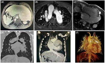
A 41-year-old male who was previously living abroad was referred to our adult CHD centre. He had a history of CHD, having underwent a Blalock–Thomas–Taussig shunt in the first days of life. In 1992 (with 15 years old), he underwent another cardiac surgery: ventricular septal defect (VSD) closure and implantation of a conduct between the subpulmonary ventricle and the pulmonary artery. As the baseline CHD was uncertain, several imaging exams were performed. The transthoracic echocardiogram showed what seemed to be a visceral and cardiac situs solitus, with mesocardia and levoapex. There was an atrioventricular discordance, but the ventriculoarterial connections were impossible to access. The morphologic left atrium was severely dilated and connected to the morphologic right ventricle (both these structures were on the left-side). The morphologic right atrium was connected to the morphologic left ventricle (positioned on the right-side). The systemic atrioventricular valve (morphologically tricuspid) had severe regurgitation, and the systolic function of the systemic right ventricle was at least moderately impaired. We proceeded the study with a cardiac magnetic resonance, to further understand the patient’s cardiac anatomy. The cardiac magnetic resonance showed abdominal situs inversus, with the liver medial and a polylobulated spleen (Fig 1a). The aorta was anterior and to the right of the pulmonary artery. Right-sided aortic arch is shown in Fig 1b. Levocardia and levoapex, with atrioventricular discordance and an ambiguous atrial situs. The left-sided atrium received the venous drainage of four pulmonary veins and was connected to the morphological systemic right ventricle (Fig 1c). The right-sided atrium received the systemic venous drainage and was connected to the subpulmonary morphological left ventricle. There was a persistence of the left superior vena cava (that drained directly in the right-sided atrium via the coronary sinus); an interrupted inferior vena cava continued via the azigos vein to the right superior vena cava. The supra-hepatic veins drained directly in the right-sided atrium. The systemic ventricle was dilated and hypertrophied, and its systolic function was moderately impaired (ejection fraction [EF] 34%). The subpulmonary ventricle was dilated and its systolic function was mildly impaired (EF 48%). A systemic atrioventricular valve insufficiency was observed. Ventriculoarterial discordance and hypoplastic main pulmonary artery were also observed. A left ventricle to the pulmonary artery conduct was observed; estimated pressure gradient is 58 mmHg, likely underestimated. A normal coronary anatomy was observed (the left main coronary artery arised from the left coronary ostium and the right coronary artery originated from the right coronary ostium). At this moment, our diagnostic hypothesis was a heterotaxy syndrome (left isomerism) with a dextro-transposition of the great arteries and pulmonary atresia, palliated with a left ventricle to pulmonary artery conduit. An angioCT scan confirmed that both lungs were bilobated and the main bronchi were hyparterial (Fig 1d); the atrial appendages morphology were similar (chicken wing) (Fig 1e); polysplenia and right upper quadrant stomach.
( a ) Cardiac magnetic resonance (CMR) image revealing an abdominal situs inversus, with the liver medial; ( b ) CMR image of the great vessels: the aorta was anterior and to the right of the pulmonary artery, with an right-sided aortic arch; ( c ) CMR image revealing the venous drainage of four pulmonary veins to the left-sided atrium, which was connected to the morphological systemic right ventricle; ( d ) CT image showing bilobated lungs and hyparterial main bronchi; ( e ) CT image confirming the similar morphology of the atrial appendages (chicken wing); ( f ) CT multiplanar reconstruction: the aorta was anterior and to the right of the pulmonary artery; persistence of the left superior vena cava and an interrupted inferior vena cava continued via the azigos vein to the right superior vena cava; ventriculoarterial discordance and hypoplastic main pulmonary artery were also observed; a calcified left ventricle to the pulmonary artery conduct was observed.

Heterotaxy syndrome is defined as “an abnormal symmetry of certain viscera and veins (lungs, liver, vena cava) and situs discordance between the various segments of the heart.” Reference Van Praagh, Sanders, Keane, Fyler, Lock and Teele2,Reference Lin, Krikov and Riehle-Colarusso3 It is an early embryonic development problem, accounting for 3–6% of all CHD. Reference Sharma, Devine, Anderson and Zuberbuhler4 It occurs in 1 per 10,000–40,000 people. Reference Lim, McCrindle and Smallhorn5 It is the cardiac condition with the highest familial recurrence rate, presenting a recurrence risk ratio of 79.1 among first-degree relatives. Reference Øyen, Poulsen, Boyd, Wohlfahrt, Jensen and Melbye6 Classically, patients were still classified as presenting with asplenia or polysplenia. Nowadays, right versus left isomerism (depending on paired bilateral atria morphology) is the most common classification, Reference Sharma, Devine, Anderson and Zuberbuhler4 as atrial isomerism is associated with morbidity and mortality. Nevertheless, this classification is not perfect, as the atrial appendage morphology can be difficult to assess, and there is not a perfect concordance between broncho-atrial relationship, atrial appendage morphology, and splenic status. Reference Yim, Nagata and Lam7 Both isomerisms can present with different CHD types, although left isomerism is more associated with cardiac abnormalities. Left isomerism is characterised by an interrupted inferior vena cava (as the left atrium lacks an inferior vena cava connection) in 80% of patients, absent coronary sinus in 30–55% of patients, and bilateral superior vena cava in 40–50% of patients. Reference Peoples, Moller and Edwards8–Reference Mishra10 A VSD or an atrioventricular canal defect is present in two-thirds of patients, transposition of the great arteries in 16%, and double-outlet ventricles in 15–37% of patients. Reference Sharma, Devine and Anderson9,Reference Yildirim, Tokel and Varan11,Reference Gilljam, McCrindle, Smallhorn, Williams and Freedom12 As for rhythm abnormalities, patients with heterotaxy syndrome can have either duplicated sinus node (right isomerism) or absent (left isomerism); this increases the likelihood of an arrhythmic event, with a prognostic impact. Reference Gilljam, McCrindle, Smallhorn, Williams and Freedom12 As for concordance between cardiac and pulmonary defects, patients with left isomerism are expected to have symmetric bilobed lungs with bilateral hyparterial bronchi. Lung lobation concordance was present in 71% of cases, and bronchial to pulmonary relationship concordance was present in 93% of cases, in a post-mortem series. Reference Louise Calder13 A central liver occurs in more than 70% of cases, and the stomach can be central-, right-, or left-sided. Reference Mishra10 Biventricular repair is only achieved in 40–50% of patients. Reference DeLeon, Gidding and Ilbawi14,Reference Hirooka, Yagihara and Kishimoto15 A common cause for reoperation is progressive systemic atrioventricular regurgitation. Reference Hirooka, Yagihara and Kishimoto15 Heterotaxy syndrome has a long-term adverse prognosis, with a recent study in a paediatric population estimating a 20-year survival rate of 54%, with a median age of death during childhood (0,47 years) Reference Banka, Adar, Schaetzle, Sleeper, Emani and Geva16 ; the survival rate and quality of life are determined by the concomitant CHD.
As the patient has heart failure with biventricular disfunction, severe systemic atrioventricular valve regurgitation, and a highly complex CHD, the patient is currently being assessed for heart transplant. The main issues to heart transplant in these patients are the development of HLA-directed antibodies (due to previous exposure to multiple blood products) and post-transplant pulmonary venous obstruction. Reference Razzouk, Gundry and Chinnock17,Reference Lamour, Addonizio and Galantowicz18
Conclusion
Heterotaxy syndrome is a rare clinical entity, with cardiac malformations being one of its major components. Heterotaxy syndrome is responsible for 3–6% of all CHD, with a wide spectrum of puzzling cardiac anomalies. Nowadays, with the development of multimodality imaging techniques, the comprehension of complex cardiac anatomies is more accurate, which can help physicians to better manage these patients. However, the long-term outcomes in this setting are poor and have not improved with the advance of medicine in recent years. Heart transplant, if feasible, is an option.
Acknowledgements
None.
Financial support
This research received no specific grant from any funding agency, commercial, or not-for-profit sectors.
Conflicts of interest
None.