I. INTRODUCTION
The photocatalysis based on semiconductor materials have aroused extensive concern on account that they can utilize the solar energy directly. Reference Lin, Li, Yu, Li and Yang1–Reference Wenderich and Mul4 Among various photocatalysts, titanium dioxide (TiO2) is considered as the most promising material in fields of photocatalytic water purification, photocatalytic water splitting into H2, CO2 photoreduction and so on due to its low-cost, chemical stability, and nontoxicity. Reference Muuronen, Parker, Berardo, Le, Zwijnenburg and Furche5–Reference Ma, Wang, Jia, Chen, Han and Li7 However, practical applications of TiO2 are severely hampered by its large band gap (3.2 eV of anatase), which can only be activated under UV irradiation. Meanwhile, TiO2 semiconductor also suffers from an easy electron–hole recombination. Reference Dahl, Liu and Yin8 In the past decades, much effort has been devoted to improving visible light absorption and decreasing the recombination of photogenerated electron–hole of TiO2 by diversified strategies, such as doping, Reference Jiménez, Bourret, Berger and McKenna9,Reference Asahi, Morikawa, Ohwaki, Aoki and Taga10 exposure of reactive facets of TiO2 Reference Yang, Sun, Qiao, Zou, Liu, Smith, Cheng and Lu11,Reference Yu, Low, Xiao, Zhou and Jaroniec12 and dye sensitization. Reference Pan, Zou, Zhang and Wang13,Reference Urbani, Grätzel, Nazeeruddin and Torres14 Anatase-type TiO2 has been investigated intensively for photocatalytic applications owing to its high photoreactivity. Recently, it has been found that low-dimensional TiO2 nanostructures, such as nanotubes, nanorods, and nanosheets, can preferentially expose their high-energy planes, for example, (001) and (010), on the crystal surface for the high photocatalytic activity and adsorption capacity of the N719 dye. Reference Wen, Itoh, Tang and Feng15–Reference Wen, Itoh, Tang and Feng17 The reported low-dimensional nanostructures of TiO2 are still limited compared with spherical TiO2 nanoparticles until now. Thus, it is essential to synthesize more low-dimensional nanostructures of TiO2 for further improvement of photocatalytic activity.
Metalloporphyrins are prominent photosensitizers for TiO2 nanomaterials due to their semiconductivity and very strong absorption in visible region. Reference Morris, Marton and Meyer18,Reference Ren, Chen and Qian19 Moreover, its capability of electron mobility can lead to a high ratio of photoinduced electron–hole separation, and thus enhanced photocatalytic efficiency. In the past years, it has been demonstrated that metalloporphyrin-sensitized TiO2 composites are predominant over the corresponding bare TiO2 toward the photocatalytic activity. Reference Zhao, Liu, Yu, Wang and Li20–Reference Wang, Yang, Li, Mele, Slota, Broda, Duan, Vasapollo, Zhang and Zhang22 However, the morphologies of reported metalloporphyrin–TiO2 composites are almost all spherical nanoparticles, and few research on the low-dimensional metalloporphyrin–TiO2 nanostructures.
Porphyrins with different peripheral groups have variational influence on electron transfer efficiency between porphyrins and TiO2. Reference Yu, Li, Sun, Jiang and Zhang23–Reference Wang, Li, Mele, Duan, Lü, Palmisano, Vasapollo and Zhang25 Usually, carboxyl group has strong affinity to the surface of TiO2, thus in favor of electron transport. In addition, the site of peripheral carboxyl group on the porphyrin ring also affects the bonding level on the surface of TiO2.
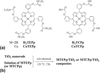
Herein, we synthesized two novel porphyrins and their copper(II) porphyrins: H2TEPp, CuTEPp, H2TCPp, CuTCPp [Fig. 1(a)]. With synthesized porphyrins, four new porphyrin-sensitized TiO2 nanorod composites (H2TEPp/TiO2, H2TCPp/TiO2, CuTEPp/TiO2, and CuTCPp/TiO2) were prepared and characterized [Fig. 1(b)]. Finally, their photocatalytic performances were evaluated by the degradation of 4-NP in aqueous solution.
(a) Chemical structures of synthesized porphyrins; (b) The preparation of MTEPp (or MTCPp)/TiO2 composites.

II. EXPERIMENTAL
A. Materials and reagents
Titanium dioxide (P25) was purchased from Acros Organics (Geel, Belgium), used as a comparison substance in photodegradation tests. Other reagents and solvents were all purchased from Tianjin Chemical Reagent Co. (Tianjin, China) and used without further purification except the pyrrole, which was distilled before use. The silica gel (100–200 mesh) for chromatographic separation was purchased from Yantai Sanhe Chemical Reagent Co. (Yantai, China).
B. Equipments
Mass spectrometry (MS) tests were carried out on a matrix assisted laser desorption/ionization time of flight mass spectrometer (MALDI-TOF MS, Krato Analytical Company of Shimadzu Biotech, Manchester, Britain). Elemental analyses (C, H, and N) were conducted by using a Vario EL-III CHNOS apparatus (Elementar Analysensysteme GmbH, Jena, Germany). 1H NMR spectra were recorded by Varian Inova 400 MHz instrument (Varian Medical Systems, Palo Alto, California) at room temperature, using DMSO as solvent and tetra-methylsilane (TMS) for reference. The Powder X-ray diffraction (XRD) measurement was performed with a Bruker D8 diffractometer (Bruker Corporation, Karlsruhe, Germany) using graphite monochromatic copper radiation (Cu K) at 40 kV, 30 mA over the 2θ range 20–80°. The morphologies of the samples were observed by scanning electron microscopy (SEM; Quanta400FEG, FEI, Hillsboro, Oregon, mag. 200,000×, HV 20.00 kV, High vacuum, WD 11.5 mm, HFW 1.49 μm) with energy-dispersive X-ray spectrometry (EDS). High-resolution transmission electron microscopy (TEM) was done using a JEOL JEM-3010 instrument (JEOL, Tokyo, Japan). FT-IR spectra were gained with samples in KBr matrix for the title complexes on ABEQUZNDX-550 series FT-IR spectrophotometer (Bruker Corporation, Karlsruhe, Germany) in the range 4000–500 cm−1. UV-vis diffuse reflectance (UV-vis-DRS) spectra were obtained on a Shimadzu UV-3100 spectrophotometer (Shimadzu, Kyoto, Japan) by using BaSO4 as a reference. The nitrogen adsorption–desorption isotherms were got using micropore surface area and the pore diameter distribution curves of the samples were calculated from pore size analyzer (Autosorb-1MP, Quantachrome, Beijing, China). Photoluminescence (PL) spectra were examined at room temperature on a Hitachi FL-4500 fluorescence spectrometer (Hitachi, Tokyo, Japan) using a Xe lamp. The photodegradation of 4-NP was conducted in Model XPA-VII photocatalytic reactor (Xujiang Electromechanical Plant, Nanjing, China) with a halogen lamp as light source. UV-vis characteristic absorption spectra of 4-NP were measured by Shimadzu UV-1800 spectrophotometer (Shimadzu, Kyoto, Japan).
C. Synthesis of porphyrins H2TEPp, H2TCPp, CuTEPp, and CuTCPp
The synthetic routes of the porphyrins are shown in Scheme S1. The detailed synthetic procedures are as follows.
1. 5,10,15,20-Tetra(3-(carboethoxymethyleneoxy)phenyl)porphyrin (H2TEPp)
The porphyrin H2TEPp was prepared by a two-step method. The first step was the synthesis of the (3-formyl-phenoxy)-acetic acid ethyl ester, according to the literature. Reference Cochran, Wu, Wang, Nanda, Saven, Therien and DeGrado26,Reference Duncan, Wu and Therien27 In the second step, (3-formyl-phenoxy)-acetic acid ethyl ester (4.0 g, 0.02 mol) was slowly added to propionic acid (80 mL) with stirring. Reference Adler, Longo and Shergails28 Successively, an equivalent amount of freshly distilled pyrrole (1.39 mL, 0.02 mol) was added dropwise after propionic acid refluxing. The reaction was proceeded at 140 °C for 1 h and then most of solvent was removed by distilling under reduced pressure. Finally, ethanol (20 mL) was added after residual solution cooling to room temperature. The mixture was put in a refrigerator for overnight. Then, the crude product was gained by filtration and purified by column chromatography on silica gel with dichloromethane as eluant. A purple solid of H2TEPp was obtained.
Yield: 12%. Mp: >250 °C. Anal. Calcd. (found) for C60H54N4O12 (Mol. Wt: 1022.37), %: C 70.43 (70.44); H 5.29 (5.32); N 5.49 (5.48). MS: m/z 1023.4 ([M + H]+) amu. UV-vis (CH2Cl2) λmax/nm, 420 (Soret band), 514, 550, 590, 643 (Q bands). 1H NMR (400 MHz, DMSO): δ (ppm) = 8.88 (s, 8H, β-H), 7.75 (d, J = 25.0, 1.0 Hz, 12H, Ar), 7.43 (d, J = 8.2, 1.8 Hz, 4H, Ar), 5.01 (s, 8H, –OCH2CO–), 4.20 (q, J = 7.1 Hz, 8H, –COOCH2–), 1.31–1.13 (m, 12H, –CH3), −2.94 (s, 2H, NH). FT-IR (KBr): v, cm−1, 3651, 2978, 1755, 1586, 1474, 1289, 1176, 1082, 922, 802, 727.
2. 5,10,15,20-Tetra(3-(carboxymethyleneoxy)phenyl)porphyrin (H2TCPp)
The porphyrin H2TCPp was prepared by hydrolysis of H2TEPp under the condition of KOH aqueous solution. H2TEPp (0.8 g, 0.78 mmol) was dissolved in THF (25 mL), followed by addition of aqueous solution (25 mL) with KOH (2.8 g, 50 mmol). Then, the mixture was refluxed for 48 h under stirring. The evaporation of THF was done after solution cooling to room temperature and then solid was dissolved with continually adding distilled water to the residual water phase. Finally, HCl (aq, 2 mol/L) was added to the solution until the precipitate was not detected. A purple solid of H2TCPp was obtained.
Yield: 81%. Mp: >250 °C. Anal. Calcd. (found) for C52H38N4O12 (Mol. Wt: 910.25), %: C 70.02 (68.57); H 4.13 (4.20); N 5.58 (6.15). MS: m/z 933.4 ([M + Na]+) amu. UV-vis (CH2Cl2) λmax/nm, 422 (Soret band), 518, 553, 595, 650 (Q bands). 1H NMR (400 MHz, DMSO): δ (ppm) = 8.91 (s, 8H, β-H), 11.05 (s, 4H, –COOH), 7.82 (s, 8H, Ar), 7.74 (s, 4H, Ar), 7.44 (s, 4H, Ar), 4.92 (s, 8H, –COOCH2–), −2.93 (s, 2H, –NH). FT-IR (KBr): v, cm−1, 3315, 3028, 1730, 1579, 1490, 1429, 1236, 1178, 1078, 920, 802, 696.
3. Cu(II) 5,10,15,20-tetra(3-(carboethoxymethyleneoxy)phenyl)porphyrin (CuTEPp)
CuTEPp was prepared in following procedure. Reference Das, Chakraborty, Sarma, Banerjee and Venakatesh29 H2TEPp (200 mg, 0.196 mmol) and Cu(Ac)2 (0.4 mg, 1.96 mmol) were dissolved in dichloromethane (50 mL) and ethanol (15 mL) separately. The synthesis was finished by mixing the above solutions with stirring for 12 h at room temperature. The degree of reaction was monitored by TCL. After filtrating the unreacted solid salt and evaporating the solvent under vacuum, the crude product was purified on silica gel column with dichloromethane as eluant. A red solid of CuTEPp was obtained.
Yield: 93%. Mp: >250 °C. Anal. Calcd. (found) for C60H52CuN4O12 (Mol. Wt: 1083.29), %: C 63.29 (66.44); H 4.50 (4.83); N 5.09 (5.17). MS: m/z 1106.3 ([M + Na]+) amu. UV-vis (CH2Cl2) λmax/nm, 414 (Soret band), 538 (Q band). FT-IR (KBr): v, cm−1, 3437, 2922, 1757, 1602, 1496, 1290, 1180, 1086, 997, 800, 700.
4. Cu(II) 5,10,15,20-tetra(3-(carboxymethyleneoxy)phenyl)porphyrin (CuTCPp)
The synthesis method of CuTCPp is similar to the process of CuTEPp, only H2TCPp replaced with H2TEPp.
Yield: 82%. Mp: >250 °C. Anal. Calcd. (found) for C52H36CuN4O12 (Mol. Wt: 971.16), %: C 64.34 (64.23); H 3.61 (3.73); N 5.62 (5.76). MS: m/z 1010.2 ([M + Na]+) amu. UV-vis (CH2Cl2) λmax/nm, 419 (Soret band), 541 (Q band). FT-IR (KBr): v, cm−1, 3490, 2910, 1730, 1596, 1490, 1427, 1182, 1080, 997, 794, 698.
D. The preparation of TiO2 nanorods
The TiO2 nanorods were prepared by two-step hydrothermal method, as follows: 1.0 g of P25 was added to 60 mL concentrated NaOH aqueous (10 mol/L) under vigorous stirring at room temperature for 4 h. Then the mixture was transferred into a Teflon-lined stainless steel autoclave and heated to 200 °C kept for 24 h. After cooling to room temperature, the product was separated by high-speed centrifuge and washed with distilled water for several times until pH was at around 7. Next, the obtained solid was dispersed into HNO3 (0.1 mol/L) for 4 h, then transferred again into a Teflon-lined autoclave to conduct a further hydrothermal reaction (180 °C, 24 h). Finally, the nanorod TiO2 was obtained after washing with distilled water and drying at 80 °C for 3 h on vacuum.
E. Preparation of porphyrin-sensitized TiO2 composites: H2TEPp/TiO2, H2TCPp/TiO2, CuTEPp/TiO2, and CuTCPp/TiO2
The composites were prepared as follows: 6 μmol of porphyrins (H2TEPp, H2TCPp, CuTEPp, and CuTCPp) were respectively dissolved in DMF (2 mL). Successively, the synthesized TiO2 nanorods (1.0 g) were added to the solution of porphyrins, respectively. The suspension was stirred for 2 h at room temperature and then done a solvothermal reaction (120 °C, 72 h). Then the mixture was centrifuged and washed with distilled water and ethanol for several times. Finally, the composite photocatalysts were obtained after drying at 80 °C for 4 h on vacuum. By measuring UV-vis absorbance value of the solution before and after adding TiO2, the actual loading amounts of porphyrins or Cu(II) porphyrins on TiO2 were calculated. They are 4.12, 5.16, 5.03, and 5.16 μmol for H2TEPp/TiO2, H2TCPp/TiO2, CuTEPp/TiO2, CuTCPp/TiO2, respectively.
F. Photocatalytic degradation
The activity and stability of photocatalysts was measured by photodegradation of 4-NP in aqueous solution.
1. Activity test of photocatalysts
The photocatalysts (10 mg) was dispersed in the 4-NP aqueous solution (50 mL, 1 × 10⁻⁴ mol/L) and then placed in a Model XPA-VII photacatalytic reactor, in which air was continuously bubbled throughout the entire reaction process. Before irrradiation under 400 W halogen lamp, the suspension was stirred for 30 min to balance the adsorption–desorption between photocatalyst and 4-NP. Then, the suspension solution was irradiated by the halogen lamp, which cooled by circulating water. The photocatalytic reaction lasted 60 min with 4 mL of the suspension suction every 6 min. The photocatalyst was separated from the suspension by centrifugation and then dried on vacuum. The photodegradation efficiency was determined by measuring the characteristic absorption of 4-NP at 317 nm using a UV-vis spectrophotometer. According to the direct proportion between absorbance and concentration, the absorption values of 4-NP at 317 nm were recorded to deduce the concentration change of 4-NP.
2. Stability test of photocatalysts
The photocatalyst (10 mg) was dispersed in the 4-NP aqueous solution (50 mL, 1 × 10⁻⁴ mol/L) with constant air bubble. The suspension was stirred and irradiated by 400 W halogen lamp. 4 mL of the solution was removed when the process lasted 90 min. The photocatalyst was collected by centrifugation and desiccation. The cycle experiments were done for six times to explore the stability of photocatalysts. The characteristic absorption of 4-NP at 317 nm was recorded to assess the efficiency of photodegradation.
III. RESULTS AND DISCUSSIONS
A. XRD analysis
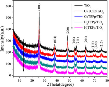
Figure 2 shows the XRD patterns for TiO2 and corresponding porphyrin-sensitized TiO2 composites. The present peaks at 25.3°, 37.8°, 48.0°, 54.0°, 62.7°, 68.8°, 70.3°, 75.1° clearly represent the formation of anatase crystallites (JCPDS no. 01-089-4921). The broadening of the XRD peaks of the anatase phase indicates that the TiO2 and composites are nano-sized. In addition, the loading of porphyrins on the surface of TiO2 doesn’t affect the crystal structure of TiO2. No apparent peaks for porphyrins were observed due to the trace amounts of loaded porphyrins which can’t be resolved by XRD. However, the existence of porphyrins can be clearly elucidated by UV-vis analysis, as discussed next.
XRD patterns of bare TiO2 and MTEPp (or MTCPp)/TiO2 composites.

B. SEM and TEM analysis
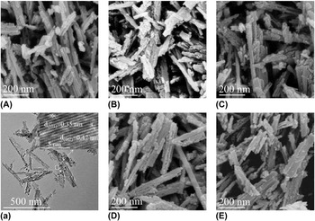
The morphologies of samples were analyzed by SEM, shown in Fig. 3. From SEM image of TiO2, it can be seen that the prepared TiO2 is irregular nanorods. Furthermore, two kinds of lattice fringes, i.e., d (101) = 0.35 nm and d (002) = 0.47 nm, were observed from TEM of TiO2 [Fig. 3(a)], indicating a (010)-oriented plane of TiO2. Thus, the TiO2 nanorods treated hydrothermally can preferentially expose the high-energy plane (010) at the crystal surface, which can dominantly enhance the activity of TiO2. As a result, an excellent photocatalytic performance of TiO2 nanorods can be expected.
SEM (A–E) images of TiO2 nanorods, H2TEPp/TiO2, H2TCPp/TiO2, CuTEPp/TiO2, CuTCPp/TiO2 composites and TEM (a) image of TiO2 nanorods.

Compared with bare TiO2, the morphologies of MTEPp (or MTCPp)/TiO2 composites are nearly invariable except increased roughness of the surface, thereby elucidating the presence of porphyrins on the surface of TiO2.
In addition, to further confirm whether porphyrins loaded on the TiO2, EDS was used to detect the existing elements in these composites (see Fig. S1). As expected, only oxygen and titanium elements were detected in bare TiO2, but carbon and nitrogen elements or carbon, nitrogen, and copper elements were also detected in these composites, except oxygen and titanium elements, which adequately demonstrated that TiO2 had been successfully sensitized by porphyrins or copper(II) porphyrin.
C. FT-IR analysis
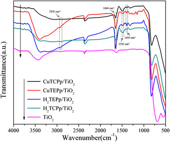
The FT-IR spectra of composite photocatalysts were measured to discuss the interaction between TiO2 nanorods and porphyrin molecules. As shown in Fig. 4, for bare TiO2, there are two sets of absorption peaks at around 3420 and 1630 cm−1, separately referring to the stretching and bending vibration of hydroxyl group, showing the existence of hydroxy on the surface of TiO2. Besides, the bands at around 542 and 689 cm−1 are the characteristic absorptions of Ti–O bond in TiO2. Furthermore, the C–H and C=C characteristic vibration bands of porphyrin ring at around 2920 and 1500 cm−1 appear in the composites (see Fig. S2). It proves that TiO2 nanorods have been sensitized by porphyrins. It is worth noticing that the C=O vibration bands of ester and carboxyl group at around 1750 and 1730 cm−1 in porphyrin molecules have all disappeared. Instead, the asymmetrical and symmetrical stretching vibration of –COO− group at around 1646 and 1400 cm−1 are found, indicating that porphyrin or copper(II) porphyrin molecules have bonded to the surface of TiO2 by –COO− group. This bonding is beneficial for the electron transfer between TiO2 and porphyrins. Therefore, it is laudable to improve the photoactivities of MTEPp (or MTCPp)/TiO2 composites.
FT-IR spectra of bare TiO2 and MTEPp (or MTCPp)/TiO2 composites.

D. UV-vis-DRS analysis
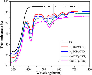
The UV-vis-DRS spectra of TiO2 nanorods and MTEPp (or MTCPp)/TiO2 composites were examined to investigate their optical response at the range of 200–800 nm. As shown in Fig. 5, there is no distinct absorption above 400 nm for bare TiO2, nevertheless, all the composites have apparent peaks at around 420 nm and in region of 450–700 nm, conforming to the characteristic absorptions of porphyrins. It illuminates that the porphyrins have loaded on the TiO2 nanorods surface with intact molecule frameworks. Therefore, the photoresponse range of TiO2 is extended from UV to the visible light region due to the participation of porphyrins on its surface, significantly enhancing visible-light-driven photocatalytic activity of TiO2 to some extent.
UV-vis-DRS spectra of TiO2 and MTEPp (or MTCPp)/TiO2 composites.

E. BET analysis
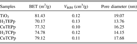
The N2 adsorption–desorption isotherms and pore size distribution curve were used to study the specific surface area and porosity of samples (see Fig. S3), revealing a typical IV isotherm. Reference Li, Zhao, Gao, Wang and Qiu30 Specially, the detailed BET surface area, pore volume, and pore diameter were exhibited in Table I, thereinto, the pore size distribution curve was got from the desorption isotherm by the Barret–Joyner–Halenda (BJH) method by using the Halsey equation. From the data, it is seen that the samples all exhibit large specific surface areas and a certain pore size distribution. Besides, the hysteresis loops appearing in the adsorption–desorption isotherms are consistent with wide pore size distributions of samples formed on account of random stacking of TiO2 nanorods, displayed in SEM images. Furthermore, compared with the bare TiO2, the specific surface areas of MTEPp (or MTCPp)/TiO2 composites decrease slightly because of the loading of porphyrins on the TiO2 surface.
The BET surface area, pore volume, pore diameter of bare TiO2, and MTEPp (or MTCPp)/TiO2 composites.

F. Fluorescence emission spectra analysis
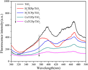
To explore the lifetime of electron–hole pairs in TiO2 and prepared MTEPp (or MTCPp)/TiO2 composites, the fluorescence emission spectra were analyzed. Reference Wang, Li, Lu, Quan and Chen31 As shown in Fig. 6, there are two absorption peaks at around 400 and 468 nm for TiO2 nanorods and composites. By the related reports, the absorption at around 400 nm can be attributed to the self-trapping of excited electronic in the octahedral TiO2, while the peak at around 468 nm was generated due to the oxygen vacancy. Reference Baiju, Zachariah, Shukla, Biju, Reddy and Warrier32 Comparing with TiO2, the absorption strengths of two peaks apparently weaken for composites, which may be the reason of photoinduced electrons on the TiO2 transferring to porphyrin molecules, resulting in decreased fluorescence emission. Therefore, there is a low ratio of photoproduced electron–hole recombination in composites. Naturally, the composites display higher photocatalytic activity than bare TiO2 as evidenced by photocatalytic measurements. In addition, the fluorescence spectra of composites reveal that the fluorescence intensities can be influenced by the structures of porphyrin molecules, and CuTCPp/TiO2 composite exhibits the most effective controlling to the recombination of electron–hole.
PL spectra of bare TiO2 and MTEPp (or MTCPp)/TiO2 composites.

G. Photocatalytic activity and stability analysis
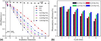
The photocatalytic activities of MTEPp (or MTCPp)/TiO2 composites were evaluated by the photodegradation of 4-NP in aqueous solution under halogen lamp irradiation. Figure 7(a) showed the photocatalytic efficiencies of different samples as compared to blank test, which contained only 4-NP (aq). As expected, both TiO2 nanorods and composites display more excellent photocatalytic activities than commercial TiO2 (P25). What is more, the introduction of porphyrins to the TiO2 nanorods observably enhances the photocatalytic activities of the composites. Significantly, CuTCPp/TiO2 composite performs the highest efficiency of all samples. The order of their photocatalytic activities is CuTCPp/TiO2 > CuTEPp/TiO2 > H2TCPp/TiO2 > H2TEPp/TiO2 > TiO2 > P25. The highly photocatalytic activity of CuTCPp/TiO2 composite can be explained from the following two reasons: One is the strong interaction of CuTCPp’s carboxyl group with the surface of TiO2. Another is the preferably match between the excitation potential of CuTCPp and the conduction band of TiO2.
(a) The photodegradation of 4-NP versus irradiation time using different photocatalysts; (b) Photocatalytic efficiency of MTEPp (or MTCPp)/TiO2 composites for 4-NP degradation after six cycles.

In addition, the photocatalytic activities of MTEPp (or MTCPp)/TiO2 composites were also compared with H2TEPp, H2TCPp, CuTEPp, and MTCPp (Fig. S4). It can be seen that the alone porphyrins or copper(II) porphyrins only exhibit the lower photocatalytic activities compared with MTEPp (or MTCPp)/TiO2 composites, which indicate a cooperation between TiO2 and MTEPp (or MTCPp) in these composites in the process of photocatalysis. Moreover, we also noticed that the prepared TiO2 nanorods showed the improvement of properties compared with spherical TiO2 nanoparticles reported before by us. Reference Yu, Li, Sun, Jiang and Zhang23,Reference Su, Li, Zhang, Yu and Yuan33 The result implies that low-dimensional TiO2 nanostructures are more superior photocatalyst due to easily exposed high-energy plane.
To evaluate stabilities of MTEPp (or MTCPp)/TiO2 composites, the recycle experiment of photodegradation 4-NP was carried out by performing similar experimental conditions. As shown in Fig. 7(b), the degradation efficiencies of all photocatalysts are close to 100% at the first run. However, the photocatalytic efficiencies were reduced marginally to 50.1% (H2TEPp/TiO2), 58.3% (H2TCPp/TiO2), 71.6% (CuTEPp/TiO2), and 75.8% (CuTCPp/TiO2) after six cycles, reflecting the relatively high stability for composite photocatalysts. The photocatalytic stability exhibits the same trend as the photocatalytic activity, which suggests the strong combination of carboxyl in CuTCPp with TiO2. Besides, the reduction of photocatalytic efficiency probably occurs because of the damage of the interaction between porphyrin molecules and TiO2 with the reaction proceeding, leading to the drain of porphyrins.
IV. CONCLUSION
In summary, four novel composites based on porphyrin-sensitized TiO2 nanorods were prepared hydrothermally and characterized by XRD, SEM, TEM, FT-IR, UV-vis DRS, nitrogen adsorption–desorption and fluorescence spectra in this paper. Characterization results indicate that the synthesized TiO2 nanorods can preferentially expose their high-energy plane (010) on the crystal surface, and the porphyrin and Cu(II) porphyrin are linked firmly to the surface of TiO2 nanorods in prepared composites. The photocatalytic activities were investigated by the photodegradation experiments of 4-NP in aqueous solution under irradiation with 400 W halogen lamp. The results show that all the prepared TiO2 nanorods and composites exhibit superior photocatalytic properties to commercial TiO2 (P25). The sensitization of porphyrins and Cu(II) porphyrins to the TiO2 nanorods observably enhances the photoactivities of composites. Particularly, CuTCPp/TiO2 composite performs supreme photocatalytic activity.
Supplementary Material
To view supplementary material for this article, please visit https://doi.org/10.1557/jmr.2017.230.
ACKNOWLEDGMENTS
The authors acknowledge the research grant provided by NNSF (No. 21671158), the Natural Science Foundation of Shaanxi Province (No. 2014JM2054), the Education Committee Foundation of Shaanxi Province (No. 15JK1736) and the NWU Scientific Research Foundation (No. 12NW24).