Introduction
Humans and other vertebrates exhibit left–right (LR) asymmetric arrangement of the internal organs, including asymmetry of unpaired organs and paired organs. The normal arrangement of internal organs along LR axis is called situs solitus (SS), such as human lungs bilobed on the left and trilobed on the right, left-pointing cardiac apex, left-sided aortic arch, stomach and spleen, right-sided liver and gallbladder and right-to-left course of the large intestine. Failure to establish normal LR asymmetry leads to internal laterality disorders, including situs inversus (SI) and heterotaxy (HTX, also named situs ambiguus) (Refs Reference Kosaki and Casey1, Reference Peeters and Devriendt2, Reference Jacobs3).
Human LR asymmetry disorders occur in different ethnic backgrounds. SI occurs in 1 out of 6000–8000 newborns (Ref. Reference Peeters and Devriendt2), and the prevalence of HTX has been estimated as 1 in 10 000 at birth (Ref. Reference Lin4). SI and HTX generally exist in different families. SS, SI and HTX phenotypes may present occasionally in the same family (Refs Reference Kosaki and Casey1, Reference Casey5).
Both SI and HTX are genetically heterogeneous disorders with reduced penetrance, presumably because of monogenic, polygenic or multifactorial causes. Genetic factors, environmental modifiers, and developmental stochasticity are proposed to be responsible for human LR asymmetry disorders. Retinoic acid (RA), a vitamin A derivative, is implicated in embryonic development. Exposure to RA in utero causes laterality defects in several vertebrates (Refs Reference Peeters and Devriendt2, Reference Sutherland and Ware6). Maternal diabetes, maternal cocaine use, parental exposure to hair dye, smoking and laboratory chemicals are also associated with HTX (Refs Reference Kuehl and Loffredo7, Reference Kuehl and Loffredo8). Nodal signal transduction pathway is recognised as a common pathway in embryonic LR development, and progress has been made in exploring the mechanism of LR asymmetry by discovery of many chromosomal abnormalities and more than 100 related genes in animal models (Ref. Reference Sutherland and Ware6). SI is usually characterised by autosomal recessive inheritance (Ref. Reference Kosaki and Casey1), while HTX displays X-linked inheritance, autosomal dominant inheritance with reduced penetrance, or autosomal recessive inheritance (Ref. Reference Sutherland and Ware6).
Balanced translocations, insertions, small deletions or duplications, inversions and more complex chromosomal rearrangements are connected with LR asymmetry disorders (Refs Reference Kosaki and Casey1, Reference Sutherland and Ware6, Reference Bisgrove, Morelli and Yost9). Since the zinc finger protein of the cerebellum 3 gene (ZIC3) was successfully identified in a patient with X-linked HTX phenotype in 1997 (Ref. Reference Gebbia10), a growing number of loci and disease genes involved in human embryonic LR development have been identified for the last 17 years. This article highlights the classification, chromosomal abnormalities, pathogenic genes and the possible mechanism of human LR asymmetry disorders.
Embryonic LR development pathway
The established process of LR asymmetry can be divided into four phases: breaking of LR symmetry at early embryonic stages; transmission of asymmetric signals from the node to lateral plate mesoderm (LPM); asymmetric gene expression in the left LPM; LR asymmetric morphogenesis of the internal organs (Refs Reference Shiraishi and Ichikawa11, Reference Vandenberg and Levin12, Reference Speder13) (Fig. 1).
Pathway of left–right (LR) development in the mouse embryo, list of genes associated with human LR asymmetry disorders and corresponding phenotypes in humans. Lrd-containing monocilia generate leftward nodal flow, and polycystin 2 containing cilia sense nodal flow and initiate an asymmetric calcium signal, which induces Nodal expression around the node. Nodal signalling is involved in asymmetric morphogenesis by inducing expression of the Nodal-responsive genes (Nodal, Lefty2 and Pitx2) in the left LPM, and expression of Lefty1 at the midline. Mutations of genes associated with ciliopathies and Nodal signal transduction pathway have been identified in human LR asymmetry disorders. Full arrows indicate a direct positive effect on gene expression, dotted arrows indicate an indirect effect and lines indicate inhibition. ASD, atrial septal defect; AVSD, atrioventricular septal defects; BBS, Bardet–Biedl syndrome; CHD, congenital heart disease; CRPT2, Carpenter syndrome 2; DEX, dextrocardia; DORV, double-outlet right ventricle; HTX, heterotaxy; JSRD, Joubert syndrome-related disorders; LI, left isomerism; NPHP, nephronophthisis; NVPs, nodal vesicular parcels; PCD, primary ciliary dyskinesia; PKD2, polycystic kidney disease 2; PS, pulmonary stenosis; RA, retinoic acid; RHPD2, renal–hepatic–pancreatic dysplasia 2; RI, right isomerism; RP, retinitis pigmentosa; SCDO4, spondylocostal dysostosis 4; SHH, sonic hedgehog; SI, situs inversus; TGA, transposition of the great arteries.

Alteration at the initial stage of embryonic LR development pathway, when the total direction of LR asymmetry being determined, might result in complete inversion of LR axis (SI). Nevertheless, disturbance at a later stage might alter only some organs, which lead to randomised arrangement of organs along LR axis (HTX) and mirror image duplication of organs (isomerism) (Ref. Reference Bisgrove, Morelli and Yost9).
Classification of LR asymmetry disorders
Human LR asymmetry disorders can be divided into SI and HTX. SI is defined as complete mirror-imaged arrangement of the internal organs along LR axis. HTX is described as abnormal arrangement of the internal thoraco-abdominal organs with respect to each other and the LR axis, but cannot be categorised as either SS or SI (Refs Reference Kosaki and Casey1, Reference Peeters and Devriendt2, Reference Jacobs3).
Isolated SI usually refers to SI without ciliary dysfunction, congenital heart disease (CHD) or other congenital malformations. Individuals with isolated SI usually are healthy, probably because of no change in their relative position and the existence of normal structure and function in organs (Ref. Reference Bisgrove, Morelli and Yost9). However, HTX is usually associated with complex cardiovascular malformations, often including isomerism. HTX phenotype is usually discovered in children with cyanotic CHDs (Ref. Reference Sutherland and Ware6). Isomerism is characterised by asymmetric defects of paired organs that generally have obvious left and right shape, and it can be separated into two classes: left isomerism (both lungs are bilobed with long hyparterial bronchi, both atria with left atrial appendages, and polysplenia) and right isomerism (both lungs with trilobed with short eparterial bronchi, both atria with right atrial appendages, and asplenia) (Refs Reference Kosaki and Casey1, Reference Peeters and Devriendt2).
No less than 59 syndromes display LR asymmetric defects (Ref. Reference Peeters and Devriendt2). Disease genes have been identified in syndromic SI, primarily in some ciliopathies, including primary ciliary dyskinesia (PCD), nephronophthisis (NPHP), polycystic kidney disease 2 (PKD2) and Bardet–Biedl syndrome (BBS) (Refs Reference Kosaki and Casey1, Reference Marion14, Reference Bataille15, Reference Otto16), and in syndromic HTX, such as VACTERL association and Carpenter syndrome 2 (CRPT2) (Refs Reference Twigg17, Reference Wessels18).
Loci and disease genes of SI
Genetic studies in isolated SI are very rare, and only a few families with SI, HTX and SS have been classified into HTX families to discuss the genetic basis (Refs Reference Kosaki and Casey1, Reference Perles19). Most of genetic studies involved in SI pay close attention to syndromic SI, especially to PCD with SI, which accounts for only 20–25% of individuals with SI (Ref. Reference Aylsworth20).
PCD with SI
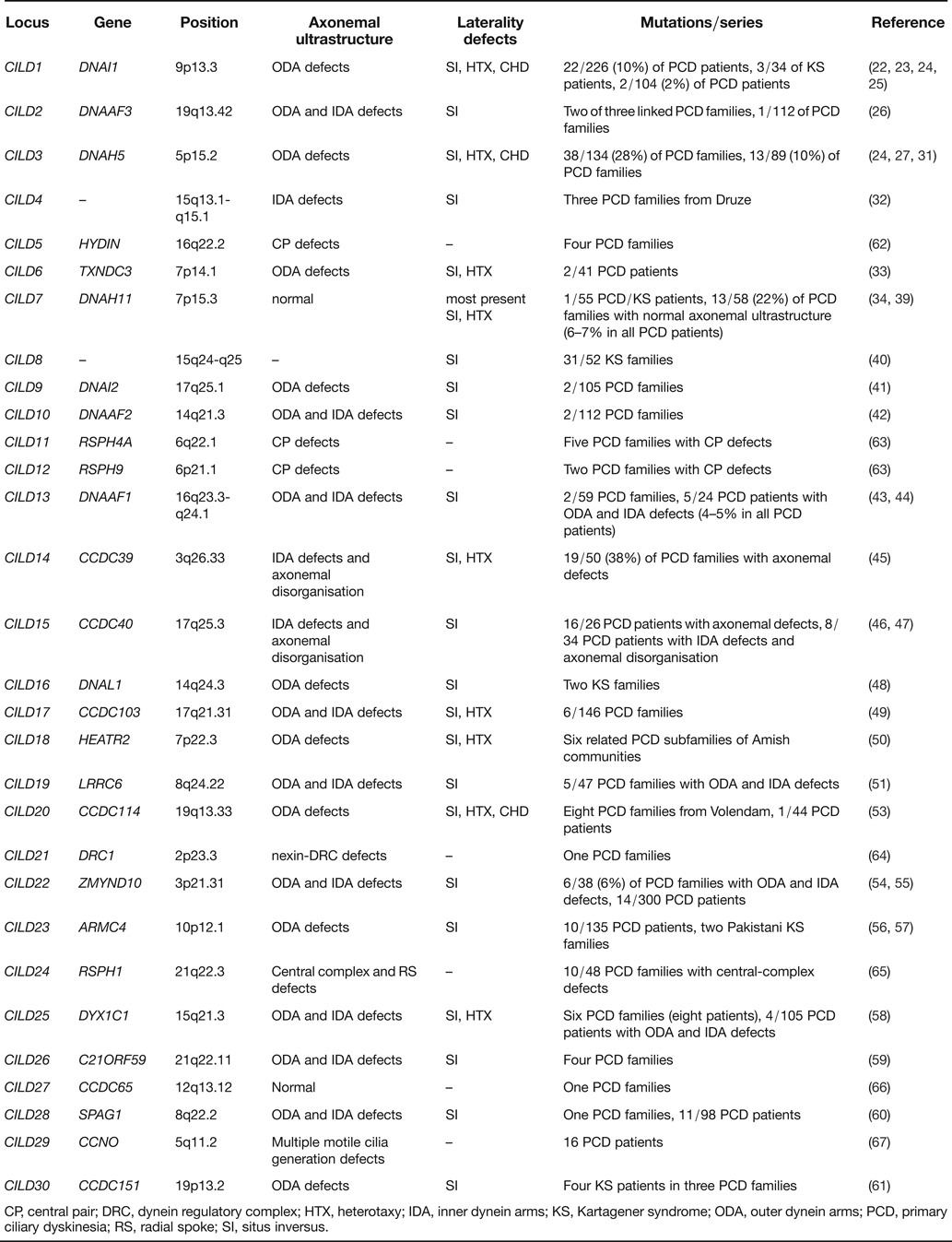
PCD is an autosomal recessive disorder of ciliary dysfunction with a prevalence of about 1 in 16 000 to 20 000, characterised by chronic sinusitis, bronchiectasis, male infertility and situs abnormalities (Ref. Reference Zariwala, Omran and Ferkol21). About 50% of PCD patients present with SI, named Kartagener syndrome (KS), which is caused by defective nodal ciliary function during embryogenesis (Ref. Reference Zariwala22). PCD can be caused by mutations in any protein or structural subunit of cilia in theory. However, most patients present with defective outer dynein arms (ODA) and/or inner dynein arms (IDA), which offer the force of ciliary beating (Ref. Reference Zariwala22). Up to now, at least 30 loci (termed ciliary dyskinesia, primary 1–30; CILD1-30) and 28 disease genes for PCD have been identified, and 23 loci and 21 genes were involved in KS (Table 1).
Genetic causes and laterality defects of PCD
CP, central pair; DRC, dynein regulatory complex; HTX, heterotaxy; IDA, inner dynein arms; KS, Kartagener syndrome; ODA, outer dynein arms; PCD, primary ciliary dyskinesia; RS, radial spoke; SI, situs inversus.
CILD1 (9p13.3) and the axonemal dynein intermediate chain 1 gene (DNAI1)
The DNAI1 gene encodes an axonemal intermediate chain of dynein. In 2001, Guichard et al. (Ref. Reference Guichard23) identified compound heterozygous DNAI1 gene defects in three of 34 KS patients and two PCD with SS. Zariwala et al. (Ref. Reference Zariwala22) identified biallelic DNAI1 mutations in 14 PCD patients (including seven KS patients). Mutations in the DNAI1 gene were also identified in nine PCD patients with HTX, and two of those had CHD (Ref. Reference Kennedy24). DNAI1 mutations occur in about 2–10% of PCD patients (KS accounts for 52–59%) and the IVS1 + 2_3insT splice mutant seems to be a common founder, which accounts for about 57% of mutant alleles (Refs Reference Zariwala22, Reference Failly25).
CILD2 (19q13.42) and the axonemal dynein assembly factor 3 gene (DNAAF3)
The DNAAF3 gene encodes a protein involved in cytoplasmic preassembly of axonemal dyneins into assembly-competent complexes. Dnaaf3 morphant zebrafish embryos presented with static cilia and LR axis patterning defects (Ref. Reference Mitchison26). Mitchison et al. (Ref. Reference Mitchison26) identified two homozygous mutations, p.L108P and p.R136X, of the DNAAF3 gene in eight PCD patients (including three KS patients), and one homozygous frameshift mutation, p.V255CfsX12, in a PCD family (one patient with SI) by screening of other 112 PCD families.
CILD3 (5p15.2) and the axonemal dynein heavy chain 5 gene (DNAH5)
The DNAH5 gene encodes a heavy chain of ODA (Ref. Reference Hornef27). Mdnah5 −/− mice display recurrent respiratory infections, ciliary immotility and randomisation of LR asymmetry (Ref. Reference Ibanez-Tallon, Gorokhova and Heintz28). In 2000, Omran et al. (Ref. Reference Omran29) mapped a recessive causing gene locus of a consanguineous PCD family (two KS in four PCD patients) to chromosome 5p15-p14 and indicated DNAH5 is likely to be an excellent candidate gene. Sequence analysis in three PCD families (four KS in seven PCD patients) identified homozygous mutations (c.5563insA, c.10555G > C and IVS74-1G > C) in the DNAH5 gene, which lead to non-functional DNAH5 proteins (Ref. Reference Olbrich30). Mutations in the DNAH5 gene were also identified in five PCD patients with HTX, and one of the patients had CHD (Ref. Reference Kennedy24). DNAH5 mutations, a major cause of PCD with ODA defects, account for 15–28% of PCD families (Refs Reference Hornef27, Reference Failly31).
15q13.1-q15.1 (CILD4)
Jeganathan et al. (Ref. Reference Jeganathan32) mapped the disease genes of three PCD families (four KS in five PCD patients) from Druze to chromosome 15q13.1-q15.1 (CILD4), but no apparent candidate genes were found in the critical region.
CILD6 (7p14.1) and the thioredoxin domain-containing protein 3 gene (TXNDC3)
The TXNDC3 gene, also known as the sperm-specific thioredoxin 2 gene (SPTRX2) or the NME8 gene, encodes a protein that plays a role in ciliary function. Duriez et al. identified a heterozygous p.L426X mutation and a heterozygous intronic variant, c.271-27C > T, of the TXNDC3 gene in a PCD patient with HTX. The heterozygous c.271-27C > T variant was also identified in a KS patient and it decreased expression of TXNDC3d7 isoform, which has a higher binding to microtubules (Ref. Reference Duriez33).
CILD7 (7p15.3) and the axonemal dynein heavy chain 11 gene (DNAH11)
The DNAH11 gene is homologous to mouse LR dynein (Lrd) gene, which is mutated in inversus viscerum (iv) mice (Refs Reference Bartoloni34, Reference Supp35). Lrd-containing, motile node monocilia generate leftward nodal flow, which transports ‘nodal vesicular parcels’ (NVPs) carrying sonic hedgehog (SHH) and RA to the left node and initiates LR asymmetry in the mouse (Refs Reference Tanaka, Okada and Hirokawa36, Reference McGrath37). Bartoloni et al. (Ref. Reference Bartoloni34) found a homozygous DNAH11 nonsense mutation, p.R2852X, in a patient with SI and cystic fibrosis. Schwabe et al. (Ref. Reference Schwabe38) reported DNAH11 compound heterozygous mutations, c.12384C > G and c.13552_13608del, in a German PCD family (one KS in six PCD patients) with normal axonemal ultrastructure. Knowles et al. (Ref. Reference Knowles39) identified biallelic DNAH11 mutations in 15 PCD patients (11 patients with SI and 1 patient with HTX) with normal axonemal ultrastructure, and these mutations may occur in 6–7% of all PCD patients.
15q24-q25 (CILD8)
Geremek et al. (Ref. Reference Geremek40) localised the pathogenic genes of 31 KS families on chromosome 15q24-q25 (CILD8) and the candidate region contains no known genes.
CILD9 (17q25.1) and the axonemal dynein intermediate chain 2 gene (DNAI2)
The DNAI2 gene encodes a protein that contributes to ODA assembly throughout the ciliary axoneme. Loges et al. (Ref. Reference Loges41) identified three recessive loss-of-function homozygous DNAI2 mutations (IVS11 + 1G > A, c.787C > T and IVS3-3 T > G) in six PCD patients (including four KS patients).
CILD10 (14q21.3) and the axonemal dynein assembly factor 2 gene (DNAAF2)
The DNAAF2 gene, the human orthologue of the medaka kintoun (Ktu) gene, encodes a cytoplasmic protein involved in pre-assembly of axonemal dynein. Homozygous p.Y243X mutation of the medaka Ktu gene leads to SI and lack of directional liquid flow in Kupffer's vesicle. Omran et al. (Ref. Reference Omran42) identified homozygous p.G406Rfs89X and p.S8X mutations of the DNAAF2 gene in 2 consanguineous PCD families (including two KS patients) with ODA and IDA defects.
CILD13 (16q23.3-q24.1) and the axonemal dynein assembly factor 1 gene (DNAAF1)
The DNAAF1 gene, also termed as the leucine-rich repeat-containing protein 50 gene (LRRC50), involves in assembly of dynein-arm complexes. Loges et al. (Ref. Reference Loges43) identified homozygous p.P451AfsX5 and p.R271X mutations of the DNAAF1 gene in two KS patients with combined ODA and IDA defects. Duquesnoy et al. (Ref. Reference Duquesnoy44) found a homozygous p.L175R mutation and compound heterozygous mutations of the DNAAF1 gene in four PCD families (three KS in five PCD patients).
CILD14 (3q26.33) and the coiled-coil domain-containing protein 39 gene (CCDC39)
The CCDC39 gene encodes an axonemal protein that contributes to the assembly of IDA and the dynein regulatory complex (DRC). The homozygous Ccdc39 p.R96X mutation leads to PCD phenotype in five Old English Sheepdogs. Fourteen recessive CCDC39 loss-of-function mutations were identified in 38% (19/50) of PCD families (41% of patients with SS, 45% with SI and 14% with HTX) (Ref. Reference Merveille45).
CILD15 (17q25.3) and the coiled-coil domain-containing protein 40 gene (CCDC40)
The CCDC40 gene encodes a protein that contributes to axonemal localisation of the CCDC39 protein and governs the assembly of DRC and IDA complex. Ccdc40 mutants in mouse and zebrafish display LR patterning defects. Becker-Heck et al. (Ref. Reference Becker-Heck46) detected loss-of-function biallelic CCDC40 mutations in 16 PCD patients (including 11 KS patients) with misplacement of central pair microtubules and defective axonemal assembly. Blanchon et al. (Ref. Reference Blanchon47) reported biallelic CCDC40 mutations (nonsense and frameshift mutations) in eight PCD patients (including four KS patients) with IDA defects and axonemal disorganisation.
CILD16 (14q24.3) and the axonemal dynein light chain 1 gene (DNAL1)
The DNAL1 gene encodes dynein light chain 1. Homozygous DNAL1 p.N150S mutation was discovered in two PCD families (including three KS patients) with ODA defects, and the mutation reduces the stability of the DNAL1 protein, and hampers its interactions with dynein heavy chain and with tubulin (Ref. Reference Mazor48).
CILD17 (17q21.31) and the coiled-coil domain-containing protein 103 gene (CCDC103)
The CCDC103 gene encodes a coiled-coil containing, dynein arm anchoring factor. Zebrafish Ccdc103 mutants display curved body axis, randomised LR asymmetry and pronephric kidney cysts. Panizzi et al. (Ref. Reference Panizzi49) reported a homozygous CCDC103 founder mutation, c.383_384insG, in three Pakistani PCD families with six PCD patients (including one SI and two HTX), and a homozygous CCDC103 p.H154P mutation in four PCD patients (including 2 SI) from two other Pakistani families and a German family.
CILD18 (7p22.3) and the HEAT repeat-containing protein 2 gene (HEATR2)
The HEATR2 gene, the human orthologue of the Chlamydomonas Htr2 gene, encodes a HEAT-containing protein that functions as the preassembly or stability of the axonemal dynein arms. Horani et al. discovered a homozygous HEATR2 p.L795P mutation in nine related PCD patients (including four SI and one HTX) with absent ODA. Silencing of Chlamydomonas homologue Htr2 gene leaded to absent ODA and decreased flagellar beat frequency (Ref. Reference Horani50).
CILD19 (8q24.22) and the leucine-rich repeat-containing protein 6 gene (LRRC6)
The LRRC6 gene, the human orthologue of the Zebrafish Lrrc6l gene, functions as a cytoplasmic preassembly player of IDA and ODA (Ref. Reference Kott51). Zebrafish Lrrc6l mutants present with pronephric cysts and LR patterning defects (Ref. Reference Serluca52). Kott et al. (Ref. Reference Kott51) found biallelic LRRC6 mutations in six PCD patients (including two KS patients) and these mutations account for 10.6% (5/47) of PCD families with absence of IDA and ODA.
CILD20 (19q13.33) and the coiled-coil domain-containing protein 114 gene (CCDC114)
The CCDC114 gene encodes a ciliary protein that functions as microtubular attachment of ODAs in the axoneme. Onoufriadis et al. (Ref. Reference Onoufriadis53) reported a homozygous founder mutation, c.742G > A, of the CCDC114 gene in 16 PCD patients (including four SI and two HTX) from Volendam, and a homozygous CCDC114 mutation, c.486 + 1G > A, in a KS patient with CHD from UK.
CILD22 (3p21.31) and the zinc finger MYND domain-containing protein 10 gene (ZMYND10)
The ZMYND10 gene encodes a cytoplasmic protein that interacts with the LRRC6 protein and contributes to proper axonemal assembly of IDA and ODA. Moore et al. (Ref. Reference Moore54) described recessive biallelic mutations of the ZMYND10 gene in 16% (6/38) of PCD families with combined IDA and ODA defects, and 60% (6/10) of ZMYND10-PCD patients have SI. Zariwala et al. (Ref. Reference Zariwala55) detected recessive biallelic ZMYND10 mutations in patients, including six PCD, seven KS and one SI without PCD.
CILD23 (10p12.1) and the armadillo repeat-containing protein 4 gene (ARMC4)
The ARMC4 gene encodes a ciliary protein, which is essential for proper targeting and anchoring of ODAs. Both zebrafish embryos with Armc4 suppression and Armc4 −/− mutant mice exhibit LR asymmetry defects. Hjeij et al. (Ref. Reference Hjeij56) identified biallelic mutations of the ARMC4 gene in 12 PCD patients (including 8 KS patients). Onoufriadis et al. (Ref. Reference Onoufriadis57) detected homozygous ARMC4 mutations, p.S892X and p.E658X, in two consanguineous Pakistani KS families, respectively.
CILD25 (15q21.3) and the DYX1C1 gene
The DYX1C1 gene encodes a new cytoplasmic axonemal assembly factor, which interacts with the DNAAF2 protein in cytoplasmic ODA and IDA assembly. Both knockdown of zebrafish Dyx1c1and Dyx1c1 −/− mutant mice produced laterality defects. Tarkar et al. (Ref. Reference Tarkar58) found recessive biallelic mutations of the DYX1C1 gene in 12 PCD patients (including five SI and two HTX) with IDA and ODA defects.
CILD26 (21q22.11) and the chromosome 21 open reading frame 59 gene (C21ORF59)
The C21ORF59 gene, the human orthologue of the Chlamydomonas FBB18 gene, encodes a flagellar matrix protein contributing to the cytoplasmic dynein arm assembly. Knockdown of the C21orf59 gene in zebrafish led to pronephric cysts, axis curvature and LR asymmetry defects. Austin-Tse et al. (Ref. Reference Austin-Tse59) reported recessive truncating mutations of the C21ORF59 gene in four unrelated PCD patients (including three KS patients).
CILD28 (8q22.2) and the sperm-associated antigen 1 gene (SPAG1)
The SPAG1 gene encodes a protein involved in the cytoplasmic assembly and/or trafficking of the axonemal dynein arms. In 2013, Knowles et al. (Ref. Reference Knowles60) identified biallelic loss-of-function mutations of the SPAG1 gene in 14 PCD patients (including eight KS patients) with defective ODA and IDA.
CILD30 (19p13.2) and the coiled-coil domain-containing protein 151 gene (CCDC151)
The CCDC151 gene encodes an ODA docking complex related protein, contributing to ODAs assembly and docking. Both zebrafish Ccdc151 −/− and mouse Ccdc151 −/− mutants display situs defects and CHD phenotypes. In 2014, two homozygous CCDC151 mutations, p.E309X and p.S419X, were identified in three PCD families (including four KS patients) (Ref. Reference Hjeij61).
However, none of the seven CILD disease genes, including the mouse hydrocephalus-inducing homologue gene (HYDIN, CILD5) (Ref. Reference Olbrich62), the chlamydomonas radial spoke head 4 homologue A gene (RSPH4A, CILD11) (Ref. Reference Castleman63), the chlamydomonas radial spoke head 9 homologue gene (RSPH9, CILD12) (Ref. Reference Castleman63), the chlamydomonas DRC subunit 1 homologue gene (DRC1, CILD21) (Ref. Reference Wirschell64), the chlamydomonas radial spoke head 1 homologue gene (RSPH1, CILD24) (Ref. Reference Kott65), the coiled-coil domain-containing protein 65 gene (CCDC65, CILD27) (Ref. Reference Horani66) and the cyclin O gene (CCNO, CILD29) (Ref. Reference Wallmeier67) were found to be associated with LR asymmetry phenotype. Whether these genes are involved in LR asymmetry disorders remains to be clarified.
Renal cystic diseases with SI
Two renal cystic diseases, NPHP and PKD2, are connected with SI. NPHP is an autosomal recessive cystic kidney disease that results in renal failure in childhood or adolescence and, may be associated with hepatic fibrosis, SI or cardiac malformations (Refs Reference Otto16, Reference Olbrich68). PKD2 is an autosomal dominant polycystic kidney disease, characterised primarily by the slow development of renal cysts resulting in kidney failure (Ref. Reference Bataille15)
Nephronophthisis type 2 (NPHP2) and the inversin gene (INVS, 9q31.1)
The INVS gene, also known as the nephrocystin 2 gene (NPHP2) or the inversion of embryonic turning gene (INV), encodes an inversin protein that localises to primary cilia of renal tubular cells and contributes to LR axis specification (Ref. Reference Otto16). The homozygous inv mice exhibit SI, renal cystic changes and reversed expression of the Nodal and Lefty genes (Ref. Reference Mochizuki69). Otto et al. (Ref. Reference Otto16) described a homozygous p.R603X mutation of the INVS gene in a Turkish patient with NPHP2, SI and ventricular septal defect (VSD). Bellavia et al. (Ref. Reference Bellavia70) also detected a homozygous INVS p.R899X mutation in two siblings with juvenile NPHP and SI.
The nephrocystin 3 gene (NPHP3, 3q22.1)
The NPHP3 gene encodes a protein that interacts with nephrocystin (Ref. Reference Olbrich68). Nphp3 ko/ko mice with complete loss-of-function alleles presented with SI, complex cardiac defects, and embryonic lethality (Ref. Reference Bergmann71). Hypomorphic NPHP3 mutations cause adolescent NPHP, tapeto-retinal degeneration and hepatic fibrosis (Ref. Reference Olbrich68), whereas NPHP3 homozygous loss-of-function mutation c.1985 + 5G > A was identified in a Vietnam girl with renal–hepatic–pancreatic dysplasia (RHPD) and SI (Ref. Reference Bergmann71). NPHP3-null mutation c.2694-2_2694-1delAG was also detected in two siblings with RHPD (a female foetus revealed SI) (Ref. Reference Fiskerstrand72). NPHP3 homozygous p.R702X mutation was discovered in 12 Amish neonates with lethal NPHP (a neonate presented SI) (Ref. Reference Simpson73). Homozygous c.3702delA mutation of the NPHP3 gene was also identified in a patient with HTX, cerebellar vermis hypoplasia and congenital contractures (Ref. Reference Chaki74).
Nephronophthisis type 16 (NPHP16) and the ankyrin repeat and sterile alpha motif domains-containing protein 6 gene (ANKS6, 9q22.33)
The ANKS6 gene encodes a protein involved in renal and cardiovascular development (Ref. Reference Hoff75). Anks6-depleted zebrafish displays laterality defects and pronephric cysts. Hoff et al. (Ref. Reference Hoff75) identified a homozygous ANKS6 truncating mutation p.Y790X in an India infant with NPHP, SI, aortic stenosis and hypertrophic obstructive cardiomyopathy.
Oculo-renal form of Joubert syndrome (JS)-related disorders and the 290-KD centrosomal protein gene (CEP290, 12q21.32)
JS is an autosomal recessive disease, characterised by ‘molar tooth sign’, hypotonia, ataxia, neonatal breathing abnormalities, oculomotor apraxia, and psychomotor delay. The oculo-renal form of JS-related disorders connects neurological features of JS with phenotypes of NPHP and retinal dystrophy (Ref. Reference Brancati76). The CEP290 gene encodes a centrosomal protein involved in ciliogenesis and ciliary trafficking (Ref. Reference Coppieters77). Brancati et al. (Ref. Reference Brancati76) identified a homozygous CEP290 c.4787_4790delTAAA mutation in a Turkish boy with oculo-renal form of JS-related disorders and SI.
PKD2 and the polycystin 2 gene (PKD2, 4q22.1)
The PKD2 gene encodes an ion channel polycystin 2 involved in LR axis determination (Ref. Reference Pennekamp78). Polycystin 2 containing, nonmotile node monocilia sense nodal flow and initiate an asymmetric calcium signal, which acts upstream of the nodal cascade (Refs Reference McGrath37, Reference Pennekamp78). Pkd2 −/− mice present with complex laterality defects (Ref. Reference Pennekamp78). Bataille et al. (Ref. Reference Bataille15) identified PKD2 mutations (a large gene deletion, a single-exon duplication and an in-frame duplication) in three patients with autosomal dominant polycystic kidney disease (ADPKD) and LR laterality defects (2 SI and 1 dextrocardia). In 2014, a novel PKD2 mutation, p.R325X, was identified in a Japanese patient with ADPKD and SI (Ref. Reference Oka, Mochizuki and Kobayashi79).
BBS with SI
BBS is a genetically heterogeneous disorder, characterised mainly by polydactyly, retinitis pigmentosa, obesity, cognitive impairment, and kidney dysfunction (Ref. Reference Marion14). SI, as a minor criterion for BBS, is detected in 1–2% of BBS patients (Ref. Reference Deffert80).
BBS type 2 (BBS2) and the BBS2 gene (16q12.2)
The BBS2 gene encodes a protein, as a component of a BBS protein complex (termed as BBSome), which is involved in ciliogenesis (Ref. Reference Nachury81). Ansley et al. detected compound heterozygous BBS2 mutations, p.R413X and p.R480X, in a sporadic BBS patient with SI (Refs Reference Deffert80, Reference Ansley82).
BBS type 8 (BBS8) and the tetratricopeptide repeat domain-containing protein 8 gene (TTS8, 14q31.3)
The TTC8 gene, also known as BBS8, encodes the BBS8 protein involved in ciliogenesis (Ref. Reference Nachury81). Ansley et al. (Ref. Reference Ansley82) identified a homozygous 3 bp-deletion mutation of the BBS8 gene in a consanguineous Pakistani family with BBS (one patient with SI).
BBS type 17 (BBS17) and the leucine zipper transcription factor-like 1 gene (LZTFL1, 3p21.31)
The LZTFL1 gene negatively regulates BBSome ciliary trafficking and shh signalling. A homozygous p.P136TfsX5 mutation of the LZTFL1 gene was identified in a BBS family with recurrent insertional polydactyly and SI, and neither LZTFL1 transcript nor protein was detected in the patient's fibroblasts (Ref. Reference Marion14).
Retinitis pigmentosa with SI
Retinitis pigmentosa is an inherited disorder, characterised by degeneration of retinal photoreceptors, night vision and visual-field constriction (Ref. Reference Davidson83).
The ADP ribosylation factor-like 2-binding protein gene (ARL2BP, 16q13)
The ARL2BP gene localises to the basal body of ciliated cells. Davidson et al. (Ref. Reference Davidson83) described a homozygous splice variant, c.101-1G > C, of the ARL2BP gene in three siblings with retinitis pigmentosa from an Arab-Muslim family (two patients with SI).
Spondylocostal dysostosis (SCDO) with SI
SCDO is a rare congenital malformation, characterised by extensive hemivertebrae, truncal shortening and abnormally aligned ribs (Ref. Reference Sparrow84).
SCDO type 4 (SCDO4) and the drosophila hairy/enhancer of split homologue 7 gene (HES7, 17p13.1)
Notch activity acts as a sensor for extracellular calcium and induces Nodal expression around the node, and the HES7 gene is the direct target of Notch signalling (Refs Reference Sparrow84, Reference Sparrow85, Reference Raya86, Reference Raya87). Sparrow et al. (Ref. Reference Sparrow85) reported a homozygous HES7 mutation, p.R137QfsX42, in seven SCDO patients from a large extended family (including three SCDO patients with SI).
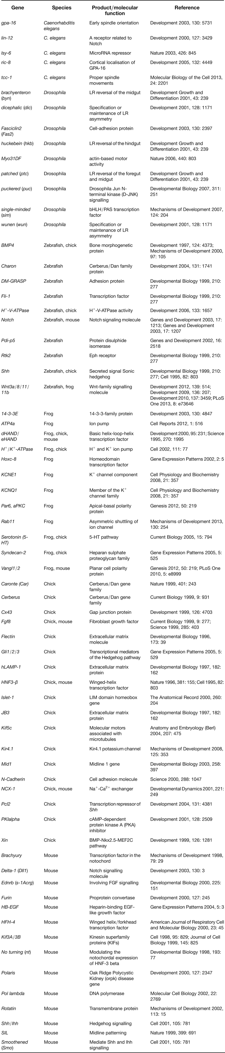
LR asymmetry defects were also associated with a range of genes in animal models, such as genes related with components of serotonin, gap junctions, motor proteins, cytoskeletal components, proton pumps, potassium channels, planar cell polarity (PCP), etc (Refs Reference Vandenberg and Levin12, Reference Speder13, Reference Levin88, Reference Blum89, Reference Pohl90, Reference Okumura91). Homologues of these genes may be potential candidate genes of SI in humans (Table 2).
The other genes involved in LR asymmetry in animal models
Loci and disease genes of HTX
HTX is usually accompanied by complex cardiovascular malformations, splenic abnormalities and intestinal malrotation (Ref. Reference Jacobs3). HTX phenotype occurs in 6.3–12.1% of PCD patients (Refs Reference Kennedy24, Reference Shapiro92).
Isolated HTX
Most of isolated HTX are sporadic, while familial clustering of HTX, occurring with X-linked, autosomal dominant or recessive inheritance, also has been described (Refs Reference Kosaki and Casey1, Reference Sutherland and Ware6). X-linked HTX results from mutations in the ZIC3 gene (Ref. Reference Gebbia10). Mutations of genes involved in Nodal signal transduction pathway have been identified in human HTX. Complex inheritance and autosomal dominant inheritance with reduced penetrance and variable expressivity are also recognised as genetic mechanisms (Ref. Reference Sutherland and Ware6). These genes include the cryptic protein gene (CFC1) (Ref. Reference Bamford93), the activin A receptor type IIB gene (ACVR2B) (Ref. Reference Kosaki94), the mouse Nodal homologue gene (NODAL) (Ref. Reference Mohapatra95), the growth/differentiation factor 1 gene (GDF1) (Ref. Reference Kaasinen96), the drosophila mothers against decapentaplegic homologue 2 gene (SMAD2) (Ref. Reference Zaidi97), the forkhead box H1 gene (FOXH1) (Ref. Reference De Luca98), the CBP/p300-interacting transactivator with GLU/ASP-rich C-terminal domain 2 gene (CITED2) (Ref. Reference Yang99), the LR determination factor 2 gene (LEFTY2, also termed as LEFTYA) (Ref. Reference Kosaki100), the NK2 homeobox 5 gene (NKX2-5) (Ref. Reference Watanabe101), and the GATA-binding protein 4 gene (GATA4) (Ref. Reference Hirayama-Yamada102). The sestrin 1 gene (SESN1) is also involved in activating Nodal signalling (Ref. Reference Peeters103). Mutations in the coiled-coil domain-containing protein 11 gene (CCDC11) (Ref. Reference Perles19) or the shroom family member 3 gene (SHROOM3) (Ref. Reference Tariq104) result in the autosomal recessive form of HTX.
HTX type 1 (HTX1, Xq26.3) and the ZIC3 gene
The ZIC3 gene activates an upstream Nodal enhancer to mediate Nodal expression at the node and is involved in node morphogenesis, LR patterning and cardiac development (Refs Reference Sutherland105, Reference Ware, Harutyunyan and Belmont106). Zic3 deleted mice present with complex cardiac defects, disturbances of laterality, neural tube defects and vertebral/rib anomalies (Ref. Reference Purandare107). In 1997, Gebbia et al. identified one frameshift, two missense and two nonsense mutations of the ZIC3 gene in familial and sporadic HTX1 patients. The frameshift mutation is also associated with SI among some heterozygous females (Ref. Reference Gebbia10). The ZIC3 p.R350 G mutation was identified in a male with SI from a family with X-linked HTX (Ref. Reference D'Alessandro, Casey and Siu108). In 2004, Ware et al. (Ref. Reference Ware109) detected five novel heterozygous ZIC3 mutations in three classic HTX kindreds and two sporadic patients with CHD. ZIC3 mutations account for about 1–5.2% of sporadic CHD and HTX (Refs Reference Ware109, Reference Cowan, Tariq and Ware110). Megarbane et al. (Ref. Reference Megarbane111) discovered a novel ZIC3 heterozygous truncation mutation in two cousins with transposition of the great arteries (TGA) and cardiac defects, and considered TGA as a LR body defect confined to the heart. Wessels et al. (Ref. Reference Wessels18) detected a six-nucleotide (GCCGCC) insertion of the ZIC3 gene in a male newborn with X-linked HTX and VACTERL association.
HTX type 2 (HTX2, 2q21.1) and the CFC1 gene
The CFC1 gene, a family member of EGF–CFC (epidermal growth factor-Cripto, Frl-1 and Cryptic), encodes an extracellular, glycosylated protein, which is part of ActRIIB/Alk4/EGF–CFC receptor complexes and functions as coreceptor for Nodal ligands (Ref. Reference Schier112). Cryptic −/− mice display right isomerism, absent LR asymmetric expression of the Nodal, Lefty2, and Pitx2 genes in the LPM (Ref. Reference Yan113). In 2000, Bamford et al. (Ref. Reference Bamford93) identified several loss-of-function CFC1 heterozygous mutations in patients with HTX. CFC1 mutations occur in about 2% (2/86) of patients with TGA or double-outlet right ventricle (DORV) (Ref. Reference Goldmuntz114).
HTX type 3 (HTX3, 6q21) and the SESN1 gene
The SESN1 gene, also known as the p53-activated gene 26 (PA26), mediates Nodal signalling and contributes to LR asymmetry determination (Ref. Reference Peeters103). Kato et al. (Ref. Reference Kato, Yamada and Niikawa115) reported a Japanese girl with HTX associated with a balanced translocation (6; 18) (q21; q21.3). The PA26 gene was disrupted by the 6q21 breakpoint and the PA26 protein expression decreased in an Epstein-Barr virus-transformed cell line (Ref. Reference Peeters116).
HTX type 4 (HTX4, 3p22.2) and the ACVR2B gene
The ACVR2B gene, also known as the ACTRIIB gene, is involved in axial patterning and LR asymmetry determination. Acvr2b −/− mice display postnatal lethality, complicated cardiac defects, right isomerism, and splenic abnormalities (Ref. Reference Oh and Li117). Kosaki et al. (Ref. Reference Kosaki94) identified two ACVR2B heterozygous missense mutations, p.R40H and p.V494I, in three patients with LR axis malformations. Ma et al. (Ref. Reference Ma118) detected a heterozygous ACVR2B p.R40H mutation in two patients and a heterozygous ZIC3 p.S402P mutation in two siblings by screening 47 HTX patients.
HTX type 5 (HTX5, 10q22.1) and the NODAL gene
The NODAL gene, a member of the transforming growth factor-beta (TGF-β) superfamily, is expressed in the mouse node and then in the LPM. Nodal signalling induces expression of the Nodal-responsive genes (Nodal, Lefty2, and Pitx2) in the LPM, and expression of the Lefty1 gene at the midline (Refs Reference Saijoh119, Reference Yamamoto120). Mice homozygous for a hypomorphic Nodal allele display severe LR defects, including right isomerism (Ref. Reference Saijoh119). Gebbia et al. (Ref. Reference Gebbia10) reported a NODAL heterozygous p.R183Q mutation in a HTX female and her unaffected mother. Mohapatra et al. (Ref. Reference Mohapatra95) identified four heterozygous missense variants, one in-frame insertion/deletion and two splice site variants of the NODAL gene in 5.2% (14/269) of patients with HTX and isolated cardiovascular malformations.
HTX type 6 (HTX6, 18q21.1) and the CCDC11 gene
The CCDC11 gene encodes a protein that involved in ciliogenesis or ciliary function. Perles et al. identified a homozygous CCDC11 splice-site mutation, c.1213 + 1G > A, in two siblings with HTX phenotype and complex heart malformation or asymptomatic SI. The mutation led to an abnormally small protein (Ref. Reference Perles19).
The chromosome 1 open reading frame 88 gene (C1ORF88, 1p13.2)
The C1ORF88 gene, the human homologue of the mouse pitchfork (Pifo) gene, regulates primary cilia disassembly and LR asymmetry. Pifo lacZ/+ mice embryos display multiple cilia defects and LR asymmetry defects. Kinzel et al. (Ref. Reference Kinzel121) discovered the same heterozygous C1ORF88 p.R80 K mutation in two cases: one neonatal lethal case with SI, cystic kidneys and liver fibrosis, and another patient with isolated DORV.
The nephrocystin 4 gene (NPHP4, 1p36.31)
The NPHP4 gene involves in embryonic LR patterning by regulating Nodal signalling. Depletion of Nphp4 in zebrafish caused heart looping defects and embryonic gut laterality defects. French et al. (Ref. Reference French122) reported two novel homozygous missense mutations, p.R1044H and p.V1236M, of the NPHP4 gene in three patients with similar laterality heart defects from a consanguineous Iranian family, and 9 heterozygous NPHP4 missense variants in 146 unrelated patients with HTX.
The UV radiation resistance-associated gene (UVRAG, 11q13.5)
The UVRAG gene is involved in organ rotation by regulating Notch endocytosis. Drosophila Uvrag mutants exhibit seriously impaired organ rotation phenotypes (Ref. Reference Lee123). The UVRAG gene was disrupted by inv (11) (q13.5; q25) in a HTX patient with strikingly increased Notch1 protein (Refs Reference Lee123, Reference Iida124).
The GDF1 gene (19p13.11)
The GDF1 gene, a member of the TGF-β superfamily, functions as a coligand of Nodal and regulates Nodal signalling (Ref. Reference Tanaka125). Gdf1 −/− mice display SI, right isomerism and cardiac defects (Ref. Reference Rankin126). Kaasinen et al. (Ref. Reference Kaasinen96) detected two truncating mutations, p.C227X and c.909insC, of the GDF1 gene in five siblings with right atrial isomerism.
The SMAD2 gene (18q21.1)
The SMAD2 gene acts as an intracellular phosphorylation target in the TGF-β signalling pathway (Ref. Reference Nakao127). Zaidi et al. (Ref. Reference Zaidi97) identified two heterozygous variants, IVS6 + 1G > A and p.W244C, of the SMAD2 gene in two patients with dextrocardia, unbalanced complete atrioventricular canal and pulmonary stenosis.
The FOXH1 gene (8q24.3)
The FOXH1 gene functions as a forkhead DNA binding transcription factor to mediate nodal signalling. Foxh1 −/− mutant mouse embryos fail to form the outflow tract and right ventricle (Ref. Reference von Both128). De Luca et al. (Ref. Reference De Luca98) detected a heterozygous FOXH1 variant, p.P21S, and a heterozygous ZIC3 mutation, p.G17C, in a familial TGA patient.
The CITED2 gene (6q24.1)
The CITED2 gene contributes to initiation or maintenance of Nodal expression in the LPM. Cited2 −/− mice die during gestation with abnormal heart looping and right isomerism (Ref. Reference Weninger129). Yang et al. (Ref. Reference Yang99) reported three heterozygous CITED2 mutations, p.G184S, p.S192 G and c.573_578del, in four patients with CHD (one patient presented with mirror image dextrocardia).
The LEFTY2 gene (1q42.12)
Both the LEFTY2 gene and the LR determination factor 1 gene (LEFTY1, also known as LEFTYB), members of TGF-β family, are implicated in LR determination (Ref. Reference Kosaki100). Lefty1 −/− mice display multiple LR positional defects, including pulmonary left isomerism. The Lefty2 gene encodes a signal for ‘leftness’, and the Lefty1 gene confines the expression of Lefty2 and Nodal to the left side (Ref. Reference Meno130). Kosaki et al. (Ref. Reference Kosaki100) identified two heterozygous mutations, p.R314X and p.S342 K, of the LEFTY2 gene in patients with LR axis defects, whose phenotypes resemble that of Lefty1 −/− mice with LR malformations.
The NKX2-5 gene (5q35.1)
The NKX2-5 gene contributes to normal heart morphogenesis. Nkx2-5 −/− mice present with early embryonic lethality with cardiac development arrested at the linear heart tube stage (Ref. Reference Lyons131). Watanabe et al. (Ref. Reference Watanabe101) reported two NKX2-5 frameshift mutations that generated two novel phenotypes: HTX and sinus venosus type ASD.
The GATA4 gene (8p23.1)
The GATA4 gene is involved in heart morphogenesis (Refs Reference Arceci132, Reference Brown133, Reference Crispino134). GATA4 protein may initiate early Nkx2-5 expression (Ref. Reference Brown133). GATA4−/− mice display embryonic lethality, double-outlet right ventricle and defects in the semilunar valves and outflow tracts (Ref. Reference Crispino134). Hirayama-Yamada et al. (Ref. Reference Hirayama-Yamada102) identified a GATA4 c.1075delG mutation in a dextrocardia patient.
The cysteine-rich protein with EGF-like domains 1 gene (CRELD1, 3p25.3)
The CRELD1 gene encodes a cell adhesion molecule, which is highly expressed in the developing heart (Ref. Reference Rupp135). Robinson et al. (Ref. Reference Robinson136) identified a heterozygous CRELD1 mutation, p.R107H, in an individual with partial atrioventricular septal defects (AVSD) and HTX.
Fakhro et al. identified 45 HTX rare genic copy number variations in 262 HTX patients. Morpholino knockdown in Xenopus of HTX candidates indicated that the never in mitosis gene A-related kinase 2 gene (NEK2, 1q32.3), the Rho-associated coiled-coil-containing protein kinase 2 gene (ROCK2, 2p25.1), the transforming growth factor-beta receptor type II gene (TGFBR2, 3p24.1), the UDP-N-acetyl-alpha-D-galactosamine: polypeptide N-acetylgalactosaminyltransferase 11 gene (GALNT11, 7q36.1) and the 188-KD nucleoporin gene (NUP188, 9q34.11) are involved in abnormal LR patterning and Pitx2 expression (Ref. Reference Fakhro137). Boskovski et al. (Ref. Reference Boskovski138) discovered Galnt11 modified Notch to coordinate cilia type and laterality.
Syndromic HTX
CRPT2 and the multiple epidermal growth factor-like domain 8 gene (MEGF8, 19q13.2)
CRPT2 is an autosomal recessive disorder, characterised by multisuture craniosynostosis and polysyndactyly of the hands and feet, accompanied by defective LR patterning and other clinical features, including obesity, umbilical hernia, cryptorchidism and CHD (Ref. Reference Twigg17). The Megf8 protein involves in LR patterning by the regulation of Nodal signalling. Megf8 Morpholino knockdown in zebrafish generates HTX (Ref. Reference Zhang139). Twigg et al. (Ref. Reference Twigg17) identified two homozygous missense mutations, p.R1499H and p.G199R, and compound heterozygous mutations, p.S2367 G and p.R448X, of the MEGF8 gene in three CRPT2 patients.
Renal–hepatic–pancreatic dysplasia type 2 (RHPD2) and the never in mitosis gene A-related kinase-8 gene (NEK8, 17q11.2)
The NEK8 gene encodes the NIMA-related serine/threonine protein kinase-8 (Ref. Reference Frank140), which acts downstream of Inv function during zebrafish pronephros morphogenesis and LR establishment (Ref. Reference Fukui141). Nek8 −/− mice present with randomisation of LR asymmetry, cardiac defects and glomerular kidney cysts, which are similar to Pkd2 −/− phenotypes (Ref. Reference Manning142). Frank et al. (Ref. Reference Frank140) detected a homozygous NEK8 mutation, p.R599X, in three foetuses from a consanguineous pedigree with RHPD2 (one patient exhibits HTX phenotype).
The erythrocyte membrane protein band 4.1-like 5 gene (EPB41L5, 2q14.2)
The EPB41L5 gene is the human homologue of the mouse Lulu/Epb41l5 gene, which contributes to proper morphogenesis of the node and midline and is involved in LR asymmetry in the early mouse embryo (Ref. Reference Lee, Migeotte and Anderson143). Kevelam et al. (Ref. Reference Kevelam144) reported a 1.3 Mb submicroscopic heterozygous deletion encompassing the EPB41L5 gene in a patient with holoprosencephaly and HTX phenotype.
The mediator complex subunit 13-like gene (MED13L, 12q24.21)
The MED13L gene, also known as PROSIT240, is involved in early heart and brain development. Muncke et al. (Ref. Reference Muncke145) identified three missense mutations, p.E251G, p.R1872H and p.D2023G, of the MED13L gene in a patient with TGA and mental retardation.
The SHROOM3 gene (4q21.1)
The SHROOM3 gene encodes an actin binding protein that acts downstream of Pitx2 in the LR development. Tariq et al. (Ref. Reference Tariq104) identified a recessive homozygous SHROOM3 mutation, p.G60 V, in a female patient with dextrocardia, TGA, pulmonic stenosis, abdominal SI, bilateral keratoconus and sensorineural hearing loss.
Chromosome 6p
Wessels et al. (Ref. Reference Wessels146) reported suggestive linkage to chromosome 6p24.3-21.2 in nine patients from a three-generation family with autosomal dominant inheritance of noncompaction cardiomyopathy, congenital heart malformations and left isomerism. Vitale et al. (Ref. Reference Vitale147) also described suggestive linkage to chromosome 6p21 in seven patients from a large five-generation family with autosomal dominant inheritance of LR asymmetry disorders.
HTX phenotype has been reported in Pitx2 −/− (Ref. Reference Lin148), Cerl2 −/− (Ref. Reference Marques149), B9d1 −/− (Ref. Reference Dowdle150), and Zic2 −/− mutant mice (Ref. Reference Barratt, Glanville-Jones and Arkell151) and homologues of these genes may be potential candidate of HTX in humans.
Conclusion
Human LR asymmetry disorders consist of SI and HTX, both of which can fall into isolated type and syndromic type. A series of genes associated with LR asymmetry disorders have been identified. Although these genes only explain part of LR patterning defects, the findings extremely help us to better understand the genetic and embryonic mechanisms of LR patterning, as well as the development of human LR asymmetry disorders. With the advent of next-generation sequencing (NGS) technology, such as genome-wide association study (GWAS), comparative genomic hybridisation (CGH) chip analysis, exome sequencing and whole-genome sequencing, more genetic mutations causing human LR asymmetry disorders will be revealed. The discovery of the pathways involved in the development of human LR asymmetry disorders will help to explain the clinical complexity and may contribute to the target therapy of these disorders.
Acknowledgements
This work was supported by the National Natural Science Foundation of China (81271921, 81101339); the Sheng Hua Scholars Program of Central South University, China (H.D.); the Research Fund for the Doctoral Program of Higher Education of China (20110162110026); and the Natural Science Foundation of Hunan Province, China (10JJ5029).
Conflict of interest
None.