Introduction
Otosclerosis is a benign, localised and hereditary disorder of bone metabolism of the otic capsule and enchondral bone, localised to the temporal bone. The condition is characterised by resorption of stable otic capsule bone in adults, followed by a reparative phase with bone deposition.Reference Chole and McKenna1,Reference McKenna, Merchant and Nadol2
Congenital ossicular fixation and defect is defined as a malformation affecting the ossicular chain that is present at birth and leads to a dysfunction of the ossicular mechanism because of immobility or discontinuity of the ossicular chain. BergstrømReference Bergstrøm3 reported that out of 687 children with congenital hearing loss, only 1.2 per cent of children were found to have isolated middle-ear anomalies. Conductive hearing loss secondary to isolated congenital middle-ear malformations is rare; the reported occurrence varies from 0.5 to 1.2 per cent.Reference Briggs and Luxford4,Reference Stewart and Downs5
Treatment options for otosclerosis include oral medications, use of hearing aids and surgical bypass of fixed stapes footplate through operative procedures, including stapedotomy or stapedectomy. Medical management is not beneficial for hearing amplification in cases of fixed footplate of the stapes.
Surgical technique for stapedectomy was first described by JJ Shea Jr in 1956 for surgical repair of otosclerosis using a vein interposition graft and was considered the pioneer of the modern surgical treatment of otosclerosis.Reference Shea6,Reference Shea7 Stapedotomy and stapedectomy, both successful procedures for hearing improvement in the treatment of otosclerosis, but there are fewer complications in stapedotomy making this procedure preferable to stapedectomy.Reference House, Hansen, Al Dakhail and House8,Reference Cheng, Agrawal and Parnes9
During the last four decades, the technique has evolved from stapedectomy to stapedotomy with or without tissue interposition. Previous reports of surgical management of otosclerosis have shown that stapedotomy gives better high-frequency gain and reduces the risk of sensorineural hearing loss when compared with stapedectomyReference Aarnisalo, Vasama, Hopsu and Ramsay10,Reference Cremers, Beusen and Huygen11 for long-term hearing improvement.
Using Medline (PubMed), a comprehensive review of the literature was performed using the key words: treatment of otosclerosis, stapedotomy, conductive hearing loss treatment and skeeter drill. Studies with small sample sizes demonstrated good results, while only one study was available with a large sample size of 2525 patients. It was a prospective study published in 2007, and it concluded that stapedotomy with vein graft interposition for otosclerotic stapes fixation is safe and successful.Reference Vincent, Sperling, Oates and Jindal12
Our study provides the second-largest case seriesReference Vincent, Sperling, Oates and Jindal12 of pre-operative and post-operative results of stapedotomy procedures performed by a single surgeon in the world at the same institution over a period of 16 years using a self-retaining ear canal retractor for exposure and skeeter drill for fenestration in the stapes footplate.
Materials and methods
This study was approved by the institutional ethics committee of Care Hospitals, Banjara Hills, Hyderabad, India. This was a retrospective study conducted on 944 patients who underwent stapedotomy surgery from 2003 to 2018. The records of all patients were reviewed to yield data on 1007 operated ears that met the inclusion criteria of: age ranging from 10 to 80 years, diagnosis of otosclerosis, congenital fixation of the stapes footplate, tympanosclerosis and osteogenesis imperfecta.
All patients had at least one year of follow up after the surgery. An assessment was performed on the basis of documented pre-operative and post-operative pure tone audiometry results. Patients with prior middle-ear exploration, prior stapes surgery and previous ear trauma were excluded from the study.
Surgical procedure
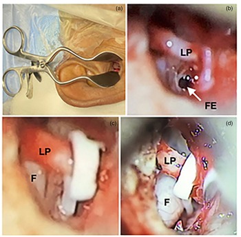
All procedures were performed through a previously well described surgical technique with a trans-canal approach using Dr A Mahadevaiah's self-retaining ear canal retractor (Figure 1) under either local (adults) or general (children) anaesthesia. After premedication and adequate local infiltration using 2 per cent lignocaine with 1:200 000 adrenaline in the external auditory canal, a posterosuperior long U-shaped tympanomeatal flap was elevated. In cases of posterosuperior meatal bony overhang, a 1 mm diameter skeeter diamond drill was used to expose the long process of incus, stapes superstructure and stapes footplate posteriorly up to the attachment of the stapedius tendon to the pyramid and superiorly identifying the horizontal segment of the facial nerve.
Photographs showing (a) Mahadevaiah's self-retaining ear canal retractor placed in the right ear, (b) right ear microscopic view demonstrating a 0.6 mm fenestra (arrow) made using skeeter drill in the stapes footplate, (c) right ear microscopic view demonstrating a Teflon piston anchored to the long process of incus and inserted into the fenestra that was made and (d) right ear microscopic view demonstrating dehiscent overhanging horizontal segment of the facial nerve in contact with shaft of Teflon piston. LP = long process of incus; FE = fenestra in the posterior half of the stapes footplate; F = horizontal segment of the dehiscent facial nerve.

The stapedius tendon was divided using the Schuknecht Sickle Knife (Surgiments, Hosur, India) or McClure Iris Scissors (Surgiments, Hosur, India). The posterior crus of the stapes was drilled using a 0.6 mm diameter diamond skeeter drill, and the same drill was used to cut the anterior crus if accessible. When the anterior crus was not visible, the incudostapedial joint was disconnected using a right-angled pick and the anterior crus was fractured. The distance between the footplate and the long process of incus was measured, and a 0.6 mm fenestra was made using the same size skeeter diamond drill, in the posterior half of the footplate along the axis of the piston going to be used.
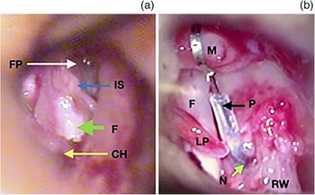
Teflon piston, of measured length and diameter (usually 0.4 × 4.5 mm) was anchored to the long process of incus and simultaneously inserted into the fenestra. In 7 cases where the long process of the incus was necrosed or the incus was absent, the malleus was utilised to anchor the 7-mm Teflon-platinum wire piston to the neck of malleus in 5 cases, and by making a groove on the malleus handle in 2 cases to anchor the piston (Figure 2). The fenestra was sealed with a small piece of soft tissue harvested from the postauricular groove. The position of the piston was confirmed by moving the shaft of the piston with a right-angled pick to make sure that the shaft was inside the fenestra. The tympanomeatal flap was replaced, and post-operative hearing assessment was performed by whispering test with occlusion of the opposite ear and by tuning fork tests. The ear canal was packed with gelatin sponge (gelfoam) smeared with neomycin ointment. The pack was removed within a week post-operatively under a microscope.
(a) Image from a 17-year-old female patient with congenital conductive hearing loss with anomalous course of the facial nerve running inferior to the stapes footplate in the left ear and (b) ossicular anomaly in the right ear with absent stapes superstructure and a dehiscent horizontal segment of the facial nerve occupying the anatomical site of the oval window. FP = footplate of the stapes, left ear; IS = incudostapedial joint; F = horizontal segment of the facial nerve (image (a) shows left ear with facial nerve inferior to the footplate, and image (b) shows right ear with completely dehiscent facial nerve); CH = chorda tympani nerve; M = neck of the malleus; P = Teflon-wire malleus to vestibule piston anchored to the neck of the malleus and inserted in to the neo-oval window created by using 0.6 mm diamond skeeter drill; LP = long process of incus and absent lenticular process; N = neo-oval window created using 0.6 mm diamond skeeter drill to see the relation and short distance to the round window; RW = round window niche

Audiometric assessments
The audiometric evaluation was reported according to the American Academy of Otolaryngology–Head and Neck Surgery guidelines and included pre-operative and post-operative air–bone gap (ABG) with pure-tone averages. Hearing thresholds were calculated over 0.5, 1, 2 and 4 kHz. Air conduction and bone conduction thresholds obtained at 1, 5 and 10 years post-operatively were used for the calculation of ABG closure. Success of the procedure was defined as a post-operative ABG of 10 dB or less at 1 year follow up.
Statistical analysis
Continuous variables were represented as mean and standard deviation where data followed normal distribution and were otherwise presented as median with range. Categorical variables were represented as frequencies and percentages. The statistical significance between the pre- and post-operative cases was assessed by the paired t-test, and analysis of variance testing was used for between group analysis. Data were analysed using RStudio® statistical software.
Results
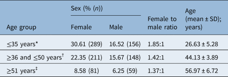
A total of 944 (881 unilateral and 63 bilateral) patients were included in the study, including 581 female and 363 male patients. The mean age was 37.79 years with a range of 10–80 years. The mean age for women was 37.63 years and was 38.02 years for men. Female to male ratio among all the patients was 1.60:1 (Table 1).
Distribution of the study population by age and sex

*n = 445; †n = 359; ‡n = 140. SD = standard deviation
Hearing outcome after one year of follow up
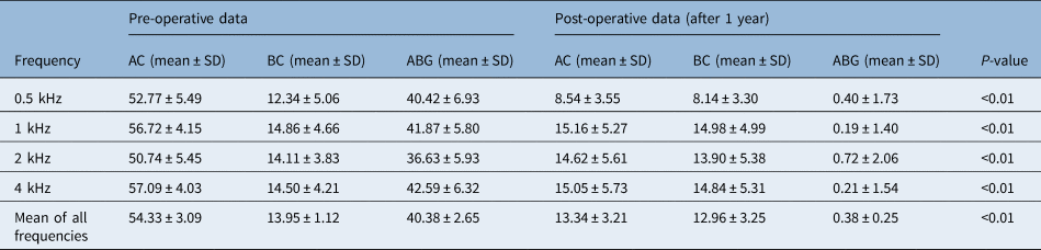
The complete data after one year of follow up was available for 1007 surgical procedures (Table 2). Out of 1007 operated ears in the study period with one year follow up, 993 (98.61 per cent) cases demonstrated negligible ABG of less than or equal to 5 dB with a mean value of 0.26 dB, whereas 12 (1.19 per cent) cases demonstrated minimal ABG of more than 5 dB but less than 10 dB with a mean value of 6.77 dB. Only 2 (0.2 per cent) cases showed ABG of more than 10 dB with a mean value of 21.25 dB. Comparison of pre- and post-surgery hearing thresholds was assessed by calculating pure-tone air conduction and bone conduction thresholds. The clinical data of 1007 stapedotomy cases demonstrated an average gain of 41.0 dB in air conduction with a mean air conduction threshold of 13.34 dB in post-operative cases (Table 3).

Pre-operative and post-operative mean air conduction, bone conduction and air–bone gap values at various hearing frequencies after one year's follow up

AC = air conduction; BC = bone conduction; ABG = air–bone gap; SD = standard deviation
Hearing outcome after five years’ follow up
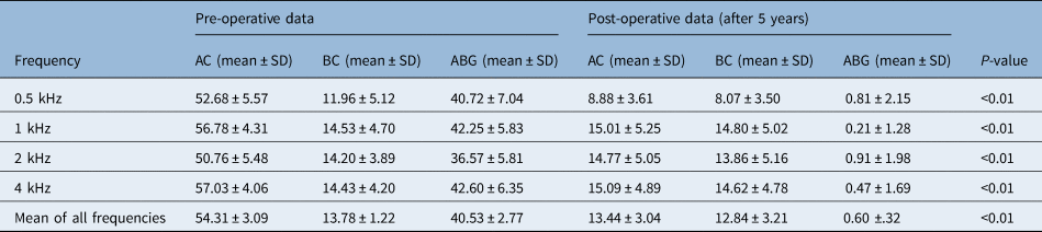
A total of 666 stapedotomy procedures were performed between 2003 and 2013 and therefore had at least 5 years of follow up post-surgery. Of these patients, telephonic assessment and follow-up audiometry data were available for only 536 patients. The average hearing threshold gain in the above-mentioned group was 40.88 dB with an average post-operative air conduction of 13.44 dB (Table 4).
Pre-operative and post-operative mean air conduction, bone conduction and air–bone gap values at various hearing frequencies after 5 years’ follow up

AC = air conduction; BC = bone conduction; ABG = air–bone gap; SD = standard deviation
Hearing outcome after 10 years’ follow up
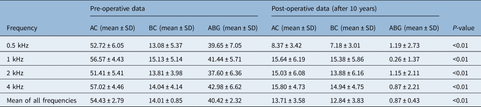
A total of 308 stapedotomy procedures were performed between 2003 and 2008 and therefore had at least 10 years of follow up post-surgery. Of these, telephonic assessment and follow-up audiometry data were available for only 156 patients. The average hearing threshold gain in this group was 40.72 dB with an average post-operative air conduction of 13.71 dB (Table 5).
Pre-operative and post-operative mean air conduction, bone conduction and air–bone gap values at various hearing frequencies after 10 years’ follow up

AC = air conduction; BC = bone conduction; ABG = air–bone gap; SD = standard deviation
Discussion
Otosclerosis is a common cause of progressive hearing impairment and is characterised by the formation of an abnormal growth of bone on the footplate of the stapes. Conductive hearing loss is commonly seen in otosclerosis, and otosclerosis might also result in sensorineural hearing loss if cochlear otosclerosis is present.Reference Uppal, Bajaj, Rustom and Coatesworth13 Surgical treatment of otosclerosis is considered the most effective option.Reference Kisilevsky, Dutt, Bailie and Halik14
In all children with congenital stapes fixation, it is imperative to consider congenital abnormalities including malformation of the labyrinth or anomalous facial nerve course. Therefore, all suspected patients should undergo imaging of the temporal bones to assess ossicular anomalies and to screen for increased risk of facial nerve injury, perilymph gush and sensorineural hearing loss. In cases of perilymph gush, abnormal communication between the perilymph space and cerebrospinal fluid has been proposed to occur by mechanisms that include a patent cochlear aqueduct or defect in the internal auditory canal or modiolus.Reference Massey, Hillman and Shelton15
A literature review by Dornhoffer et al. on stapes gusher in congenital fixation of stapes suggests that it is an abnormality in the internal auditory canal that leads to perilymph gush in the above-mentioned cases. They found that pre-existing sensorineural hearing loss suggests underlying otic capsule abnormalities and places patients at increased risk of perilymph gush and sensorineural hearing loss when the footplate is manipulated.Reference Dornhoffer, Helms and Hoehmann16
In our series of patients, perilymph gush was encountered in six cases, out of which five cases of flooding stopped spontaneously after waiting for a few minutes. But in a 23-year-old female patient with congenital fixation of stapes, the gush was severe and could not be stopped even after waiting for half an hour. The perilymph gush was controlled by using the soft tissue taken from the postauricular groove. It was held over the fenestra that was made and was pushed into the vestibule by the shaft of the Teflon piston anchored to the long process of the incus. There was no identifiable cause reported in the pre-operative temporal bone high-resolution computed tomography scan. Post-operative hearing showed improvement in hearing from a pre-operative 60 dB mixed hearing loss to 40 dB.
Osteogenesis imperfecta is a generalised disorder of the connective tissue, resulting in bone fragility. The classic triad of osteogenesis imperfecta includes spontaneous fractures, blue sclera and hearing lossReference Sillence, Senn and Danks17 known as Van der Hoeve syndrome.Reference Mukherjee, Sadhu, Ghakraborty and Saha18 Among all patients with osteogenesis imperfecta, 45 to 80 per cent experience hearing loss,Reference Pedersen19–Reference Kuurila, Kaitila, Johansson and Grénman21 which may be conductive, sensorineural or mixed. Treatment options for hearing improvement in these patients include the use of hearing aids or stapes surgery for conductive hearing loss and cochlear implants or bone-anchored hearing aids for sensorineural hearing loss.Reference Pillion, Vernick and Shapiro22 Our series included two female patients with osteogenesis imperfecta, one with bilateral conductive hearing loss and another with bilateral mixed hearing loss, and both had successful surgery.
There are many different methods used to perform stapes surgery. In recent years, stapedotomy has become popular and is increasingly replacing stapedectomy as the preferred procedure of choice.Reference Marchese, Paludetti, De Corso and Cianfrone23 Air-bone gap can be closed or reduced with stapes bypass surgery, with improvement in overall hearing thresholds and in some cases reducing the need for amplification devices and relieving tinnitus associated with otosclerosis.Reference Sparano, Leonetti, Marzo and Kim24,Reference Ayache, Earally and Elbaz25
Kylén et al.Reference Kylén, Stjernvall and Arlinger26 investigated the factors enhancing the intensity of drill-generated noise and found that the size of the burr was the most important factor. As the size becomes smaller, the noise intensity decreases, and the diamond burrs cause less noise.
Michaelides and Kartush,Reference Michaelides and Kartush27 who measured the intensity of sounds in the temporal bone caused by various instruments used in surgery, recorded a less than 100 dB sound intensity when using a microdrill, potassium titanyl phosphate laser and carbon dioxide laser, whereas sound intensity was recorded to be between 118 dB and 132 dB with an air-powered drill and Erbium laser.
The microdrill has low noise intensity, low torque, good accuracy and needs only a very small duration of contact, particularly with the thin grey footplates to make the fenestra, without causing acoustic trauma.
Stapedotomy provides excellent results for correcting conductive hearing loss in patients with otosclerosis. As demonstrated in our study, appreciable improvement in air conduction thresholds can be observed with a mean gain in air conduction of 41.0 dB, mean post-operative air conduction threshold of 13.34 dB and mean post-operative ABG of 0.38 dB in one year follow-up cases. These results are consistent in long-term follow up with a mean gain in air conduction of 40.88 and 40.72 in 5-year and 10-year follow up, respectively.
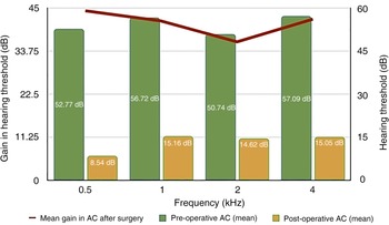
Hearing improvement patterns in our study demonstrated that improvement in air conduction was dependent on the pre-operative ABG, such that the greater the pre-operative ABG, the larger the improvement in air conduction thresholds after surgery (Figure 3). These findings are generally consistent with previously published reports. Stapedotomy is highly successful in terms of hearing improvements for the majority of treated patients. However, one of the known potential risks following stapes surgery is sensorineural hearing loss, especially at higher frequencies, although the incidence is negligible in our experience. In stapedotomy, there is low manipulation and minimal trauma to the inner ear, resulting in a lower incidence of sensorineural hearing loss compared with stapedectomy. It is anticipated that less than 1 per cent of cases undergoing stapedotomy will develop sensorineural hearing loss post-operatively.Reference Alzhrani, Mokhatrish, Al-Momani, Alshehri, Hagr and Garadat28 In our study, the rate of development of high tone sensorineural hearing loss was only 0.49 per cent (5 cases) at 1 year follow up, which is in accordance with previous studies.
Pattern of hearing outcome in post-operative cases one year after surgery. AC = air conduction

In this study, out of 1007 operated ears after one year's follow up, 993 cases (98.61 per cent) demonstrated negligible ABG, 12 cases (1.19 per cent) showed an ABG of more than 5 dB but less than 10 dB and only 2 cases (0.2 per cent) showed a post-operative ABG of more than 20 dB. The size of the pre-operative ABG is likely to contribute to the lack of improvement in the air conduction for these patients. Additionally, consistent with these results, some studies reported less benefit from stapes surgery at higher frequencies.Reference Bauchet, Martin, Rubinstein and Hirsch29,Reference Strömbäck, Köbler and Rask-Andersen30 This has been attributed to the effect of surgical trauma, perilymph leakage and the noise generated by surgical drilling on the sensory receptor cells.
Theoretically, the use of connective tissue seal may prevent perilymph leakage and restore the stiffness of the annular ligament. In this study, soft tissue from postauricular sulcus was used, whereas Shah et al.Reference Shah and Ganapathy31 used absorbable gelatin sponge or fat as a sealing material.
In the study by Shah et al.,Reference Shah and Ganapathy31 bone conduction showed improvement in the 0.6 mm diameter piston group and worsening in 0.4 mm diameter piston group, although in our study only a 0.4 mm diameter piston was used. The mean bone conduction improved significantly. Before surgery, the mean bone conduction was at 13.95 dB, whereas 1 year after surgery the mean bone conduction level was at 12.96 dB.
A study by De Souza and GlasscockReference De Souza and Glasscock32 concluded that there was less chance of a perilymph fistula in stapedotomy compared with stapedectomy. This is reflected in our study, where there are no cases of perilymph fistula after stapedotomy surgery.
Prevention of accidents in stapedotomy surgery is very crucial for successful outcome. Using Dr Mahadevaiah's self-retaining ear canal retractor for all the 1007 cases left both hands free to work together with excellent field of exposure.
In cases of posterosuperior bony overhang, there is a need to drill the posterosuperior meatal wall for exposure. Here the tympanomeatal flap that is raised should be long enough to cover the anticipated bony defect, otherwise it may result in a perforated tympanic membrane. Secondly, the origin of the chorda tympani nerve varies and can be very inferior or superior and sometimes arises in the bony external auditory canal; this should be known to prevent damage. Drilling the bony overhang or curetting increases the risk of damage to the chorda tympani nerve or accidental dislocation of the incudostapedial joint. If the incus is dislocated or absent, the only option is to use a malleus to vestibule piston, which is a longer piston and should be available to complete the procedure. When there was good visibility and accessibility of the footplate of the stapes, a controlled fenestra was made using a 0.6 mm diamond skeeter drill before dislocation of incudostapedial joint, which helps to prevent a floating footplate, particularly in cases of thin footplates. We strongly believe that the incudostapedial joint, which gives stability and prevents a floating footplate, should never be disconnected before drilling, cutting or weakening the posterior crus.
It is safer to use a manual perforator than a drill to make the fenestra in cases of dehiscent overhanging horizontal segment of the facial nerve if there is limited visibility of the footplate. But in our series of patients, skeeter drill was used successfully to make the fenestra (Figure 1) at the junction of the posteroinferior edge of the narrow visible footplate and promontory, and the same technique was also used in cases of floating footplates.
• Skeeter drill with a diamond burr of 0.6 mm is very safe for making the fenestra in stapedotomy
• This technique shows excellent long-term hearing results with more than 98.5 per cent success rate
• The skeeter drill is an excellent tool to prevent floating footplate by preserving the incudostapedial joint and drilling the posterior crus
• Minor bleeding from the footplate can be successfully controlled using the dry skeeter diamond drill
• Skeeter drill can be safely used over laser in cases with a dehiscent overhanging horizontal segment of the facial nerve
• In cases of thick otosclerotic focus, biscuit type footplates and obliterative otosclerosis, skeeter drill is very useful
In a 17-year-old female with congenital conductive hearing loss where there was dehiscent horizontal segment of the facial nerve completely occupying the oval window with no lenticular process of incus and no stapes superstructure, a 7-mm long Teflon-wire piston was anchored to the neck of the malleus, a neo-oval window was created just below the facial nerve and the piston was inserted (Figure 2) into the vestibule with successful hearing of 20 dB maintained for over 10 years so far. Interestingly, the second ear of this patient showed a facial nerve inferior to the footplate of stapes (Figure 2) where she also had successful stapedotomy.
Competing interests
None declared